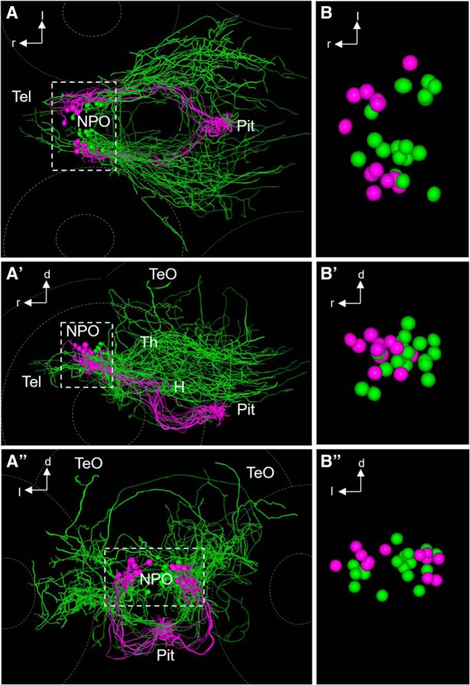
Figure 10.
Comparison of hypophysiotropic and encephalotropic cell types after registration. A–A″, Comparison of encephalotropic (green) and hypophysiotropic (magenta) innervation of Oxt-producing cells, dorsal (A), lateral (A′), and frontal (A″) views. A 3D rotation of this dataset is provided as Movie 3. B–B″, Magnified view of reconstructed soma centers after registration, dorsal (B), lateral (B′), and frontal (B″) views. Three additional somata of encephalotropic oxytocinergic cells were added, since the soma was reconstructable. Note that the two morphological subtypes appear to possess segregated somata, suggesting a functionally relevant regionalization within the cluster of Oxt-producing cells. Hypophysiotropic cells reside in a more rostral part of the NPO compared with encephalotropic cells.

