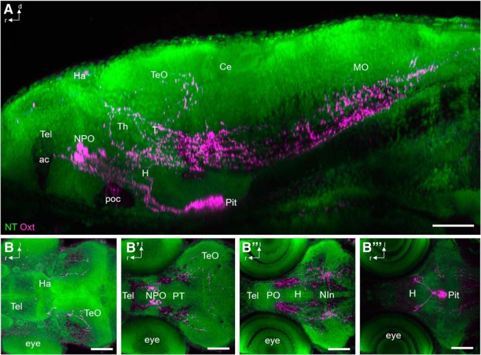
Figure 2.
Innervated regions of Oxt-positive neurons at 6 dpf. A, Lateral view of immunostained Oxt-carrying fibers (magenta) in combination with NT (green) to identify brain regions at 6 dpf (n = 12). Fibers reach the pituitary through the hypothalamohypophyseal tract, but also innervate the caudal Tel and TeO. Spinal projections pass the metencephalon and myelencephalon ventrally. B–B″′, Serial horizontal substacks (dorsal to ventral) show innervation of the TeO (B), the caudal Tel (B′), hypothalamic and tegmental regions (B″), and the pituitary (B″′). The fibers mostly traverse within Nissl-negative regions, which correspond to white matter. Scale bars: 100 µm. ac, anterior commissure; Ha, habenula; MO, medulla oblongata; NIn, interpeduncular nucleus; poc, postoptic commissure.

