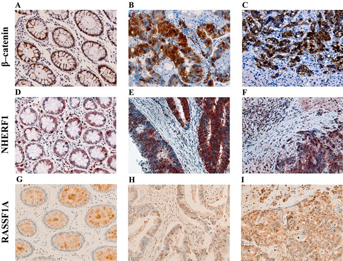
Figure 1. Representative images of the immunoreactivity of β-catenin, NHERF1 and RASSF1A.

Membrane and cytoplasmic localization of β-catenin in ANT A., membrane, cytoplasmic and nuclear expression in T B., membrane, cytoplasmic and nuclear β-catenin staining in LM C. NHERF1 immunoreactivity is present at the apical membrane, in cytoplasm and nucleus in ANT D., while in T E. and LM F. it becomes mostly cytoplasmic and nuclear. Heterogeneous cytoplasmic staining intensity of RASSF1A in ANT G., granular cytoplasmic staining in T H., cytoplasmic and nuclear immunoreactivity in LM I. (original magnification x200).
