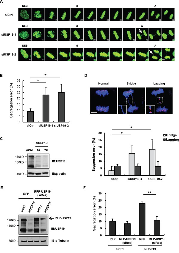
Figure 1. Knockdown USP19 induces the formation of anaphase bridge.

A. Selected frames from time-lapse movies of representative HeLa/GFP-H2B cells transfected with control siRNA (siCtrl) or USP19 siRNA (siUSP19). Arrows denote segregation error. The time on the images is in minutes. NEB, nuclear envelope breakdown; M, metaphase; A, anaphase. Scale bar, 5 μm. B. The percentage of segregation errors in control and USP19 knockdown cells during anaphase. Data are representative of three independent experiments, error bars indicate S.D. C. Immunoblot analyzes knockdown efficiency of USP19. D. HCT116 cells were transfected with control or USP19 siRNA, then synchronized in mitosis by thymidine block and release. The cells through mitosis were immunostained with anti-centromere antibodies (ACA; for kinetochores; red) and DAPI (for chromosomes; blue). Upper panels shows example images of normal segregation, anaphase bridges and lagging chromosomes in anaphase. Scale bar, 5 μm. Lower panels is quantification of mitotic cells with anaphase bridges or lagging chromosomes in control and USP19 knockdown cells. *p<0.05, **p<0.01. E, F. Complementation of RFP-USP19 in knockdown HeLa/GFP-H2B cells rescues chromosome segregation errors. The USP19 knockdown cells were transfected with USP19 siRNA-resistant expression construct or RFP vector. (E) The expression of siRNA-resistant RFP-USP19 was detected by Immunoblot. (F) The data show the percentage of mitotic cells with segregation errors during anaphase in RFP-positive cells, data are shown as mean ± S.D. *p<0.05, **p<0.01.
