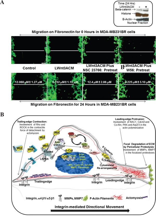
Figure 8. Effect of RAC1 inhibitors, NSC 23766 and W56 on LWnt3A-stimulated fibronectin-mediated migration of brain metastasis-specific TNBC cells.

A. EGFP-tagged brain metastasis-specific TNBC cells, MDA-MB231BR were allowed to migrate on fibronectin for 24 hours under LWnt3A stimulation in the presence or absence of RAC1 inhibitors. Zero hour control for each scratch was presented as the internal control. Activation of the canonical Wnt signaling pathway in MDA-MB231BR cells under LWnt3A stimulation (Inset). Wnt signaling is stimulated in TNBC cells with LWnt3ACM (+) at 24 hours, and is assessed based on the level of beta-catenin accumulation compared to the non-stimulated(-)-condition. Histones were used as a nuclear marker. Beta-actin expression was used as total protein loading control. B. Schematic representation of the mode of involvement of WP via activation of RAC1 in the integrin-mediating directional movement of tumor cells through actin cytoskeletal organization.
