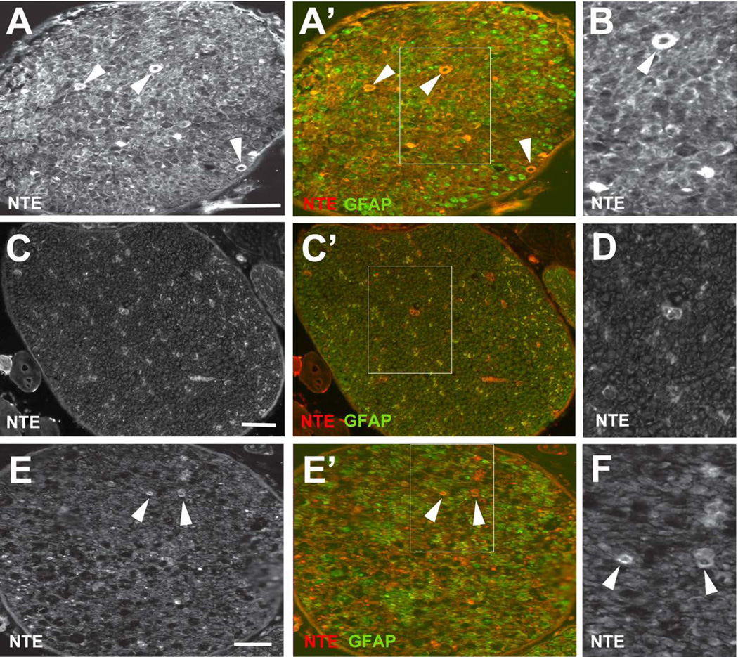
Figure 6.
NTE is upregulated after axotomy. (A, A’) 4d after nerve crush, NTE levels are increased in the nerve with strong staining detectable in some ring-shaped structures (arrowheads). (B) Magnified view of the boxed region in A’. (C, C’) NTE staining in the uninjured sciatic nerve 4d after nerve crush. (D) Magnified view of the boxed region in C’. (E, E’) 11d after nerve injury, an increase in NTE levels is still detectable, including in the ring shaped structures (arrowheads) but the NTE levels have decreased compared to 4d post-injury. (F) Magnified view of the boxed region in E’.NTE in red, GFAP in green. The nerve crush was performed at PND28. Scale bar=50 µm.

