

Abstract
Extensive portomesenteric thrombosis presents a technical challenge in liver transplantation. Establishing portal inflow in living donor liver transplantation (LDLT) is indispensable to ensure regeneration of the graft. The use of a pericholedochal varix for inflow has been described only in a few case reports. Described herein is one such instance in the setting of LDLT, highlighting the nuances of this procedure in the light of available literature.
Abbreviations: CECT, contrast-enhanced computed tomography; DDLT, deceased donor liver transplantation; LDLT, living donor liver transplantation
Keywords: living donor liver transplantation, portal vein thrombosis, pericholedochal varix
Management of extensive portomesenteric thrombosis (Yerdel Grade IV)1 in liver transplant recipients poses a challenge and requires careful operative planning. In living donor liver transplantation (LDLT), establishment of portal inflow to the partial graft is mandatory to ensure its regeneration. Hence, portocaval hemitransposition or portal arterialization is not a tenable option. Renoportal anastomosis can be done if there are significant portosystemic shunts draining into the renal vein. Jump grafts can be useful if there is a patent vein in the portal circulation, which can provide adequate inflow. If expertise is available, multivisceral transplantation circumvents the problem of portal inflow elegantly; however, this comes at the cost of increased morbidity due to small bowel transplantation. In very select cases, a large varix can be used to provide inflow, and thus it avoids more complex procedures. Presented herein is a patient who underwent LDLT with portal inflow derived from a pericholedochal varix.
Case Report
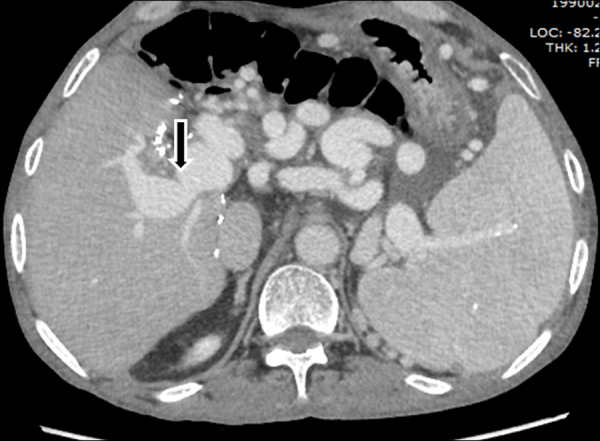
A 52-year-old male with hepatitis B virus-related chronic liver disease (Model for end-stage liver disease score 16; Child score 11), decompensated with ascites, spontaneous bacterial peritonitis, and hepatic encephalopathy, was evaluated for LDLT. He had large high-risk esophageal varices for which band ligation was done. Preoperative contrast-enhanced computed tomography (CECT) scan revealed extensive portomesenteric thrombosis. There was a large pericholedochal varix and extensive perigastric collaterals (Figure 1). There were no major shunts draining into the renal vein. At surgery, there was massive bleeding while dissecting the gastrohepatic omentum, requiring multiple transfusions. After hepatectomy, a thrombectomy was attempted; however, portal flow was not satisfactory. The pericholedochal varix was sufficiently thick walled to hold sutures and had good flow. The patient received a modified right hemiliver graft (weight 746 g; graft recipient weight ratio 1.08); anastomosis between the varix and donor portal vein was done with 6.0 polypropylene continuous sutures without any interposition graft, leaving a growth factor. Right hepatic artery of recipient was used for arterial anastomosis. A Roux en Y hepaticojejunostomy was done as it was not possible to safely dissect the bile duct. Intraoperative Doppler confirmed good portal flow (416 ml/100 g of liver). Outflow was triphasic. Arterial waveform was normal with resistive index of 0.68. The portal pressure distal to the anastomosis 1 h after reperfusion was 17 mmHg. No inflow modulation was performed. The patient made a satisfactory recovery; however, postoperative ascites took 40 days to settle. Follow-up Doppler done at 7 months revealed a patent anastomosis (Figure 2) with a portal flow of 3.7 l/min. A CECT scan done 14 months postoperatively showed a patent anastomosis, partial resolution of collaterals, and no ascites (Figure 3). Till date, at 39 months follow-up, the patient remains free of symptoms/signs of portal hypertension.
Figure 1.

Coronal contrast-enhanced computed tomogram of the abdomen in portal venous phase. Arrows depict thrombosis in the entire portal vein and extending into the superior mesenteric vein. Splenic vein is patent. Arrowhead points to the large pericholedochal varix. Also seen are large perigastric collaterals, splenomegaly, and ascites.
Figure 2.

Doppler done 7 months post liver transplant depicting patent anastomosis between the pericholedochal varix and donor portal vein. Also seen is the patent inferior vein.
Figure 3.
Axial contrast-enhanced computed tomography scan in portal venous phase done 14 months after liver transplantation. The varix–portal vein anastomosis is depicted by the arrow. Residual collaterals are still present.
Discussion
Establishment of portal inflow through a pericholedochal varix was reported as early as 1986 in deceased donor liver transplantation (DDLT).2 Since then, till date there are around 10 patients reported in DDLT, the largest series being that that of 5 patients in whom an iliac vein conduit was utilized, with 100% 3-month patency.3 In LDLT, the Asan group has reported two patients with detailed description of operative technique and good outcomes.4 They advocate dissection, isolation of the left hepatic artery, en mass clamping of the hilum to minimize bleeding, and preparation the varix by using an interposition vein graft. There are some differences in the index patient reported here. The left hepatic artery could not be used as there were extensive perigastric collaterals. En massing clamping would have been a good option; but at that time, we attempted thrombectomy and dissection of the hilum to isolate the right artery. We did not use an interposition graft as the varix reached the neo hilum easily, was more than 1 cm in diameter, and held sutures reasonably well. There has been a report from India5 on the use of an infracolic collateral for portal inflow in LDLT in the setting of portal biliopathy. An interposition graft from the donor internal juglar vein and explanted liver middle hepatic vein was used to achieve adequate length with good results.
The Asan group also recommends selecting patients with low volume ascites and low risk varices, indicating effective decompression of the portal system. However, the index patient had significant portal hypertension as evidenced by banded esophageal varices, large perigastric collaterals, and ascites, which took some time to resolve after surgery. Fortunately, the patient did not bleed postoperatively and remains well on follow-up. We have done this procedure only in one patient out of 308 LDLTs over the last 5 years.
To summarize, use of a pericholedochal varix can be a good option in grade IV portomesenteric thrombosis in LDLT. With proper planning and availability of expertise, these patients can be offered the life-saving option of transplantation.
Authors’ Contribution
Study conception: Viniyendra Pamecha; data collection: Shridhar Vasantrao Sasturkar and Piyush Kumar Sinha; manuscript drafting: Kishore GS Bharathy; critical revision of the manuscript for important intellectual content: Viniyendra Pamecha, Senthil Kumar, and Kishore G.S. Bharathy.
Conflicts of Interest
The authors have none to declare.
References
- 1.Yerdel M.A., Gunson B., Mirza D. Portal vein thrombosis in adults undergoing liver transplantation: risk factors, screening, management, and outcome. Transplantation. 2000;69:1873–1881. doi: 10.1097/00007890-200005150-00023. [DOI] [PubMed] [Google Scholar]
- 2.Busuttil R.W., Hiatt J.R. Restoration of portal flow with a pericholedochal varix in adult living donor liver transplantation for patients with total portosplenomesenteric thrombosis. Liver Transpl. 2014;20:1530–1531. doi: 10.1002/lt.24040. [DOI] [PubMed] [Google Scholar]
- 3.Alexopoulos S.P., Thomas E., Berry E., Whang G., Matsuoka L. The portal vein-variceal anastomosis: an important technique for establishing portal vein inflow. Clin Transplant. 2014;28:52–57. doi: 10.1111/ctr.12278. [DOI] [PubMed] [Google Scholar]
- 4.Moon D.B., Lee S.G., Ahn C.S. Restoration of portal flow using a pericholedochal varix in adult living donor liver transplantation for patients with total portosplenomesenteric thrombosis. Liver Transpl. 2014;20:612–615. doi: 10.1002/lt.23850. [DOI] [PubMed] [Google Scholar]
- 5.Gupta S., Singhal A., Goyal N., Vij V., Wadhawan M. Portal biliopathy treated with living-donor liver transplant: index case. Exp Clin Transplant. 2011;9:145–149. [PubMed] [Google Scholar]