Abstract
Introduction
It is not known whether the age-related increase in fatigability of fast dynamic contractions in lower limb muscles also occurs in upper limb muscles. We compared age-related fatigability and variability of maximal-effort repeated dynamic contractions in the knee extensor and elbow flexor muscles; and determined associations between fatigability, variability of velocity between contractions and functional performance.
Methods
35 young (16 males; 21.0±2.6 years) and 32 old (18 males; 71.3±6.2 years) adults performed a dynamic fatiguing task involving 90 maximal-effort, fast, concentric, isotonic contractions (1 contraction/3 s) with a load equivalent to 20% maximal voluntary isometric contraction (MVIC) torque with the elbow flexor and knee extensor muscles on separate days. Old adults also performed tests of balance and walking endurance.
Results
Old adults had greater fatigue-related reductions in peak velocity compared with young adults for both the elbow flexor and knee extensor muscles (P<0.05) with no sex differences (P>0.05). Old adults had greater variability of peak velocity during the knee extensor, but not during the elbow flexor fatiguing task. The age difference in fatigability was greater for the knee extensor muscles (35.9%) compared with elbow flexor muscles (9.7%, P<0.05). Less fatigability of the knee extensor muscles was associated with greater walking endurance (r=−0.34, P=0.048) and balance (r=−0.41, P=0.014) among old adults.
Conclusions
An age-related increase in fatigability of a dynamic fatiguing task was greater for the knee extensor compared with the elbow flexor muscles in males and females, and greater fatigability was associated with lesser walking endurance and balance.
Keywords: aging, sex, females, knee extensors, elbow flexors, concentric contractions
1. Introduction
Advanced age is associated with reductions in muscle strength and power, and these age-related reductions are typically greater in knee extensor muscles compared to other muscles such as the finger flexor or elbow flexor muscles (Frontera and others 2000; Hunter and others 2000; Nogueira and others 2013; Raj and others 2010). Although young adults are stronger than old adults for both upper and lower limb muscles, age-related reductions in maximal muscle power are greater than age-related reductions in maximal isometric strength (Raj and others 2010; Thom and others 2007; Thompson and others 2013; Valour and others 2003). Reduced physical activity that often accompanies aging can accelerate the age-related decline in strength and power (Doherty 2003; Hunter and others 2016). For example, reductions in muscle strength and power with advanced age are larger in old adults who are sedentary than those who engage in regular physical activity [e.g. (Buchman and others 2007; Hunter and others 2000)]. Disuse is thought, in part, to be responsible for the greater age-related reduction in strength and power of lower limb muscles compared with the upper limbs because of a greater decline in lower limb use among old adults (Degens and Korhonen 2012; Hunter and others 2000; Janssen and others 2000; Venturelli and others 2014).
Age-related declines in strength and power are associated with poor physical function (Chandler and others 1998; Justice and others 2014; Macaluso and others 2003), and predictive of morbidity and mortality (Metter and others 2004). The ability to perform activities of daily living repeatedly or for an extended period of time, such as walking, stair climbing and even carrying loads can be limited by fatigability of the limb muscles (Justice and others 2014), further exacerbating age-related reductions in strength and power required for activities of daily living. Fatigability is the reduction in motor performance that occurs with the onset of muscle contraction, and in the laboratory setting is assessed as the exercise-induced reduction in maximal muscle torque, power or velocity (Enoka and Duchateau 2016; Gandevia 2001). Studies that examine age-differences in fatigability of human muscles have historically focused on isometric contractions (Christie and others 2011; Kent-Braun 2009) and more recently dynamic tasks that involve measuring the reduction in power or velocity primarily of lower limb muscle groups [e.g., (Callahan and Kent-Braun 2011; Dalton and others 2012)]. Old adults (~60–75 years) are usually less fatigable than young adults (18–40 years) during isometric contractions (Christie and others 2011; Kent-Braun 2009), although this appears to reverse in very old adults (>75 years) at least for the ankle dorsiflexor muscles (Justice and others 2014) and slow-to-moderate velocity contractions (72–87 years) (Baudry and others 2007). The age differences in fatigability appear to be minimal for slow-to-moderate velocity shortening contractions with the knee extensor muscles (Callahan and others 2009; Lindstrom and others 1997) and elbow flexor muscles (Yoon and others 2015; Yoon and others 2013). However, in previous studies involving repeated fast velocity contractions performed with maximal effort (isokinetic and isotonic) in the knee extensor and ankle plantar and dorsiflexor muscles, the reductions in power were greater for old adults compared with young adults (Callahan and Kent-Braun 2011; Dalton and others 2010b; Dalton and others 2012; McNeil and Rice 2007; Petrella and others 2005) with minimal data for the upper limb.
Impaired motor performance with advanced age also includes increased variability in force or velocity during a motor task (e.g., Enoka and others 2003) and between trials for a given motor task (e.g., Christou 2011). This large age-related variability [e.g. (Marmon and others 2011; Martinikorena and others 2016)] can lead to less predictable and less accurate performance (Almuklass and others 2016), further compromising motor function in old adults. Fatigue induced by exercise may further exacerbate the age-related increases in variability of a motor task. One study showed that during a dynamic fatiguing task with the knee extensor muscles, old females (65–85 years) with impaired mobility exhibited greater torque variability compared with healthy old (not mobility impaired) and healthy young females (Kent-Braun and others 2014). Less is known about the age-related variability of contraction velocity of upper extremity muscles during a fatiguing task which may potentially impair functional performance.
The primary purposes of this study were to 1) compare the reduction in peak velocity and maximal voluntary isometric contraction (MVIC) torque during a fatiguing task involving high-velocity concentric contractions with a load equivalent to 20% MVIC for both the elbow flexor and knee extensor muscles in young and old adults, and 2) compare the variability in peak velocity during the dynamic fatiguing task in the elbow flexor and knee extensor muscles in young and old adults. We hypothesized that old adults would be more fatigable and more variable in peak velocity between contractions than young adults for a high-velocity fatiguing task with the elbow flexor and knee extensor muscle groups. Because age-related reductions of maximal force and power are greater for the lower limb than the upper limb [e.g. (Raj and others 2010)], we hypothesized that the age difference in fatigability and variability in velocity during the fatiguing task would be greater for the knee extensors muscles than the elbow flexor muscles. Given the sex-based differences in fatigability during isometric and slow dynamic contractions (Hunter 2016a; Hunter 2016b) we also determined whether aging influenced fatigability of males and females differently for the fast dynamic contractions in the knee extensor and elbow flexor muscles.
Finally, there is limited understanding of the importance of fatigability with dynamic contractions and the variability within a fatiguing task on functional performance tasks among old adults (Enoka and Duchateau 2016; Hunter and others 2016), and also limited knowledge on the ecological validity of our laboratory measures of fatigability (Justice and others 2014) and variability within a fatiguing task. Thus, an additional purpose was to determine the association between fatigability in old adults, the variability in peak velocity between contractions during the dynamic fatiguing task and measures of motor function important to daily living (balance and walking endurance). We hypothesized that greater fatigability and variability of contractions of the knee extensor muscles would be associated with lower physical function and physical activity. Determining the relationship between laboratory measures of fatigability and measures of functional endurance (e.g. walking test) can aid the NIH Toolbox goal to provide a standard set of measures (Reuben and others 2013).
2. Materials & Methods
Thirty-five young (18–31 years, 21.0 ± 2.6 years; 16 males and 19 females) and 32 old (60–85 years, 71.3 ± 6.3 years; 18 males and 14 females) adults participated in the study. A subset of these data that include the young males and females has been previously reported (Senefeld and others 2013). Participants were healthy, community dwelling and ambulatory males and females with no known neurological disease or contraindications to exercise. Participants were screened and excluded for depression (geriatric depression scale score > 5) (Snowdon 1990), and for use of medication affecting the central nervous system and hormonal status (e.g. hormone-replacement therapies and thyroid-stimulating hormones). Participants were also excluded if they demonstrated increased risk of falling (Berg Balance Score < 41) (Berg and others 1992). All participants provided written informed consent and the protocol was approved by the Marquette University Institutional Review Board in accordance with the Declaration of Helsinki.
Each participant attended an introductory session followed by two experimental sessions that involved a dynamic fatiguing task with either the right knee extensor or elbow flexor muscles. The order of experimental sessions was counterbalanced and separated by 2–7 days.
During the introductory session, participants were familiarized with the testing equipment and procedures; and performed maximal voluntary isometric contractions (MVICs) and maximal voluntary concentric contractions (MVCCs) with both knee extensor and elbow flexor muscles. The MVCCs were performed with a load equivalent to 20% MVIC and participants were asked to exert maximal effort to achieve a peak angular velocity with that load. Participants also completed a questionnaire to estimate physical activity (Kriska and others 1990). The physical activity questionnaire involved recall of occupational and leisure physical activity over the previous 12 months and each activity was weighted to estimate of the metabolic cost of the activity (METs). Participants were provided with a list of 37 activities (with space for additional activities) and asked to provide frequency, quantity and intensity of activities over the previous 12-month period. METs were also able to be estimated for occupational physical activity based on occupational history and questions. Thus, the weekly metabolic equivalents (MET hour week−1) was calculated from the occupational and leisure physical activity.
During the introductory session, old adults only, performed a standard assessment of balance (Berg and others 1992) and a six-minute walk test to assess functional endurance using the lower limb muscles (Enright 2003). The six-minute walk was performed on an indoor course, and participants were encouraged to walk as quickly as possible for six minutes. The six-minute walk test is highly correlated with the NIH toolbox measure of functional endurance, the two-minute walk test, (r = 0.97, Bohannon and others 2014); and performance on the six-minute walk test is predictive of morbidity and mortality [e.g. (Boxer and others 2010; Newman and others 2003)].
2.1 Experimental Set up
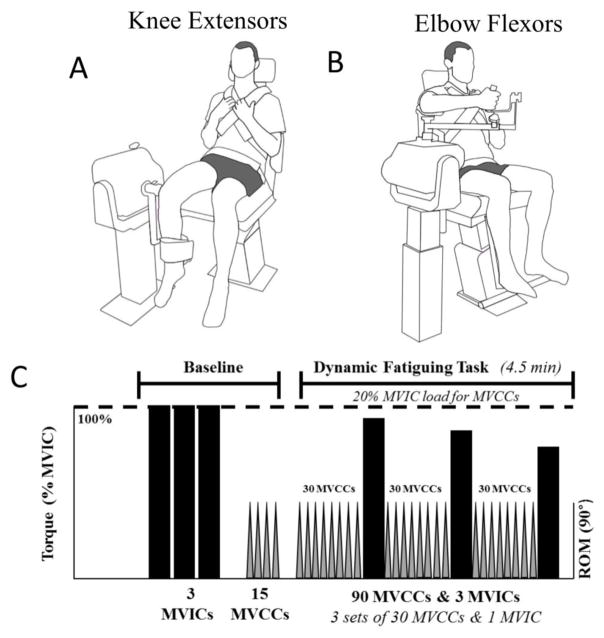
During the experimental sessions (sessions 2 and 3), participants performed a 4.5-minute dynamic fatiguing task with either the right elbow flexor muscles or knee extensor muscles. MVIC and MVCCs were performed before, during and after each dynamic fatiguing task (see Experimental Protocol and Figure 1 for greater detail). All contractions were performed while the participant was seated in a chair connected to a dynamometer [Biodex System 4-Pro dynamometer (Biodex Medical, Shirley, NY)]. Each participant was seated with 90° of hip flexion and secured with padded straps across the shoulders and the waist to minimize ancillary movements during leg and arm contractions. The right limb (either leg or arm) was positioned such that the axis of rotation of the joint was aligned with the axis of rotation of the dynamometer at the joint position used for MVICs (Figures 1A & 1B). The experimental protocol was similar for the knee extensor and elbow flexor muscle group sessions. However, MVICs were performed at different joint angles for the knee extensor and elbow flexor muscle groups and according to the optimal joint angle of the length-tension relationship (Singh and Karpovich 1966; Smidt 1973). During the knee extensor session, participants performed MVICs at 75° of knee flexion (Smidt 1973), with 0° of knee flexion considered to be full knee extension. MVCCs with the knee extensor muscles were performed through 90° range of motion, between 90° of flexion to 0° of knee flexion. During the elbow flexor session, the right arm of the participant was positioned midway between abduction and adduction of the shoulder, such that the arm and forearm were parallel with the ground, and the humerus was supported by an arm rest. The forearm was near full supination while the participant grasped a handle (Figure 1B). Each participant performed MVICs at 90° of elbow flexion (Singh and Karpovich 1966), and 0° of elbow flexion was considered to be full elbow extension. Concentric elbow flexion was performed through a 90° range of motion, from 55° of flexion to 145° of elbow flexion. The experimenters verbally encouraged participants to contract through the entire range of motion and the 90° range of motion was maintained by a large majority of participants for the knee extensor session and all participants for the elbow flexor session. There were a small number of participants which demonstrated a reduction in range of motion (<15°); however, there were similar numbers of young (n = 3) and old adults (n= 3) who demonstrated the reduction in range of motion.
Figure 1.
A–B: Experimental setup and participant positioning for the testing of the knee extensor muscles (A) and elbow flexor muscles (B). C: Experimental protocol for both sessions with the knee extensor and elbow flexor muscles. From left to right the participants performed the following: 1) Baseline: maximal voluntary isometric contraction (MVIC) shown as the black bars and maximal voluntary concentric contraction (MVCC) shown as grey bars; and 2) Dynamic Fatiguing Task: 3 subsequent sets of 30 MVCCs and 1 MVIC for a total of 90 MVCCs and 3 MVICs.
Mechanical recordings of torque, velocity and position from the dynamometer were digitized using a Power 1401 analog-to-digital converter and Spike 2 software [Cambridge Electronics Design (CED), Cambridge, UK] with a sampling rate of 1,000 Hz.
2.2 Experimental Protocol
Each experimental session with either the elbow flexor muscles or knee extensor muscles involved: (a) measurement of MVIC torque of the agonist muscles; and (b) a dynamic fatiguing task performed with a load equivalent to 20% of MVIC. See Figure 1C. Participants were strongly, verbally encouraged to achieve maximal force during all MVICs and peak velocity during all MVCCs throughout the protocol. During each dynamic contraction cycle (3 s), participants contracted their muscles (knee extensors or elbow flexors) as quickly as possible across 90° range of motion. Upon completion of this contraction, the participant relaxed the muscles and the limb was passively moved to the initial position by the dynamometer. Following is greater detail of each procedure.
MVIC: At the beginning of the experimental session, participants performed at least three MVICs of the agonist muscle group (knee extensor muscles or elbow flexor muscles) with 60 s rest between each trial. Each MVIC involved an increase to maximal torque over 1–2 s followed by a 3 s hold. If two of the three MVICs were not within 5% then additional MVICs were performed until this was achieved. Ninety percent of the participants achieved the criteria for the MVIC within three attempts and no participant needed more than 4 attempts.
MVCC and Dynamic Fatiguing Task: Prior to the dynamic fatiguing task, each participant performed a set of 15 MVCCs with a load equivalent to 20% of MVIC (baseline MVCCs). After a 2 min rest period, the dynamic fatiguing task was performed with 20% MVIC load. This load allowed most participants to achieve the full range of motion throughout the fatiguing task which is not always possible with higher loads in some older subjects, as we observed during pilot testing and has been reported previously in plantar flexor muscles (Dalton and others 2010b). The 20% MVIC load in the isotonic contraction mode represents near maximal power in vivo, for young and old adults that occurs in a range of ~20–35% MVIC (Dalton and others 2015; Valour and others 2003).
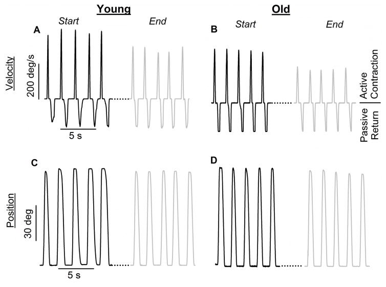
The dynamic fatiguing task involved three sets of 30 MVCCs with one MVCC every 3 s. Each set of 30 contractions was immediately followed by one MVIC for a 3-s duration. For performance of each 3-s MVIC during the dynamic fatiguing task, the dynamometer was changed from isotonic to isometric testing mode and then back to isotonic testing mode (~6-s duration in total) prior to starting the next 30 MVCCs. See Figures 2A–D for representative data traces of MVICs and MVCCs performed during the dynamic fatiguing task.
Figure 2. Representative velocity (A, B) and position (C, D) data from a young and old male subject for the knee extensor muscles.
A–B: Velocity traces of maximal voluntary concentric contractions (MVCCs) at the start (black line) and end (grey line) of the dynamic fatiguing task. C–D. Position traces of MVCCs at the start (black line) and end (grey line) of the dynamic fatiguing task.
2.3 Data Analysis
The torque during each MVIC was quantified as the average value over a 0.5-s interval over the peak torque of the contraction. The peak velocity during dynamic contractions was quantified as the peak velocity of the concentric phase of each individual contraction, and this peak value was then averaged over five consecutive contractions. Fatigability of dynamic contractions was quantified as the percentage reduction in the mean peak velocity of five consecutive MVCCs and relative to the mean peak velocity of the five fastest consecutive MVCCs performed prior to the dynamic fatiguing task (baseline). Fatigability of the MVIC was quantified as the percentage reduction in the MVIC from baseline to those during the fatiguing task. The variability of the peak velocity (MVCCs) during the concentric phase of each set of five consecutive contractions was calculated as the coefficient of variation (CV) using the following formula: [(standard deviation of peak velocity)/(mean peak velocity) × 100].
2.4 Statistical Analysis
Values are reported as means ± standard deviation (SD) in the text and tables and displayed as means ± standard error (SE) in the figures. A mixed-design repeated-measure analysis of variance (ANOVA) across time was used to compare the dependent variables including measures of fatigability (relative reduction in maximal torque and peak velocity) between the two experimental sessions (elbow flexor vs knee extensor). Separate repeated-measures ANOVAs across time (for the fatiguing task) with age-group (young vs old) and sex (male vs female) as between-subject factors were used to compare the dependent variables of MVIC torque (% of baseline), MVCC peak velocity (% of baseline), and CV of peak velocity. One-way ANOVA was used to compare baseline measures between all groups (i.e. young and old males and females). For each ANOVA the sphericity of data was verified and technical corrections were performed if necessary. If needed post hoc analysis with Bonferroni corrections were used when F test was significant. Pearson correlation coefficients (r) were used to determine associations. Linearity of correlations was verified with visual inspection. Shapiro-Wilk’s tests and visual inspection of histograms, normal Q-Q plots and box plots demonstrated one outlier in the six-minute walk test (P < 0.001) and one outlier in self-reported physical activity (P < 0.001), both of which were subsequently removed from the analysis. Significance was determined at P < 0.05 and all the analysis were performed in International Business Machines Corporation (IBM) statistical package for social sciences (SPSS) version 22.
3. Results
3.1 Knee Extensor Muscles
MVIC Torque
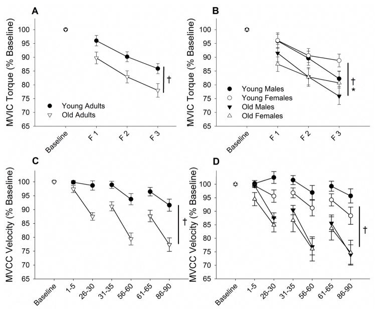
At baseline, MVIC torque of the knee extensor muscles, was lower in old adults than young adults (age effect, P < 0.001; 16% age difference) and males were stronger than females (sex effect, P < 0.001; 24% sex difference; age × sex, P = 0.33; Table 1; Figure 2). Mean MVIC torque declined during the dynamic fatiguing task for both age groups (time effect, P < 0.001). When compared with baseline MVIC, old adults had greater reductions in MVIC torque than young by the end of the dynamic fatiguing task (21.1 ± 9.2% vs 13.7 ± 11.0%, respectively; age effect, P = 0.04; Figure 3A). Further, males had greater decrements in MVIC torque than females (time × sex, P = 0.002) and similarly across the two age groups (time x age, P = 0.33; time × age × sex, P = 0.76; Figure 3B).
Table 1.
Baseline characteristics, maximal force and peak velocity for the knee extensors (KE) and elbow flexors (EF) for young and old males and females. Data are mean (SD) for values of torque and peak velocity before and after the dynamic fatiguing tasks. Sex difference between young males and females are denoted (*, P < 0.05). Age difference, with sexes combined, are denoted (†, P < 0.05). Sex differences between old males and females are denoted (#, P < 0.05). BMI, body mass index; METS, metabolic equivalent; hr, hour; EF, elbow flexor; KE, knee extensor; MVCC, maximal voluntary concentric contraction; MVIC, maximal voluntary isometric contraction; Nm, newton meters; deg·s−1, degrees·second−1. Note that for the Berg balance and six-minute walk tests, two outliers (old females) were removed from correlation analyses.
| Young
|
Old
|
||||
|---|---|---|---|---|---|
| Variable | Units | Females n = 19 |
Males n = 16 |
Females n = 14 |
Males n = 18 |
| Age † | Years | 20.6 (1.5) | 21.6 (3.6) | 72.0 (7.3) | 71.3 (5.1) |
| Height *† | m | 1.68 (0.08) | 1.83 (0.06) | 1.62 (0.05) | 1.76 (0.06) |
| Weight *† | kg | 61.8 (6.7) | 72.2 (7.9) | 66.7 (13.4) | 81.7 (12.7) |
| BMI † | kg·m−2 | 22.0 (2.6) | 21.5 (2.1) | 25.4 (4.2) | 26.4 (3.8) |
| Physical Activity † | MET·hr·week−1 | 85.5 (48.5) | 90.2 (45.4) | 32.7 (21.8) | 51.8 (45.0) |
| Berg Balance (max=56) | AU | -- | -- | 53.5 (4.5) | 55.1 (1.2) |
| Six-Minute Walk | m | -- | -- | 519 (106) | 547 (56) |
|
| |||||
| Before the fatiguing Exercise | |||||
|
| |||||
| KE MVIC Torque *†# | Nm | 161.3 (36.1) | 199.1 (51.2) | 107.9 (33.4) | 166.8 (49.6) |
| KE MVCC Velocity †# | deg·s−1 | 404 (36) | 391 (49) | 277 (60) | 344 (46) |
| EF MVIC Torque *† | Nm | 57.5 (8.3) | 86.0 (13.6) | 47.4 (22.1) | 70.1 (20.0) |
| EF MVCC Velocity *† | deg·s−1 | 306 (40) | 375 (44) | 294 (72) | 348 (43) |
|
| |||||
| After the fatiguing Exercise | |||||
|
| |||||
| KE MVIC Torque †# | Nm | 141.8 (29.8) | 162.7 (42.7) | 89.4 (32.3) | 128.1 (34.8) |
| KE MVCC Velocity † | deg·s−1 | 357 (65) | 374 (55) | 213 (74) | 259 (55) |
| EF MVIC Torque *# | Nm | 40.7 (7.6) | 60.3 (10.4) | 35.5 (21.3) | 53.8 (20.1) |
| EF MVCC Velocity * | deg·s−1 | 266 (47) | 315 (50) | 246 (90) | 267 (56) |
Figure 3. Fatigability of the knee extensor muscles during a dynamic fatiguing task of young and old males and females.
Shown are means (±SEM) of data normalized to the baseline values for young adults (filled circles) and old adults (open triangles) for torque of maximal voluntarily isometric contraction (MVIC) torque (A & B) and peak velocity from maximal voluntary concentric contractions (MVCC) (C & D). In panels A and B, MVICs are shown at baseline and the end of the three sets of 30 dynamic contractions (F1, F2 and F3 respectively shown on the x axis). In panels C & D, MVCC data is the average of sets of 5 contractions at the start and end of each set of 30 contractions in each set, with the contraction numbers shown on the x axis. A and C show the comparison of young and old (males and females in each group combined). Old adults had greater reductions in MVIC torque and MVCC velocity than young adults (†, P < 0.05). B and D show the comparison of young and old males and females separately. Males (young and old) had greater reductions in MVIC torque compared to females (*, P < 0.05).
Dynamic Contractions
At baseline, young adults had higher peak velocities during the MVCC with the knee extensor muscles than the old adults (age effect, P < 0.001; Table 1). Males also had higher peak velocities relative to females (sex effect, P = 0.021) but an interaction (sex × age, P < 0.01) revealed this sex difference was only significant within the old adults (P = 0.001) and not the young adults (P = 0.37), because the older males had higher peak knee extensor velocities than old females (Table 1).
All groups had decrements in peak velocity (MVCC) performed during the dynamic fatiguing task (time effect, P < 0.001), however, old adults had greater peak velocity reductions during the MVCC than young adults for both males and females (time × age, P < 0.001; time × sex, P = 0.13; Figures 3C & 3D).
Variability of Peak Velocity
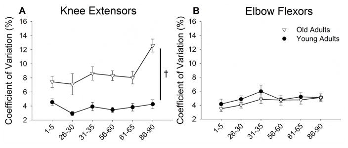
During the dynamic fatiguing task, the variability (CV) of the peak velocity was greater for the old adults than the young (7.8 ± 0.4% vs 3.8 ± 0.3%, respectively; age effect, P < 0.001; post-hoc pairwise comparison, P < 0.001, Figure 4A). The CV of peak velocity increased during the dynamic fatiguing task (time effect, P < 0.001) but only for the old adults (6.7 ± 0.7% to 12.4 ± 0.8%) and did not change for young adults (4.5 ± 0.6% to 4.2 ± 0.7%, time × age, P < 0.001). Furthermore, there were no differences between the sexes in CV of peak velocity for the knee extensor muscles and no interaction between age groups (sex effect, P = 0.71; age × sex, P = 0.22).
Figure 4. Variability in the peak velocity of the knee extensor and elbow flexor muscles during a dynamic fatiguing task by young and old adults.
The coefficient of variation of peak velocity for each set of five consecutive maximal voluntary concentric contractions (MVCC) during a dynamic fatiguing task with the contraction numbers shown on the x axis. Old adults a larger coefficient of variation of peak velocity during MVCCs with the knee extensor muscles during the fatiguing task (†, P < 0.05), with no differences for the elbow flexor muscles.
3.2 Elbow Flexor Muscles
MVIC Torque
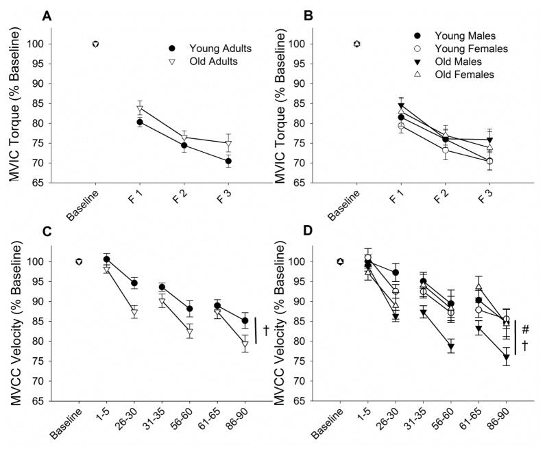
At baseline, MVIC torque of the elbow flexor muscles, was lower in the old adults than young adults (12% age-related difference; age effect, P = 0.002; Table 1). Further, males (young and old) were stronger than females by 31% (sex effect, P < 0.001). All groups (young and old males and females) demonstrated declines in MVIC torque during the dynamic fatiguing task (time effect, P < 0.001; Figure 5B), and each age group had similar reductions in torque from baseline (29.5% for young and 24.6% for old; time × age, P = 0.13; Figure 5A). There was no sex difference in the rate of decline of MVIC (time × sex, P = 0.87; time × age × sex, P = 0.61).
Figure 5. Fatigability of the elbow flexor muscles during a dynamic fatiguing task of young and old males and females.
Shown are means (±SEM) of data normalized to the baseline values for young adults (filled circles) and old adults (open triangles) for torque of maximal voluntarily isometric contraction (MVIC) torque (A & B) and peak velocity from maximal voluntary concentric contractions (MVCC) (C & D). MVCC data is the average of sets of 5 contractions. A and C show the comparison of young and old (males and females in each group combined). Old adults had greater reductions in MVCC velocity than young adults (†, P < 0.05). B and D show the comparison of young and old males and females separately. Old males had greater reductions in peak velocity compared with old females (#, P < 0.05).
Dynamic Contractions
At baseline, there were no differences between the age groups in peak velocity of the elbow flexor muscles during the MVCCs (age effect, P = 0.11; Table 1). Males however, had higher peak velocities during elbow flexion than females (sex effect, P < 0.001) for both the young adults (18.3% sex difference) and old adults (14.6% sex difference; age × sex, P = 0.53; Table 1).
There was a reduction in peak elbow flexion velocity (MVCCs) during the fatiguing contraction from baseline to the end of the fatiguing task (time effect, P < 0.001). The old adults, however, had greater reductions in peak velocity during the MVCC than the young adults (20.5% vs 14.9% respectively; time × age, P = 0.016; Figure 5C). Post-hoc analysis revealed that old males had larger reductions in peak velocity than old females (23.9% vs 14.5%, respectively; time × sex × age, P = 0.004; pairwise comparison, P = 0.009; Figure 5D).
Variability of Peak Velocity
During the dynamic fatiguing task, the CV of the peak velocity of the elbow flexor muscles was not different between young and old adults (4.9 ± 0.3% vs 4.5 ± 0.4%, respectively; age effect, P = 0.44; Figure 4B). Furthermore, the CV of peak velocity did not change during the dynamic fatiguing task (time effect, P = 0.12) for the young or old adults (time × age, P = 0.85).
3.3 Muscle Group Comparison
The reductions of maximal torque (MVIC) and peak velocity (MVCC) during the dynamic fatiguing task differed for the knee extensor and elbow flexor muscle groups. The elbow flexor muscles demonstrated greater reductions compared with the knee extensor muscles in MVIC torque (27.4 ± 11% vs 16.2 ± 10%, respectively; time × session, P < 0.001) and peak velocity (17.3 ± 12% vs 14.1 ± 14%, respectively; time × session, P = 0.001). Additionally, the difference between old and young adults in the reduction of MVIC torque was greater for the knee extensors than the elbow flexors (session × age, P = 0.014; session × sex, P = 0.151). Similarly, the age-related difference in the reduction of MVCC velocity was greater for the knee extensors than the elbow flexors (session × sex, P = 0.004; session × sex, P = 0.233).
The CV of peak velocity (variability) during the dynamic fatiguing task was greater for the knee extensors than the elbow flexors (5.8 ± 0.2% vs 4.7± 0.3%, respectively; P = 0.004), and the age-related difference was greater for the knee extensor muscles compared with the elbow flexor muscles (session × age, P < 0.001) with no difference across the sessions for the males and females (session × sex, P = 0.275).
3.4 Associations Between fatigability and Physical Function
Reported physical activity levels were higher among young adults relative to old adults (age effect, P < 0.001) with no sex-related differences (sex effect, P = 0.229; sex × age, P = 0.535; Table 1). Multiple significant Pearson correlations were observed between baseline function and fatigability of the knee extensor and elbow flexor muscles with measures of motor function and physical activity in old adults (Table 2). The baseline strength (MVIC) and peak velocity (MVCC) of the knee extensors and baseline peak velocity of the elbow flexors were positively associated with physical activity scores, balance test performance and walk test performance. Those individuals who had higher peak velocity of the knee extensor muscles (MVCC) had greater levels of physical activity, had higher balance scores, and walked further in the six-minute walk test. Furthermore, individuals with a higher peak velocity (MVCC) of the elbow flexor muscles were more physically active, had better balance and walked further in the six-minute walk test. See Table 2.
Table 2.
Correlations between reported physical activity, Berg Balance scores, and six-minute walk test scores with variables of muscle function (left column) for the knee extensor muscles (KE) and elbow flexor muscles (EF). Variables muscle function are baseline maximal voluntary concentric contraction (MVCC), baseline maximal voluntary isometric contraction (Baseline MVIC), the fatigue-related reduction (↓) in MVIC torque, the fatigue-related reduction (↓) in MVCC, and the variability (coefficient of variation, CV) of MVCC in the last 5 contractions (contractions 86 to 90). Old adults (n = 30) only performed the balance and six-minute walk test, and both old and young adults completed the physical activity questionnaire (n = 67). Significant Pearson correlations (r) are highlighted with an asterisk (P < 0.05).
| Physical Activity (n = 67) | Berg Balance (n = 30) | Six-Minute Walk (n = 30) | ||||
|---|---|---|---|---|---|---|
| r-value | p-value | r-value | p-value | r-value | p-value | |
| Baseline KE MVCC | 0.556* | >0.001 | 0.334 | 0.050 | 0.393* | 0.019 |
| Baseline KE MVIC | 0.448* | >0.001 | 0.326 | 0.056 | 0.425* | 0.011 |
| ↓ KE MVCC | −0.464* | >0.001 | −0.412* | 0.014 | −0.337* | 0.048 |
| ↓ KE MVIC | −0.305* | 0.010 | −0.553* | 0.001 | −0.536* | 0.001 |
| KE MVCC CV | 0.071 | 0.726 | 0.278 | 0.160 | 0.418* | 0.030 |
|
| ||||||
| Baseline EF MVCC | 0.283* | 0.019 | 0.409* | 0.018 | 0.356* | 0.042 |
| Baseline EF MVIC | 0.311* | 0.009 | 0.267 | 0.133 | 0.219 | 0.220 |
| ↓ EF MVCC | −0.190 | 0.122 | 0.017 | 0.928 | 0.041 | 0.825 |
| ↓ EF MVIC | −0.159 | 0.192 | −0.292 | 0.099 | −0.477* | 0.005 |
| EF MVCC CV | −0.036 | 0.848 | −0.239 | 0.195 | −0.142 | 0.447 |
Fatigability and Function
Functional performance scores were also associated with fatigability of the knee extensor muscles such that the percentage reduction in MVIC torque and peak velocity during the MVCCs, were negatively associated with physical activity scores, balance and the six-minute walk (Table 2). Thus, a greater reduction of MVCC in the knee extensor muscles was associated with lower levels of physical activity, balance, and walking endurance. In contrast, fatigability of the elbow flexor muscles was not associated with physical activity, balance or walking endurance, with the exception that the reduction in elbow flexor MVIC torque was negatively associated with six-minute walk test distance.
Variability and Function
The CV of peak velocity of the knee extensor muscles during the last 5 contractions of the dynamic fatiguing task was associated with performance of the six-minute walk, the reduction in MVCC of the knee extensor muscles (r = 0.88, P < 0.001), and the reduction in MVIC torque of the knee extensor muscles (r = 0.61, P = 0.001). The CV of peak velocity of the elbow flexor muscles during the MVCCs during the last 5 contractions was associated with the reduction in peak velocity of the elbow flexor muscles (r = 0.39, P = 0.03), but no other variables (Table 2).
4. Discussion
A novel finding of this study was that the age-related increase in fatigability, assessed as the reduction in peak velocity during a maximal effort, fast velocity fatiguing task, was greater for the knee extensor muscles than the elbow flexor muscles. The reduction in peak velocity however, for the knee extensor and elbow flexor muscles, did not differ between males and females within both age groups. The greater age-related fatigability of the lower limb during the dynamic task was associated with lower functional performance including balance and a standard walking test of endurance (six-minute walk test) which is tightly correlated with the NIH toolbox measure of functional endurance (Bohannon and others 2014). Furthermore, old adults demonstrated greater variability in peak velocity of the maximal voluntary concentric contractions during the dynamic fatiguing task with the knee extensor muscles but not for the elbow flexor muscles, and this variability in peak velocity of the lower limb was associated with greater fatigability and less distance walked during the six-minute walk test.
4.1 Fatigability and Aging
We demonstrated an age-related difference in fatigability for both upper and lower limb muscles during high velocity, fatiguing contractions, but this age difference was greater for the knee extensor than elbow flexor muscles. Most studies to date have focused on age-related differences in fatigability during dynamic contractions of the lower limb including the knee extensors and ankle plantar and dorsiflexor muscles [e.g. (Callahan and Kent-Braun 2011; Dalton and others 2015; Dalton and others 2012; McNeil and Rice 2007)] with no knowledge of whether age differences exist in the upper limb muscle groups. We demonstrated that fatigability of the knee extensor muscles during a high-velocity, concentric task was greater for old adults than young adults (~36% difference), with no sex differences in the reduction of peak velocity. The peak velocities during knee extension of the young males and females in this study were faster than the old males and females (see Table 1). In a previous study old females were more fatigable than young females when contractions were constrained to 270 deg·s−1 (72% vs 48% reduction in torque, respectively) (Callahan and Kent-Braun 2011). Furthermore, studies of age-related differences in fatigability of the knee extensor muscles involving different contraction speeds, demonstrated that active old males had similar fatigability compared with active young males for contractions at 60 deg·s−1 (~53% reduction in torque) (Dalton and others 2012). However, these old males were more fatigable during the middle and latter portions of a 180 deg·s−1 velocity fatiguing task, and throughout a high velocity (>350 deg·s−1) fatiguing task (old vs young: 35% vs 23% reduction) (Dalton and others 2012). Thus, for slow-to-moderate velocity fatiguing tasks with the knee extensor muscles, the age difference was minimal, and for isometric contractions old adults are usually less fatigable than young (Callahan and others 2009; Callahan and Kent-Braun 2011; Dalton and others 2010a; Griffith and others 2010). The mechanism(s) for the large age difference in fatigability at high velocities in the lower limb are not fully understood but may involve an inability to adequately activate the muscle among old adults during a fatiguing contraction (Wallace and others 2016) and contractile mechanisms (Callahan and Kent-Braun 2011; Dalton and others 2010b). Contractile mechanisms contributing to the age difference in fatigability possibly involve impaired cross-bridge cycling which is important for maintenance of contraction velocity (Allen and others 2008; Kent-Braun and others 2012) and a greater ATP cost during contraction among old adults compared with young adults (Layec and others 2014).
Importantly, we also found that the elbow flexor muscles of old adults were more fatigable than young for the high-velocity dynamic contractions. The age difference however, was considerably less for the elbow flexor muscles than for the knee extensor muscles (9.7% vs 35.9% age difference in peak velocity reduction). The smaller differences in fatigability for the upper limb muscles compared with lower limbs in our study were due to the large differences between limbs within the young adults rather than old adults. The young adults had greater fatigability in the elbow flexor muscles (15%) than for the knee extensor muscles (7%), but the old adults fatigued by similar relative magnitudes during the dynamic contractions for both the elbow flexor muscles (21%) and knee extensor muscles (23%). Thus, we showed that the knee extensor muscles demonstrated a greater age difference in fatigability than the elbow flexor muscles. The limb differences between young and old adults may be related to activity levels. Self-reported physical activity levels were less in the old adults compared with young adults, possibly more reflective of differences in the lower limb than upper limb activity (Kern and others 2001; Theou and others 2013). Regardless, age differences in fatigability of the lower limb, persist even between young and old adults who were matched for physical activity levels (Callahan and Kent-Braun 2011; Dalton and others 2012) indicating that age differences in activity levels probably do not entirely explain the age difference in fatigability. Other possible mechanisms, that have yet to be fully explored, for the age difference in fatigability between the knee extensor and elbow flexor muscles includes activation of the motor units, anatomy and contractile function, providing considerable opportunities for future studies.
For the elbow flexor muscles, old adults are less fatigable during isometric fatiguing contractions and this is a robust finding even in strength-matched young and old males (Hunter and others 2004; Hunter and others 2005; Hunter and others 2008; Yoon and others 2008; Yoon and others 2012). For a fatiguing task with slow contractions at 60 deg·s−1, the age-related fatigue resistance observed during isometric contractions was diminished for both males and females possibly due to contractile mechanisms (Yoon and others 2015; Yoon and others 2013). We showed in this current study that old adults are more fatigable than young adults for fast velocity dynamic contractions. Thus, as for lower limb muscles, the age-difference in fatigability of the elbow flexor muscles appears to be dependent on contraction velocity.
We found that the reduction in the MVIC torque for both muscle groups was greater than the relative decline in peak velocity (MVCC). However, there have been disparate findings regarding the reduction of MVIC torque and power or velocity during a dynamic contraction after a dynamic fatiguing task. Previous studies for example, have demonstrated similar reductions in MVIC torque and power for the elbow flexor muscles among young and old males (Yoon and others 2013), greater reductions in power compared to MVIC torque for the plantar flexor muscles for young and old males (Dalton and others 2010b), and greater reductions in velocity compared to MVC for the soleus and triceps brachii muscles among a cohort of young males (Cheng and Rice 2010). There are many methodological differences (e.g. contraction type (isotonic vs. isokinetic), relative intensity (applied speed or external load), sex of participants, age of participants, etc.) which makes interpretation of these collective findings difficult. However, observations from single-fiber and whole-muscle animal preparations, demonstrate that the reduction in stimulated peak force is greater than the change in maximal shortening velocity (Kent-Braun and others 2012), suggesting that contractile mechanisms are involved in the larger reductions of isometric torque. The decline in MVIC torque for the knee extensor muscles was greater for the old adults than the young, although the males from both age groups had greater reductions in MVIC torque than the females immediately upon completion of the dynamic contractions. This age and sex difference did not occur in the elbow flexor muscles. The cause for the greater reduction in MVIC torque in both young and old males compared with the females after the fatiguing task (specific to the knee extensor muscles) is not clear. It could involve sex differences in the muscle size, myosin heavy chain composition of the muscles and substrate utilization (Hunter 2016a; Hunter 2016b) but this would need to be specific to the knee extensor muscles. There was no age-related difference in the reduction of MVIC torque for the elbow flexors, but old adults had greater reductions in peak velocity compared with young adults, and these discrepant results demonstrate that the mode of testing of fatigability is very specific to the task and muscle group involved (Hunter 2016b).
4.2 Age-Related Variability of Contraction Velocity with Fatigue
A novel finding of this study was the age-related difference in variability of peak velocity during the fatiguing contraction of the knee extensor muscles (Figure 4), with no age differences in the elbow flexor muscles. The variability (CV) between peak contraction velocities during the dynamic fatiguing task was greater in the old adults than the young at the start and end of the fatiguing task with the knee extensors with a large increase by the end of the dynamic task but only in the old adults. The CV was associated with greater fatigability (both peak velocity and torque reductions, with young and old included) and associated with the six-minute walk distance among the old adults. Large age-related variability during a dynamic fatiguing task with the ankle dorsiflexor muscles had been previously observed in mobility-impaired old females compared with age-matched females (not mobility impaired) and healthy young females (Kent-Braun and others 2014). The old adults in the current study were not mobility impaired, but there was a difference between age groups for the knee extensors. The mechanisms for the age-related variability in peak velocity during the fatiguing task and between limbs are largely unknown.
Peak velocity during a dynamic contraction is reliant on the ability to optimally activate the motor units of the agonist muscles and the consequential speed of cross-bridge attachment and kinetics with adequate availability of calcium in the cell (Gandevia and others 1998; Kent-Braun and others 2012). During isometric contractions, variability in force is primarily modulated by behavior of the motor units (Negro and others 2009; Tracy and others 2005), so that age-related changes in motor unit behavior impact the control of force among old adults (Hunter and others 2016). Thus, it is possible that variability in peak velocity could arise from impaired and variable activation of motor units that are needed for rapid force development (Klass and others 2008). Although levels of self-reported activity were not associated with CV in the knee extensor muscles or elbow flexor muscles, one possible mechanism for the limb differences in variability is the greater use of the upper limb muscles compared with lower limbs in old adults (as discussed in fatigability and aging). Other explanations for the age and muscle group differences in variability involve co-activation and co-ordination differences between limbs (Christou 2011; Tracy and Enoka 2002; Tracy and others 2007) possibly influenced by muscle group differences in motor unit innervation ratio, anatomy, motor unit properties and their synaptic inputs [e.g. (Hamilton and others 2004)].
Lastly, while variability and fatigability were associated, the mechanisms for the age differences during the fatiguing task may be independent as evidenced by the different time courses and the muscle group differences. Accordingly, the mean of the peak velocity of the average of five contractions between the start and end of the 90 contractions in the dynamic task (fatigability) declined across both age groups. However, the CV of peak velocity (across five contractions) did not change across the 90 contraction fatiguing task for the young or old adults in the elbow flexors muscle nor the knee extensor muscles in the young.
4.3 Fatigability And Function: Associations
An important aim of this study was to examine the association between baseline strength and peak velocity of contraction, self-reported physical activity levels, fatigability of leg and arm muscles in old adults and measures of motor function important to daily living (balance and walking speed). First, the baseline strength and peak velocity with the 20% MVIC load for both muscle groups was associated with physical activity, balance, and endurance walk performance, with few exceptions. Similarly, associations were previously observed with the ankle dorsiflexor muscle strength and endurance walk and sit-to-stand performance (Justice and others 2014). Second, the greater fatigability of knee extensor muscles was associated with lower walking endurance and lower balance scores in old adults. Similarly, fatigability of the ankle dorsiflexors muscles was associated with the 500-m walk test and sit-to-stand test (Justice and others 2014). Strength, peak contraction velocity of knee extensor and elbow flexor muscles, and fatigability of the knee extensor muscles were associated with physical activity levels, suggesting that physical activity could modulate reduced muscle function that occurs with aging. Importantly, greater fatigability of the elbow flexor muscles was not associated with these lower extremity functional tests, with the exception that the reduction in elbow flexor MVIC torque was negatively associated with six-minute walk test distance. These results demonstrate that laboratory measurements of fatigability of the lower extremity but not upper extremity during a dynamic fatiguing task is related to standard tests for functional endurance (walking test) and balance performed with the lower limb muscles.
4.4 Conclusion
Fatigability of the knee extensor and elbow flexor muscles for a high-velocity, dynamic fatiguing task was greater among old adults than young adults, and this age difference was larger for the knee extensor muscles than the elbow flexor muscles. Variability of the peak velocity during the dynamic fatiguing task was also larger among old adults compared with young adults, but only in the knee extensor muscles. Increased fatigability and the variability of contraction velocity of the knee extensor muscles was associated with worse physical function among old adults including reduced walking endurance (fatigability and variability) and lower balance scores (fatigability). Lower physical activity levels were also associated with increased fatigability of the knee extensors and may exacerbate the age differences in fatigability and physical function.
Highlights.
Old adults were more fatigable than young during a high velocity task.
Age differences in fatigability were greater for knee extensors than elbow flexors.
Old were more variable in peak velocity than young during the knee extensor task.
Fatigability of knee extensors was associated with walking endurance and balance.
Acknowledgments
Funding
This work was supported by a National Institute of Aging award (R15AG039697) to S.K.H.
Abbreviations
- ANOVA
Analysis of Variance
- CV
Coefficient of Variation
- deg
degrees
- EF
Elbow Flexor
- hr
hour
- KE
Knee Extensor
- MET
Metabolic Equivalents
- min
minutes
- MVCC
Maximal Voluntary Concentric Contraction
- MVIC
Maximal Voluntary Isometric Contraction
- s
seconds
Footnotes
Conflict of Interest: The authors have no conflict of interest.
Disclosure of Funding: This research was supported by a National Institute of Aging award [R15AG30730] to SKH.
Publisher's Disclaimer: This is a PDF file of an unedited manuscript that has been accepted for publication. As a service to our customers we are providing this early version of the manuscript. The manuscript will undergo copyediting, typesetting, and review of the resulting proof before it is published in its final citable form. Please note that during the production process errors may be discovered which could affect the content, and all legal disclaimers that apply to the journal pertain.
References
- Allen DG, Lamb GD, Westerblad H. Skeletal muscle fatigue: cellular mechanisms. Physiological Reviews. 2008;88:287–332. doi: 10.1152/physrev.00015.2007. [DOI] [PubMed] [Google Scholar]
- Almuklass AM, Price RC, Gould JR, Enoka RM. Force steadiness as a predictor of time to complete a pegboard test of dexterity in young men and women. Journal of Applied Physiology. 2016;120:1410–1417. doi: 10.1152/japplphysiol.01051.2015. [DOI] [PubMed] [Google Scholar]
- Baudry S, Klass M, Pasquet B, Duchateau J. Age-related fatigability of the ankle dorsiflexor muscles during concentric and eccentric contractions. European Journal of Applied Physiology. 2007;100:515–525. doi: 10.1007/s00421-006-0206-9. [DOI] [PubMed] [Google Scholar]
- Berg KO, Wood-Dauphinee SL, Williams JI, Maki B. Measuring balance in the elderly: validation of an instrument. Canadian Journal of Public Health = Revue canadienne de sante publique. 1992;83(Suppl 2):S7–11. [PubMed] [Google Scholar]
- Bohannon RW, Bubela D, Magasi S, McCreath H, Wang YC, Reuben D, Rymer WZ, Gershon R. Comparison of walking performance over the first 2 minutes and the full 6 minutes of the Six-Minute Walk Test. BMC Research Notes. 2014;7:269. doi: 10.1186/1756-0500-7-269. [DOI] [PMC free article] [PubMed] [Google Scholar]
- Boxer R, Kleppinger A, Ahmad A, Annis K, Hager D, Kenny A. The 6-minute walk is associated with frailty and predicts mortality in older adults with heart failure. Congestive Heart Failure. 2010;16:208–213. doi: 10.1111/j.1751-7133.2010.00151.x. [DOI] [PMC free article] [PubMed] [Google Scholar]
- Buchman AS, Wilson RS, Boyle PA, Tang Y, Fleischman DA, Bennett DA. Physical activity and leg strength predict decline in mobility performance in older persons. Journal of the American Geriatrics Society. 2007;55:1618–1623. doi: 10.1111/j.1532-5415.2007.01359.x. [DOI] [PubMed] [Google Scholar]
- Callahan DM, Foulis SA, Kent-Braun JA. Age-related fatigue resistance in the knee extensor muscles is specific to contraction mode. Muscle & Nerve. 2009;39:692–702. doi: 10.1002/mus.21278. [DOI] [PMC free article] [PubMed] [Google Scholar]
- Callahan DM, Kent-Braun JA. Effect of old age on human skeletal muscle force-velocity and fatigue properties. Journal of Applied Physiology. 2011;111:1345–1352. doi: 10.1152/japplphysiol.00367.2011. [DOI] [PMC free article] [PubMed] [Google Scholar]
- Chandler JM, Duncan PW, Kochersberger G, Studenski S. Is lower extremity strength gain associated with improvement in physical performance and disability in frail, community-dwelling elders? Archives of Physical Medicine and Rehabilitation. 1998;79:24–30. doi: 10.1016/s0003-9993(98)90202-7. [DOI] [PubMed] [Google Scholar]
- Cheng A, Rice C. Fatigue-Induced Reductions of Torque and Shortening Velocity Are Muscle Dependent. Medicine and Science in Sports and Exercise. 2010;42:1651–1659. doi: 10.1249/MSS.0b013e3181d6c5b5. [DOI] [PubMed] [Google Scholar]
- Christie A, Snook EM, Kent-Braun JA. Systematic review and meta-analysis of skeletal muscle fatigue in old age. Medicine and Science in Sports and Exercise. 2011;43:568–577. doi: 10.1249/MSS.0b013e3181f9b1c4. [DOI] [PMC free article] [PubMed] [Google Scholar]
- Christou EA. Aging and variability of voluntary contractions. Exercise and Sport Sciences Reviews. 2011;39:77–84. doi: 10.1097/JES.0b013e31820b85ab. [DOI] [PMC free article] [PubMed] [Google Scholar]
- Dalton BH, Harwood B, Davidson AW, Rice CL. Recovery of motoneuron output is delayed in old men following high-intensity fatigue. Journal of Neurophysiology. 2010a;103:977–985. doi: 10.1152/jn.00908.2009. [DOI] [PubMed] [Google Scholar]
- Dalton BH, Power GA, Paturel JR, Rice CL. Older men are more fatigable than young when matched for maximal power and knee extension angular velocity is unconstrained. Age (Dordrecht, Netherlands) 2015;37:9790. doi: 10.1007/s11357-015-9790-0. [DOI] [PMC free article] [PubMed] [Google Scholar]
- Dalton BH, Power GA, Vandervoort AA, Rice CL. Power loss is greater in old men than young men during fast plantar flexion contractions. Journal of Applied Physiology. 2010b;109:1441–1447. doi: 10.1152/japplphysiol.00335.2010. [DOI] [PubMed] [Google Scholar]
- Dalton BH, Power GA, Vandervoort AA, Rice CL. The age-related slowing of voluntary shortening velocity exacerbates power loss during repeated fast knee extensions. Experimental Gerontology. 2012;47:85–92. doi: 10.1016/j.exger.2011.10.010. [DOI] [PubMed] [Google Scholar]
- Degens H, Korhonen MT. Factors contributing to the variability in muscle ageing. Maturitas. 2012;73:197–201. doi: 10.1016/j.maturitas.2012.07.015. [DOI] [PubMed] [Google Scholar]
- Doherty TJ. Invited review: Aging and sarcopenia. J Appl Physiol. 2003;95:1717–1727. doi: 10.1152/japplphysiol.00347.2003. [DOI] [PubMed] [Google Scholar]
- Enoka RM, Christou EA, Hunter SK, Kornatz KW, Semmler JG, Taylor AM, Tracy BL. Mechanisms that contribute to differences in motor performance between young and old adults. Journal of Electromyography and Kinesiology : official journal of the International Society of Electrophysiological Kinesiology. 2003;13:1–12. doi: 10.1016/s1050-6411(02)00084-6. [DOI] [PubMed] [Google Scholar]
- Enoka RM, Duchateau J. Translating Fatigue to Human Performance. Medicine and Science in Sports and Exercise. 2016;48:2228–2238. doi: 10.1249/MSS.0000000000000929. [DOI] [PMC free article] [PubMed] [Google Scholar]
- Enright PL. The six-minute walk test. Respiratory Care. 2003;48:783–785. [PubMed] [Google Scholar]
- Frontera WR, Hughes VA, Fielding RA, Fiatarone MA, Evans WJ, Roubenoff R. Aging of skeletal muscle: a 12-yr longitudinal study. Journal of Applied Physiology. 2000;88:1321–1326. doi: 10.1152/jappl.2000.88.4.1321. [DOI] [PubMed] [Google Scholar]
- Gandevia SC. Spinal and supraspinal factors in human muscle fatigue. Physiological Reviews. 2001;81:1725–1789. doi: 10.1152/physrev.2001.81.4.1725. [DOI] [PubMed] [Google Scholar]
- Gandevia SC, Herbert RD, Leeper JB. Voluntary activation of human elbow flexor muscles during maximal concentric contractions. The Journal of Physiology. 1998;512(Pt 2):595–602. doi: 10.1111/j.1469-7793.1998.595be.x. [DOI] [PMC free article] [PubMed] [Google Scholar]
- Griffith EE, Yoon T, Hunter SK. Age and load compliance alter time to task failure for a submaximal fatiguing contraction with the lower leg. Journal of Applied Physiology. 2010;108:1510–1519. doi: 10.1152/japplphysiol.01396.2009. [DOI] [PubMed] [Google Scholar]
- Hamilton AF, Jones KE, Wolpert DM. The scaling of motor noise with muscle strength and motor unit number in humans. Experimental Brain Research. 2004;157:417–430. doi: 10.1007/s00221-004-1856-7. [DOI] [PubMed] [Google Scholar]
- Hunter SK. The Relevance of Sex Differences in Performance Fatigability. Medicine and Science in Sports and Exercise. 2016a;48:2247–2256. doi: 10.1249/MSS.0000000000000928. [DOI] [PMC free article] [PubMed] [Google Scholar]
- Hunter SK. Sex differences in fatigability of dynamic contractions. Experimental Physiology. 2016b;101:250–255. doi: 10.1113/EP085370. [DOI] [PMC free article] [PubMed] [Google Scholar]
- Hunter SK, Critchlow A, Enoka RM. Influence of aging on sex differences in muscle fatigability. Journal of Applied Physiology. 2004;97:1723–1732. doi: 10.1152/japplphysiol.00460.2004. [DOI] [PubMed] [Google Scholar]
- Hunter SK, Critchlow A, Enoka RM. Muscle endurance is greater for old men compared with strength-matched young men. Journal of Applied Physiology. 2005;99:890–897. doi: 10.1152/japplphysiol.00243.2005. [DOI] [PubMed] [Google Scholar]
- Hunter SK, Pereira HM, Keenan KG. The aging neuromuscular system and motor performance. Journal of Applied Physiology. 2016;121:982–995. doi: 10.1152/japplphysiol.00475.2016. [DOI] [PMC free article] [PubMed] [Google Scholar]
- Hunter SK, Thompson MW, Adams RD. Relationships among age-associated strength changes and physical activity level, limb dominance, and muscle group in women. The Journals of Gerontology Series A, Biological sciences and medical sciences. 2000;55:B264–273. doi: 10.1093/gerona/55.6.b264. [DOI] [PubMed] [Google Scholar]
- Hunter SK, Todd G, Butler JE, Gandevia SC, Taylor JL. Recovery from supraspinal fatigue is slowed in old adults after fatiguing maximal isometric contractions. Journal of Applied Physiology. 2008;105:1199–1209. doi: 10.1152/japplphysiol.01246.2007. [DOI] [PubMed] [Google Scholar]
- Janssen I, Heymsfield SB, Wang ZM, Ross R. Skeletal muscle mass and distribution in 468 men and women aged 18–88 yr. Journal of Applied Physiology. 2000;89:81–88. doi: 10.1152/jappl.2000.89.1.81. [DOI] [PubMed] [Google Scholar]
- Justice JN, Mani D, Pierpoint LA, Enoka RM. Fatigability of the dorsiflexors and associations among multiple domains of motor function in young and old adults. Experimental Gerontology. 2014;55:92–101. doi: 10.1016/j.exger.2014.03.018. [DOI] [PMC free article] [PubMed] [Google Scholar]
- Kent-Braun JA. Skeletal muscle fatigue in old age: whose advantage? Exercise and Sport Sciences Reviews. 2009;37:3–9. doi: 10.1097/JES.0b013e318190ea2e. [DOI] [PMC free article] [PubMed] [Google Scholar]
- Kent-Braun JA, Callahan DM, Fay JL, Foulis SA, Buonaccorsi JP. Muscle weakness, fatigue, and torque variability: effects of age and mobility status. Muscle & Nerve. 2014;49:209–217. doi: 10.1002/mus.23903. [DOI] [PMC free article] [PubMed] [Google Scholar]
- Kent-Braun JA, Fitts RH, Christie A. Skeletal muscle fatigue. Comprehensive Physiology. 2012;2:997–1044. doi: 10.1002/cphy.c110029. [DOI] [PubMed] [Google Scholar]
- Kern DS, Semmler JG, Enoka RM. Long-term activity in upper- and lower-limb muscles of humans. Journal of Applied Physiology. 2001;91:2224–2232. doi: 10.1152/jappl.2001.91.5.2224. [DOI] [PubMed] [Google Scholar]
- Klass M, Baudry S, Duchateau J. Age-related decline in rate of torque development is accompanied by lower maximal motor unit discharge frequency during fast contractions. Journal of Applied Physiology. 2008;104:739–746. doi: 10.1152/japplphysiol.00550.2007. [DOI] [PubMed] [Google Scholar]
- Kriska AM, Knowler WC, LaPorte RE, Drash AL, Wing RR, Blair SN, Bennett PH, Kuller LH. Development of questionnaire to examine relationship of physical activity and diabetes in Pima Indians. Diabetes Care. 1990;13:401–411. doi: 10.2337/diacare.13.4.401. [DOI] [PubMed] [Google Scholar]
- Layec G, Trinity JD, Hart CR, Kim SE, Groot HJ, Le Fur Y, Sorensen JR, Jeong EK, Richardson RS. In vivo evidence of an age-related increase in ATP cost of contraction in the plantar flexor muscles. Clinical Science. 2014;126:581–592. doi: 10.1042/CS20130442. [DOI] [PubMed] [Google Scholar]
- Lindstrom B, Lexell J, Gerdle B, Downham D. Skeletal muscle fatigue and endurance in young and old men and women. The Journals of Gerontology Series A, Biological sciences and medical sciences. 1997;52:B59–66. doi: 10.1093/gerona/52a.1.b59. [DOI] [PubMed] [Google Scholar]
- Macaluso A, Young A, Gibb KS, Rowe DA, De Vito G. Cycling as a novel approach to resistance training increases muscle strength, power, and selected functional abilities in healthy older women. Journal of Applied Physiology. 2003;95:2544–2553. doi: 10.1152/japplphysiol.00416.2003. [DOI] [PubMed] [Google Scholar]
- Marmon AR, Pascoe MA, Schwartz RS, Enoka RM. Associations among strength, steadiness, and hand function across the adult life span. Medicine and Science in Sports and Exercise. 2011;43:560–567. doi: 10.1249/MSS.0b013e3181f3f3ab. [DOI] [PubMed] [Google Scholar]
- Martinikorena I, Martinez-Ramirez A, Gomez M, Lecumberri P, Casas-Herrero A, Cadore EL, Millor N, Zambom-Ferraresi F, Idoate F, Izquierdo M. Gait Variability Related to Muscle Quality and Muscle Power Output in Frail Nonagenarian Older Adults. Journal of the American Medical Directors Associaton. 2016;17:162–167. doi: 10.1016/j.jamda.2015.09.015. [DOI] [PubMed] [Google Scholar]
- McNeil C, Rice C. Fatigability Is Increased With Age During Velocity-Dependent Contractions of the Dorsiflexors. Journal of Gerontology. 2007;62A:624–629. doi: 10.1093/gerona/62.6.624. [DOI] [PubMed] [Google Scholar]
- Metter EJ, Talbot LA, Schrager M, Conwit RA. Arm-cranking muscle power and arm isometric muscle strength are independent predictors of all-cause mortality in men. Journal of Applied Physiology. 2004;96:814–821. doi: 10.1152/japplphysiol.00370.2003. [DOI] [PubMed] [Google Scholar]
- Negro F, Holobar A, Farina D. Fluctuations in isometric muscle force can be described by one linear projection of low-frequency components of motor unit discharge rates. The Journal of Physiology. 2009;587:5925–5938. doi: 10.1113/jphysiol.2009.178509. [DOI] [PMC free article] [PubMed] [Google Scholar]
- Newman AB, Haggerty CL, Kritchevsky SB, Nevitt MC, Simonsick EM, Health ABCCRG. Walking performance and cardiovascular response: associations with age and morbidity--the Health, Aging and Body Composition Study. The Journals of Gerontology Series A, Biological sciences and medical sciences. 2003;58:715–720. doi: 10.1093/gerona/58.8.m715. [DOI] [PubMed] [Google Scholar]
- Nogueira FR, Libardi CA, Vechin FC, Lixandrao ME, de Barros Berton RP, de Souza TM, Conceicao MS, Cavaglieri CR, Chacon-Mikahil MP. Comparison of maximal muscle strength of elbow flexors and knee extensors between younger and older men with the same level of daily activity. Clinical Interventions in Aging. 2013;8:401–407. doi: 10.2147/CIA.S41838. [DOI] [PMC free article] [PubMed] [Google Scholar]
- Petrella JK, Kim JS, Tuggle SC, Hall SR, Bamman MM. Age differences in knee extension power, contractile velocity, and fatigability. J Appl Physiol. 2005;98:211–220. doi: 10.1152/japplphysiol.00294.2004. [DOI] [PubMed] [Google Scholar]
- Raj IS, Bird SR, Shield AJ. Aging and the force-velocity relationship of muscles. Experimental Gerontology. 2010;45:81–90. doi: 10.1016/j.exger.2009.10.013. [DOI] [PubMed] [Google Scholar]
- Reuben DB, Magasi S, McCreath HE, Bohannon RW, Wang YC, Bubela DJ, Rymer WZ, Beaumont J, Rine RM, Lai JS, Gershon RC. Motor assessment using the NIH Toolbox. Neurology. 2013;80:S65–75. doi: 10.1212/WNL.0b013e3182872e01. [DOI] [PMC free article] [PubMed] [Google Scholar]
- Senefeld J, Yoon T, Bement MH, Hunter SK. Fatigue and recovery from dynamic contractions in men and women differ for arm and leg muscles. Muscle & Nerve. 2013;48:436–439. doi: 10.1002/mus.23836. [DOI] [PMC free article] [PubMed] [Google Scholar]
- Singh M, Karpovich PV. Isotonic and isometric forces of forearm flexors and extensors. Journal of Applied Physiology. 1966;21:1435–1437. doi: 10.1152/jappl.1966.21.4.1435. [DOI] [PubMed] [Google Scholar]
- Smidt GL. Biomechanical analysis of knee flexion and extension. Journal of Biomechanics. 1973;6:79–92. doi: 10.1016/0021-9290(73)90040-7. [DOI] [PubMed] [Google Scholar]
- Snowdon J. Validity of the Geriatric Depression Scale. Journal of the American Geriatrics Society. 1990;38:722–723. doi: 10.1111/j.1532-5415.1990.tb01438.x. [DOI] [PubMed] [Google Scholar]
- Theou O, Edwards D, Jones GR, Jakobi JM. Age-Related Increase in Electromyography Burst Activity in Males and Females. Journal of Aging Research. 2013;2013:8. doi: 10.1155/2013/720246. [DOI] [PMC free article] [PubMed] [Google Scholar]
- Thom JM, Morse CI, Birch KM, Narici MV. Influence of muscle architecture on the torque and power-velocity characteristics of young and elderly men. European Journal of Applied Physiology. 2007;100:613–619. doi: 10.1007/s00421-007-0481-0. [DOI] [PubMed] [Google Scholar]
- Thompson BJ, Ryan ED, Sobolewski EJ, Conchola EC, Cramer JT. Age related differences in maximal and rapid torque characteristics of the leg extensors and flexors in young, middle-aged and old men. Experimental Gerontology. 2013;48:277–282. doi: 10.1016/j.exger.2012.10.009. [DOI] [PubMed] [Google Scholar]
- Tracy BL, Enoka RM. Older adults are less steady during submaximal isometric contractions with the knee extensor muscles. Journal of Applied Physiology. 2002;92:1004–1012. doi: 10.1152/japplphysiol.00954.2001. [DOI] [PubMed] [Google Scholar]
- Tracy BL, Maluf KS, Stephenson JL, Hunter SK, Enoka RM. Variability of motor unit discharge and force fluctuations across a range of muscle forces in older adults. Muscle & Nerve. 2005;32:533–540. doi: 10.1002/mus.20392. [DOI] [PubMed] [Google Scholar]
- Tracy BL, Mehoudar PD, Ortega JD. The amplitude of force variability is correlated in the knee extensor and elbow flexor muscles. Experimental Brain Research. 2007;176:448–464. doi: 10.1007/s00221-006-0631-3. [DOI] [PubMed] [Google Scholar]
- Valour D, Ochala J, Ballay Y, Pousson M. The influence of ageing on the force-velocity-power characteristics of human elbow flexor muscles. Experimental Gerontology. 2003;38:387–395. doi: 10.1016/s0531-5565(02)00265-6. [DOI] [PubMed] [Google Scholar]
- Venturelli M, Morgan GR, Donato AJ, Reese V, Bottura R, Tarperi C, Milanese C, Schena F, Reggiani C, Naro F, Cawthon RM, Richardson RS. Cellular aging of skeletal muscle: telomeric and free radical evidence that physical inactivity is responsible and not age. Clinical Science. 2014;127:415–421. doi: 10.1042/CS20140051. [DOI] [PMC free article] [PubMed] [Google Scholar]
- Wallace JW, Power GA, Rice CL, Dalton BH. Time-dependent neuromuscular parameters in the plantar flexors support greater fatigability of old compared with younger males. Experimental Gerontology. 2016;74:13–20. doi: 10.1016/j.exger.2015.12.001. [DOI] [PubMed] [Google Scholar]
- Yoon T, De-Lap BS, Griffith EE, Hunter SK. Age-related muscle fatigue after a low-force fatiguing contraction is explained by central fatigue. Muscle & Nerve. 2008;37:457–466. doi: 10.1002/mus.20969. [DOI] [PubMed] [Google Scholar]
- Yoon T, Doyel R, Widule C, Hunter SK. Sex differences with aging in the fatigability of dynamic contractions. Experimental Gerontology. 2015;70:1–10. doi: 10.1016/j.exger.2015.07.001. [DOI] [PMC free article] [PubMed] [Google Scholar]
- Yoon T, Schlinder-Delap B, Hunter SK. Fatigability and recovery of arm muscles with advanced age for dynamic and isometric contractions. Experimental Gerontology. 2013;48:259–268. doi: 10.1016/j.exger.2012.10.006. [DOI] [PMC free article] [PubMed] [Google Scholar]
- Yoon T, Schlinder-Delap B, Keller ML, Hunter SK. Supraspinal fatigue impedes recovery from a low-intensity sustained contraction in old adults. Journal of Applied Physiology. 2012;112:849–858. doi: 10.1152/japplphysiol.00799.2011. [DOI] [PMC free article] [PubMed] [Google Scholar]


