Abstract
Case summary
A 14-year-old female spayed cat was referred for recurrent otitis externa and unusual proliferative lesions in both ear canals. The affected pinnae and external ear canals were covered with large reddish-to-dark-brown verrucous and necrotic tissue. Friable material and exudates occluded both ear canals. Proliferative lesions developed in both ears 2–3 weeks before referral. The histopathological diagnosis from two biopsies obtained from the friable materials with endoscopic biopsy forceps was proliferative and necrotising otitis externa (PNOE). Treatment was initiated with once-daily application of a potent topical glucocorticoid (mometasone furoate) to both ears. Although the auricle and vertical ear canals responded well, no improvement was seen in the horizontal part of the ear canal after 9 weeks. Therefore, oral triamcinolone (0.9 mg/kg q24h) was added for 1 week, and was then tapered (q48h) for 3 weeks. Most lesions resolved, and after a further 2 weeks of prednisolone (2 mg/kg q48h) there was complete resolution. No recurrence was observed during a 2 year follow-up period.
Relevance and novel information
PNOE commonly occurs in kittens, but it can develop in older cats. To our knowledge, the PNOE in this case is the oldest age of onset reported. This condition is rare and was only described recently, and therapeutic options appear limited. According to previously published reports, steroid therapy is ineffective, and tacrolimus is the only treatment known to achieve resolution. However, oral and topical glucocorticoids were beneficial in this case.
Introduction
Proliferative and necrotising otitis externa (PNOE) has been described as a condition affecting kittens,1 although Mauldin et al documented the condition in four cats ranging in age from 6 months to 5 years.2
Several published reports suggest that tacrolimus, an immunosuppressive agent with a similar mechanism of action to ciclosporin, is the only effective treatment for PNOE.1–4 In contrast, glucocorticoid therapy has been reported to be only partially effective, at best.2
We report a case of presumed PNOE in an elderly cat. To our knowledge, this is the oldest cat reported with this disease, and in this case oral and topical glucocorticoid therapy achieved complete resolution of lesions, which has also not been reported previously.
Case description
A 14-year-old female spayed Chinchilla Persian cat was referred for evaluation of otitis externa and unusual proliferative lesions in both ear canals. There was a history of mild recurrent otitis since adoption as a kitten. Prior to referral, relapse of otitis externa had occurred, but an underlying cause was not found. A food elimination trial attempt failed because of limited food preferences. Injections of methylprednisolone acetate and cefovecin were given about 1 month before referral, but no response was noted. Proliferative lesions developed on both ears 2–3 weeks before referral.
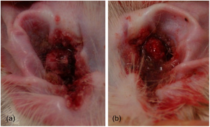
On examination at referral, the cat seemed slightly depressed and mild inappetence was reported. Both ear canals were obstructed and covered with reddish-to-dark-brown verrucous and necrotic tissue (Figure 1). The lesions were friable and bled easily. It could not be determined how deeply the lesions spread into the ear canals. The ears appeared to be moderately pruritic but were not tender on palpation.
Figure 1.
The ears at first presentation, also showing spontaneous haemorrhage: (a) right pinna; (b) left pinna
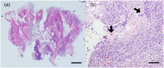
The results of haematology and biochemical tests were within normal limits. Total thyroxine was normal. Ear cytology revealed a mild, mixed yeast (Malassezia species) and bacterial infection. Aerobic cultures yielded Pasteurella multocida, while dermatophyte culture was negative. Clinically, lesions appeared to be restricted to the external ear canals – skull radiography showed no evidence of otitis media and there were no neurological signs relating to middle- or inner-ear disease. A sample for pathological examination was obtained from the friable proliferating tissue, which broke off easily during gentle manipulation. Histopathological examination showed various degenerative changes, but severe parakeratotic hyperkeratosis with neutrophilic crusts occupied most of the specimen. The epidermis and the outer root sheaths of the hair follicles showed marked papillomatous hyperplasia (Figure 2a). A number of pyknotic and hypereosinophilic keratinocytes (apoptotic cells) were observed in the non-degenerated areas (Figure 2b). These findings were compatible with a diagnosis of PNOE and a presumptive diagnosis was made based on both the clinical and histopathological features.
Figure 2.
Histopathology of the tissue easily removed by gentle manipulation. (a) At a low magnification degenerative changes, such as necrosis and vacuolar and hydropic degeneration, are seen, but there are areas of slight or no degeneration that allow observation of pathological findings. Severe papillomatous hyperplasia is seen in the epidermis, and the external root sheaths of hair follicles are preserved. Haematoxylin and eosin, bar = 500 μm. (b) At a higher magnification, the epidermis shows parakeratotic hyperkeratosis, with infiltrating cells that are mainly neutrophils and lymphocytes. Apoptotic keratinocytes are also observed (arrows). Haematoxylin and eosin, bar = 50 μm
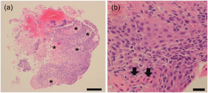
Treatment was initiated with once-daily application of a potent topical glucocorticoid (mometasone furoate) to the lesions of both ears, using an otic lotion that also contained gentamicin and clotrimazole furoate along with mometasone furoate (Mometaotic Otic Suspension; Intervet). Oral itraconazole (5 mg/kg q24h for 3 weeks) was added to enhance fungal treatment of the Malassezia species. On re-examination after 9 weeks, the proliferative tissue had resolved from both pinnae and vertical ear canals, and the frequency of head shaking had markedly decreased. However, erythema and erosions could still be detected by hand-held otoscope examination in the ear canals, and severe proliferative lesions were still evident in both horizontal ear canals, with neither tympanic membrane being visualised. Microscopic examination of smears from both ears were negative for bacteria and yeasts. Repeat skin biopsy was performed as before, with pathological findings the same as described previously,3 further supporting a diagnosis of PNOE (Figure 3). Oral triamcinolone (Ledercort tablets; Alfresa Pharma) was administered for 1 week at 0.9 mg/kg q24h, and was then tapered to alternate-day administration at the same dose for a further 3 weeks. Previous topical therapy (Mometaotic Otic Suspension) was continued. After 4 weeks there was marked improvement of both ears, with the tympanic membrane being clearly visualised and only a small residual lesion apparent in the right ear canal. Treatment was changed to prednisolone (2 mg/kg on alternate days) for 2 weeks along with a weaker steroid otic lotion (triamcinolone acetonide), after which no lesions were observed. Prednisolone was gradually tapered and stopped after 2 weeks. During the treatment, repeat haematology and biochemical tests were within normal limits except for a slightly elevated triglyceride concentration at 82 mg/dl (reference interval 7–77 mg/dl). Treatment was stopped, and there has been no recurrence of lesions during a follow-up period of 2 years.
Figure 3.
Histopathology of the biopsy specimen obtained with endoscopic forceps. (a) Marked hyperplasia of the hair follicles (asterisk) accompanied by epidermal parakeratosis and erosion. Haematoxylin and eosin stain, bar = 100 μm. (b) At a higher magnification, keratinocytes with swollen pale eosinophilic cytoplasm and vesicular nuclei (pyknosis) are seen (arrows). Bar = 20 μm
Discussion
PNOE was initially reported as a disease that typically affected kittens.1,4 Subsequently, Mauldin et al described the onset of this condition in 3- and 4-year-old cats.2 In contrast to these cases, the present case apparently developed lesions of PNOE at the age of 14 years – the oldest to be reported as far as the authors could determine – emphasising that PNOE may have a wide age range, even if it is predominantly a juvenile disease.
The pathogenesis of PNOE is still unclear, but concurrent bacterial or yeast otitis is common in the all published reports.1–4 In the current case, the cat had a long-term history of recurrent otitis externa, and some underlying causes cannot be excluded.
While feline eosinophilic granuloma complex was not compatible, based on the pathological findings, underlying atopy or food hypersensitivity could not be excluded, and a drug reaction to the cefovecin injected prior to development of the proliferative lesions cannot be excluded. However, the long-term resolution of clinical disease after cessation of therapy suggests an underlying hypersensitivity disorder may be less likely and cefovecin was administered again after the completion of treatment, with no recurrence of the lesion.
Topical glucocorticoid therapy only achieved partial improvement in the present case, in common with previously published reports. The addition of oral triamcinolone resulted in complete resolution of lesions, in contrast to previous reports where systemic glucocorticoid therapy was ineffective.2–4 This may be owing to differences in the potency of the glucocorticoids used, or individual differences of the patients.
Tacrolimus, a drug that specifically decreases T-cell signalling pathways has been successful in all previously published reports.1–4 Videmont and Pin hypothesised that tacrolimus may inhibit T-cell-induced keratinocyte apoptosis in PNOE.4 However, the inhibition of keratinocyte proliferation and the anti-inflammatory effect of glucocorticoids could also explain their benefit.5 In the present case, the severity and extent of the lesions may also have limited penetration of topical therapy alone (such as tacrolimus).
Conclusions
This is the first reported case of late-onset PNOE, and it suggests that this condition is not age-restricted.1 As combination therapy with steroid lotion and oral triamcinolone was successful, tacrolimus may not be the only medication able to achieve complete resolution of PNOE.
Footnotes
Funding: The authors received no financial support for the research, authorship, and/or publication of this article.
Conflict of interest: The authors declared no potential conflicts of interest with respect to the research, authorship, and/or publication of this article.
Accepted: 22 December 2016
References
- 1. Gross TL, Ihrke PJ, Walder EJ, et al. Necrotizing diseases of the epidermis. In: Gross TL. (ed). Skin diseases of the dog and cat. 2nd ed. Oxford: Blackwell Publishing, 2005, pp 79–91. [Google Scholar]
- 2. Mauldin EA, Ness TA, Goldschmidt MH. Proliferative and necrotizing otitis externa in four cats. Vet Dermatol 2007; 18: 370–377. [DOI] [PubMed] [Google Scholar]
- 3. Borio S, Massari F, Abramo F, et al. Proliferative and necrotising otitis externa in a cat without pinnal involvement: video-otoscopic features. J Small Anim Pract 2013; 15: 353–356. [DOI] [PMC free article] [PubMed] [Google Scholar]
- 4. Videmont E, Pin D. Proliferative and necrotizing otitis in a kitten: first demonstration of T-cell-mediated apoptosis. J Small Anim Pract 2010; 51: 599–603. [DOI] [PubMed] [Google Scholar]
- 5. Sanders S, Busam KJ, Halpern AC, et al. Intralesional corticosteroid treatment of multiple eruptive keratoacanthomas: case report and review of a controversial therapy. Dermatol Surg 2002; 28: 954–958. [DOI] [PubMed] [Google Scholar]


