Abstract
Case summary
A 10-year-old castrated male domestic shorthair cat was presented for assessment of a gingival mass surrounding the left maxillary third and fourth premolar teeth. The mass was surgically removed by means of a marginal rim excision, and the tissue was submitted for histological assessment. It was identified as a benign cementoblastoma (true cementoma). There was proliferation of mineralized eosinophilic material with multiple irregularly placed lacunae and reversal lines, reminiscent of cementum. The cat recovered uneventfully from the anesthesia, and there was no evidence of tumor recurrence 6 months after surgery.
Relevance and novel information
Cementoblastomas (true cementomas) in domestic animals are rare, with just a few reports in ruminants, monogastric herbivores and rodents. Cementoblastoma is considered a benign tumor that arises from the tooth root. The slow, expansive and constant growth that characterizes these masses may be accompanied by signs of oral discomfort and dysphagia. This case report is intended to increase knowledge regarding this tumor in cats and also highlights the importance of complete excision of the neoplasm. To our knowledge, there are no previous reports in the literature of cementoblastoma in the cat.
Introduction
Cementoblastoma or true cementoma is a rare benign mesenchymal odontogenic tumor arising from cementoblasts. This tumor is characterized by the formation of an expansive mass of cementum-like tissue intimately associated to the root of a tooth.1,2 In humans, the cortical expansive action of the cementoblastoma has been occasionally associated with low-grade intermittent pain. Radiographically, the cementoblastoma is observed as a well-defined radiopaque mass surrounded by a radiolucent uniform halo, corresponding to the space of the periodontal ligament. Surgical excision, including extraction of the affected tooth is indicated to avoid its recurrence.3–5 This case report presents the clinical, radiographic and histological findings, as well as the surgical approach for excision of a cementoblastoma in a 10-year-old castrated male domestic shorthair cat.
Case description
A 10-year-old castrated male domestic shorthair cat was referred to the Dentistry and Oral Surgery Service of the Matthew J Ryan Veterinary Hospital of the University of Pennsylvania for evaluation of a gingival mass discovered as an incidental finding during a wellness examination 1 month prior to presentation. No obvious signs of oral discomfort were reported. A biopsy performed prior to presentation showed no evidence of malignancy. An odontogenic tumor was suggested at that time.
No previous pertinent medical history was reported, and the patient was not receiving any medications at the time. Physical examination, complete blood count and serum biochemistry profile revealed no abnormalities. Given the previous histopathology results, further staging was not pursued.
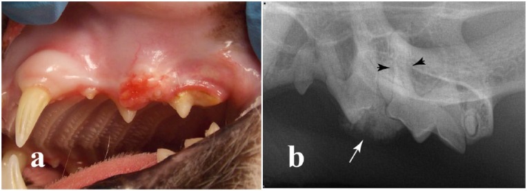
An approximately 1 cm in diameter, raised, erythematous, firm, ulcerated gingival mass was observed on the buccal aspect of the left maxillary third and fourth premolar teeth (Figure 1a). Moderate discomfort was noted on palpation of the mass during the awake oral examination. Gingivitis and calculus accumulation were more evident on these teeth when compared with the contralateral maxillary premolar teeth.
Figure 1.
(a) A 10-year-old domestic shorthair cat presented with an erythematous, firm ulcerated gingival mass on the buccal aspect of the left maxillary third and fourth premolar teeth (teeth 207 and 208). (b) The corresponding intraoral dental radiograph showed a well circumscribed, radiopaque mass (arrow) affecting the interproximal region between teeth 207 and 208; some tooth resorption and loss of the periodontal ligament space (arrowheads) are evident around the mesiobuccal root of tooth 208
The cat underwent general anesthesia for periodontal probing, dental charting, intraoral radiographic assessment and excision of the gingival mass. A left maxillary nerve block was performed for augmented pain control, using 0.4 ml (0.26 mg/kg) bupivacaine (Marcaine; Hospira). Periodontal probing of teeth adjacent to the gingival mass showed moderated build up of dental calculus and gingivitis. Periodontal pockets or malocclusion were not observed. The rest of the oral examination was unremarkable. Dental radiographs showed a well-defined mineralized mass at the alveolar margin overlying the distal aspect of the left maxillary third premolar and the mesial aspect of the maxillary fourth premolar. Some tooth resorption and loss of the periodontal ligament space were noticed (Figure 1b).
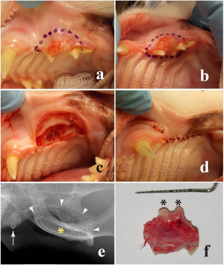
The oral cavity was rinsed with chlorhexidine gluconate 0.12% solution prior to the mass removal. Then, a rim excision was performed, including 5 mm normal-looking tissue away from the gross and radiographic margins of the mass. A full-thickness incision was made with a #15 scalpel blade. Alveolar and buccal mucosa was raised with a periosteal elevator to expose the underlying bone. Osteotomy of the maxilla was performed with a long #700 carbide bur in a sterile high-speed dental handpiece, taking care not to enter the nasal cavity or the infraorbital canal. The bony tissue was irrigated with sterile saline solution during the osteotomy.
The specimen was separated from the maxilla. Remaining root tips of the third and fourth premolar teeth were extracted using a winged dental elevator. Sharp alveolar bone edges were smoothed with a #22 round diamond bur, which was also used to debride the apical area of these sockets. Afterwards the wound was first rinsed with 0.12% chlorhexidine and then rinsed with sterile saline. A buccal flap was sutured to palatal mucosa with 5-0 poliglecaprone 25 (Monocryl; Ethicon) in a simple interrupted pattern (Figure 2). The cat recovered from the anesthesia uneventfully. Intravenous fluid therapy was maintained for the first 12 h after the surgery, at which time the cat ate soft food and drank water. Postoperatively, amoxicillin-clavulanate (62.5 mg PO q12h for 7 days; Clavamox [Zooetis]) was given orally, while buprenorphine hydrochloride (Buprenex; Reckitt Benckiser) (0.01 mg/kg SL q12h for 5 days) and robenacoxib (Onsior; Novartis) (1 tablet orally q24h for 3 days) were given to provide postoperative analgesia. Chlorhexidine gluconate (Oral Health Tooth Gel; Crosstex) was applied on the oral cavity twice a day for 2 weeks for antiseptic purpose, while soft food was maintained for the same period of time, until the surgical wound healed properly.
Figure 2.
(a) Buccal and (b) ventral views to the left maxillary area in a 10-year-old domestic shorthair cat. A surgical marker pen was used to outline the planned incisions. (c) Intraoperative view of the affected area after excision of the mass. (d) Closure buccal flap was sutured over the wound, using an absorbable monofilament in a simple interrupted pattern. (e) A postoperative intraoral radiograph was obtained, showing the affected area after rim excision (arrowheads), the left maxillary second premolar (arrow) and the zygomatic arch (asterisk). (f) The excised tissue was submitted for histopathological assessment. The asterisks show some intact roots of the third and fourth premolar teeth. Remaining root tips were extracted following excision of the mass. Because this was a clean-contaminated procedure, no surgical drapes were used. However, aseptic technique, including disposable synthetic or reusable cloth drapes may be used to minimize contamination6,7
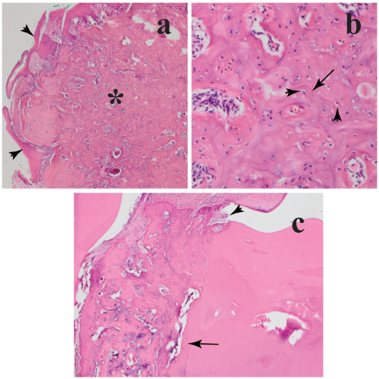
Histopathological examination showed proliferation of mineralized eosinophilic material with multiple, irregularly placed lacunae and reversal lines (Figure 3), reminiscent of cementum, between the two premolar teeth. The mass was adhered to the distal root of the left maxillary third premolar near the cementoenamel junction, contiguous with and focally replacing the normal cementum layer. The periodontal ligament region was expanded by the mass. The mass extended from the periodontal ligament region into the overlying gingiva, forming a well circumscribed lesion. Histopathology confirmed complete removal of the mass. No recurrence of the tumor was noted by the owner 6 months after surgical excision.
Figure 3.
(a) Photomicrograph of the submitted specimen showing proliferation of mineralized eosinophilic material (asterisk) that expands the gingiva (arrowheads) (×4 magnification, hematoxylin and eosin). (b) There are multiple irregularly placed lacunae (arrowheads) and reversal lines (arrow) (×20 magnification, hematoxylin and eosin). (c) The mass is focally adhered to a tooth root near the cementoenamel junction (arrowhead), contiguous with the normal cementum layer (arrow) (×4 magnification, hematoxylin and eosin)
Discussion
Odontogenic tumors have been rarely reported in cats. They include the peripheral odontogenic fibroma, ameloblastoma, ameloblastic fibroma, amyloid-producing odontogenic tumor and feline inductive odontogenic tumor.8–10 Odontogenic tumors may be locally invasive; however, the metastatic potential is low. Local recurrence after surgical excision is dependent on the type of tumor.2,11
Cementoblastoma or true cementoma is considered to be a benign odontogenic neoplasm originating from the tooth mesenchyme.2 This is a rare neoplasm in animals, with few reports in horses, hamsters, a bovine, a Dama gazelle and a red deer.12–18 In people, cementoblastoma is also a rare neoplasm that affects males more often than females, with the mandibular first molar the most commonly affected tooth.3 It may involve deciduous and permanent teeth, affecting people aged between 8 and 44 years.19,20 In animals, owing to the scarcity of reports, there are no statistical data.
In people, although more frequently found in the permanent molars, the cementoblastoma has also been reported in impacted or unerupted teeth.3,18 The reason for the molar teeth predilection in people is unknown. In animals, this pathological condition has been described on incisor, premolar and molar teeth.12,15,17,18
Cementoblastoma can be associated with intermittent pain due to the progressive expansion of cortical bone in the area of involved teeth.2 Although no signs of oral pain were reported before the initial physical examination, discomfort was evident during palpation of the site. The greater amount of calculus accumulation on teeth of the upper jaw affected by the neoplasm may be related to a reduction of masticatory action on that side of the mouth as a result of pain.
Although cementoblastomas are considered to be benign tumors, complete excision of the neoplasm is recommended, including the affected teeth, by means of a peripheral osteotomy to prevent tumor recurrence. 3–5 In people, tumor recurrence occurred between 4 and 24 months when the affected tooth or teeth were not included in the surgical excision.3,21 As the previous biopsy report suggested an odontogenic tumor, a rim excision was recommended in the present cat. This technique allowed preservation of the integrity of the nasal cavity, the infraorbital canal and the dorsal portion of the maxilla without causing any cosmetic concern. As reported previously, rim incision is a suitable surgical technique for odontogenic tumors.21–23
Radiation therapy has been used for other types of odontogenic tumors in cats (inductive fibroameloblastoma and amyloid-producing odontogenic tumor) as an adjuvant in cases where the neoplasm could not be completely excised;18 however, there are no scientific reports regarding the use of radiation therapy for treatment of cementoblastoma in animals.
Radiographically, the appearance of cementoblastoma corresponds to a densely radiopaque mass attached to the apical or lateral aspect of the root. Tooth resorption and obliteration of the periodontal ligament space, as well as a surrounding radiolucent halo and sclerotic margins, can be observed.2,4,24 Dental radiographs permitted the initial approach to the mass in this case; however, the value of this diagnostic imaging technique is limited because the overlap of anatomic structures results in poor highlighting of the radiographic features of a cementoblastoma. More advanced imaging techniques such as computed tomography scan may provide better anatomical detail and structural resolution by avoiding superimposition of dentoalveolar structures.25,26
Histologically, the main characteristics of cementoblastomas are the presence of abundant mineralized material with cores of bland spindle-shaped cells and occasional cementoblasts.2,13 A major differential diagnosis for cementoblastoma (true cementoma) includes cemento-osseous dysplasia, which is referred to as a reactive, non-neoplastic lesion characterized by a periapical fibro-osseous proliferation that contains variable amounts of cementum or bone.2 Late-stage, sclerotic cemento-osseous dysplasia lesions have been described; however, they are usually smaller and not symptomatic for the patient unless they become necrotic and secondarily inflamed. There was no evidence of necrosis or secondary inflammation in the sections evaluated of the specimen submitted from the present cat.2
Conclusions
To our knowledge, this is the first report of a cementoblastoma in a cat. In the presence of a radiopaque oral mass in a cat associated with a tooth root, cementoblastoma must be included in the differential diagnoses. Marginal rim excision involving the affected teeth should be encouraged to avoid local recurrence of the neoplasm, as well as to preserve anatomical structures.
Acknowledgments
We would like to thank M Leanne Lilly, DVM, for her help and assistance during the editing of this case report.
Footnotes
Funding: The authors received no financial support for the research, authorship, and/or publication of this article.
Conflict of interest: The authors declared no potential conflicts of interest with respect to the research, authorship, and/or publication of this article.
References
- 1. Head KW, Else RW, Dubielzig RR. Tumors of the alimentary tract. In: Meuten DJ. (ed). Tumors in domestic animals. 4th ed. Ames, IA: Iowa State Press, 2002, pp 407–408. [Google Scholar]
- 2. Regezi JA, Sciubba JJ, Jordan RCK. Odontogenic tumors. In: Oral pathology clinical pathology correlations. 5th ed. St Louis, MO: Saunders Elsevier, 2008, pp 261–281. [Google Scholar]
- 3. Brannon RB, Fowler CB, Carpenter WM, et al. Cementoblastoma: an innocuous neoplasm? A clinicopathologic study of 44 cases and review of the literature with special emphasis on recurrence. Oral Surg Oral Med Oral Pathol Oral Radiol Endod 2002; 93: 311–320. [DOI] [PubMed] [Google Scholar]
- 4. Neves FS, Falcão AF, Dos Santos JN, et al. Benign cementoblastoma: case report and review of the literature. Minerva Stomatol 2009; 58: 55–59. [PubMed] [Google Scholar]
- 5. Sharma N. Benign cementoblastoma: a rare case report with review of literature. Contemp Clin Dent 2014; 5: 92–94. [DOI] [PMC free article] [PubMed] [Google Scholar]
- 6. Reiter AM. Equipment for oral surgery in small animals. Vet Clin North Am Small Anim Pract 2013; 43: 587–608. [DOI] [PubMed] [Google Scholar]
- 7. Terpak CH, Verstraete FJM. Instrumentation, patient positioning and aseptic technique. In: Verstraete FJM, Lommer MJ. (eds). Oral and maxillofacial surgery in dogs and cats. Edinburg: Saunders Elsevier, 2012, pp 55–67. [Google Scholar]
- 8. Boehm B, Breuer WHW. Odontogenic tumours in the dog and cat [article in German]. Tierarztl Prax Ausg K Kleintiere Heimtiere 2011; 39: 305–312. [PubMed] [Google Scholar]
- 9. Poulet FM, Valentine BA, Summers BA. A survey of epithelial odontogenic tumors and cysts in dogs and cats. Vet Pathol 1992; 29: 369–380. [DOI] [PubMed] [Google Scholar]
- 10. Gardner DG. Ameloblastomas in cats: a critical evaluation of the literature and the addition of one example. J Oral Pathol Med 1998; 27: 39–42. [DOI] [PubMed] [Google Scholar]
- 11. Wiggs RB, Lobprise HB. Domestic feline oral and dental disease. In: Wiggs RB, Lobprise HB. (eds). Veterinary dentistry principles and practice. Philadelphia, PA: Lippincott–Raven, 1997, pp 510–511. [Google Scholar]
- 12. Andrews AH. A cemental abnormality of the bovine molar tooth. Vet Rec 1973;49: 318–319. [PubMed] [Google Scholar]
- 13. Martin HD, Turner T, Kollias GV, et al. Cementoblastoma in a dama gazelle. J Am Vet Med Assoc 1985; 187: 1246–1247. [PubMed] [Google Scholar]
- 14. Ernst H, Kunstyr I, Rittinghausen S, et al. Spontaneous tumours of the European hamster (Cricetus cricetus L). Z Versuchstierkd 1989; 32: 87–96. [PubMed] [Google Scholar]
- 15. Kreutzer R, Wohlsein P, Staszyk C, et al. Dental benign cementomas in three horses. Vet Pathol 2007; 44: 533–536. [DOI] [PubMed] [Google Scholar]
- 16. Schaaf KL, Kannegieter NJ, Lovell DK. Calcified tumours of the paranasal sinuses in three horses. Aust Vet J 2007; 85: 454–458. [DOI] [PubMed] [Google Scholar]
- 17. Levine DG, Orsini JA, Foster DL, et al. What is your diagnosis? Benign true cementoma (benign cementoblastoma). J Am Vet Med Assoc 2008; 233: 1063–1064. [DOI] [PubMed] [Google Scholar]
- 18. Kierdorf U, Bridault A, Witzel C, et al. Cementoblastoma in a red deer (Cervus elaphus) from the Late Pleistocene of Rochedane, France. Int J Paleopathol 2015; 8: 42–47. [DOI] [PubMed] [Google Scholar]
- 19. Ohki K, Kumamoto H, Nitta Y, et al. Benign cementoblastoma involving multiple maxillary teeth: report of a case with a review of the literature. Oral Surg Oral Med Oral Pathol Oral Radiol Endod 2004; 97: 53–58. [DOI] [PubMed] [Google Scholar]
- 20. Solomon MC, Rehani S, Valiathan M, et al. Benign cementoblastoma involving multiple deciduous and permanent teeth of the maxilla – a case report. Oral Maxillofac Pathol J 2012; 3: 258–263. [Google Scholar]
- 21. Murray RL, Aitken ML, Gottfried SD. The use of rim excision as a treatment for canine acanthomatous ameloblastoma. J Am Anim Hosp Assoc 2010; 46: 91–96. [DOI] [PubMed] [Google Scholar]
- 22. Moore AS, Wood CA, Engler SJ, et al. Radiation therapy for long-term control of odontogenic tumours and epulis in three cats. J Feline Med Surg 2000; 2: 57–60. [DOI] [PMC free article] [PubMed] [Google Scholar]
- 23. Verstraete JM. Mandibulectomy and maxillectomy. Vet Clin Small Anim 2005; 35: 1009–1039. [DOI] [PubMed] [Google Scholar]
- 24. Baghdady M. Principles of radiographic interpretation. In: White SC, Pharoah MJ. (eds). Oral radiology principles and interpretation. 7th ed. St Louis, MO: Elsevier Mosby, 2014, pp 380–381. [Google Scholar]
- 25. Forrest LJ, Schwarz T. Oral cavity, maxilla and dental apparatus. In: Schwarz T, Saunders J. (eds). Veterinary computed tomography. 1st ed. Chichester: Wiley–Blackwell, 2011, pp 111–123. [Google Scholar]
- 26. Ghirelli CO, Villamizar LA, Pinto AC. Comparison of standard radiography and computed tomography in 21 dogs with maxillary masses. J Vet Dent 2013; 30: 72–76. [DOI] [PubMed] [Google Scholar]


