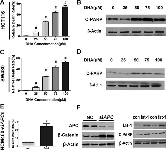
Figure 4. DHA promotes colorectal tumor cell apoptosis in vitro.

DHA promotes colorectal tumor cell apoptosis in vitro. (A) HCT116, (C) SW480, or (E) NCM460 cells transfected with APC siRNA were serum-starved and cultured with or without the indicated concentrations of DHA (dissolved in basic medium) for 12 h, and subsequently trypsinized to make single cell suspensions for trypan blue staining. Cell death was quantified using an automatic cell counter (BioRad) according to the manufacturer's instructions. Data from three repeat counts were averaged. Bars indicate mean ± SEM; #p < 0.01. (B) HCT116, (D) SW480, or (F) NCM460 cells transfected with APC siRNA, serum-starved, and cultured for 12 h in the presence of the indicated concentrations of DHA. Proteins were extracted for the immunoblot analysis of cleaved-PARP (C-PARP).
