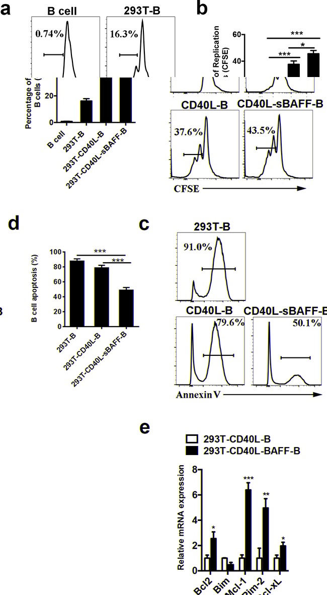
Figure 2. The 293T-CD40L-sBAFF cell line had the capacity to induce B cell growth and prevent apoptosis.

a, b. Cell division of 293T-B cells, CD40L-B cells and CD40L-sBAFF-B cells were measured by CFSE dilution with flow cytometry analysis, in comparison with non-activated cells (B cells) on day 7 after co-culture with feeder cells. c, d. Cell apoptosis of 293T-B cells, CD40L-B cells and CD40L-sBAFF-B cells were measured by Annexin V staining by flow cytometry on day 20 after co-culture with feeder cells. e. The mRNA expression of pro-apototic and anti-apoptotic factors in B cells. Data represent mean ± SD (error bars) for a representative experiment of 4 independent experiments with 4 healthy donors. The paired t-test and one-way ANOVA were used. P < 0.05 indicates statistically significance difference. * indicates P < 0.05; ** indicates P < 0.01; *** indicates P < 0.001.
