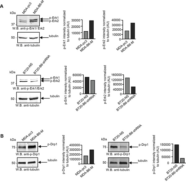
Figure 3. EPHB6 activates ERK kinases and DRP1 in cancer cells.

A. The indicated cells were lysed, cell lysates were resolved by SDS PAGE and phosphorylation of ERK1 and ERK2 kinases was analysed by Western blotting with anti-phospho-ERK (anti-p-ERK1/ERK2). ERK phosphorylation was quantitated by densitometry, measurements were normalized to tubulin controls and are presented in arbitrary units (AU). B. DRP1 phosphorylation on the activating residue, Ser616, was analysed by Western blotting with anti-phospho-DRP1 (anti-p-DRP1) as in (A). All experiments were performed at least three times.
