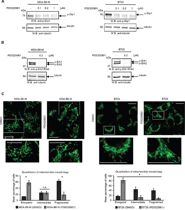
Figure 4. ERK activity is required for the EPHB6 effect on mitochondrial fragmentation.

A. Cells were treated for 24 h with a MEK inhibitor, PD0325901, at the indicated concentrations or with a DMSO volume matching the highest PD0325901 concentration (−) and DRP1 phosphorylation was assessed by Western blotting. B. To confirm the efficiency of MEK inhibition, cells were treated with 0.2 μM PD0325901 or a matching volume of DMSO (−) for 2 h and the phosphorylation status of ERK kinases was examined by Western blotting. C. Cells pre-treated for 24 h with 0.2 μM PD0325901 or DMSO were loaded with 70 nM MitoTracker Green for 20 min. Live cell imaging was performed using a LSM 700 confocal microscope. Selected areas are shown at higher magnifications. Scale bar, 20 μm. Quantification and analysis of mitochondrial morphology was done as in Figure 2A. All experiments were performed at least three times. * p<0.05, Student's t-test. n.s., statistically not significant.
