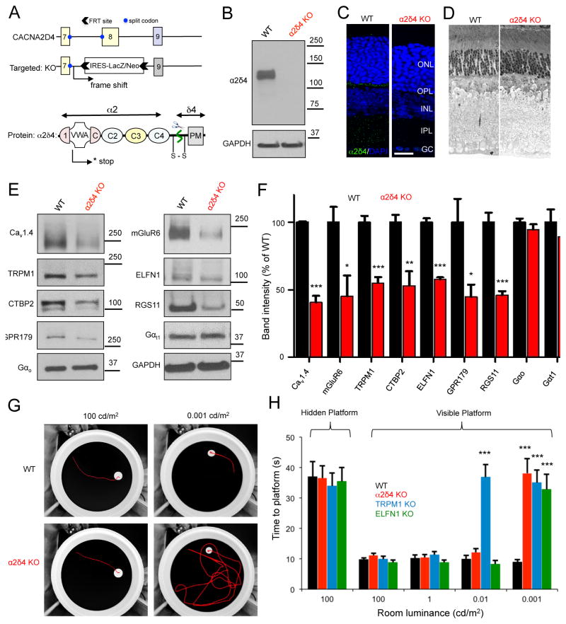
Figure 2. Generation and characterization of knockout mice with complete elimination of α2δ4 protein.
A, Scheme of the targeting strategy and predicted consequences on disrupting the protein expression. Domain organization of α2δ4 is based on high-resolution structure of α2δ1 (Wu et al., 2016). B, Loss of α2δ4 protein expression as revealed by Western blot analysis of total retina lysates. C, Absence of α2δ4 immunostaining in the OPL layer of α2δ4 knockout retinas (scale bar, 25μm). D, Analysis of the retina morphology by toluidine blue staining of ultra-thin retina cross-sections (retinas used were from 6–10 week old mice). E, Western blot analysis of protein levels in α2δ4 KO retinas in comparison to WT littermates. F, Quantification of changes in protein levels by densitometry. Band intensities were normalized to WT. Error bars are SEM values, *p<0.05, **p<0.01, ***p<0.001, n=3–5 mice, t-test. G, Analysis of mouse vision in a visually guided behavioral task at photopic (100cd/m2) and scotopic conditions (0.001 cd/m2). Representative tracks of mice swimming to visible escape platform are shown. H, Quantification of mouse escape time in water maze task (panel G) at various luminance levels. Error bars are SEM, ***p<0.001, 2-way ANOVA with Bonferroni post test, n=4–5 mice per genotype.

