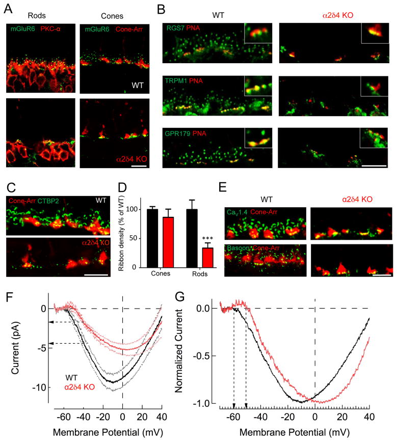
Figure 5. Deficits in structural and functional architecture of rod photoreceptor ribbon synapses associated with α2δ4 loss.
A, Loss of mGluR6 postsynaptic targeting to the dendritic tips in ON-RBCs, but not for ON-CBCs in α2δ4 knockout retinas. B, Effect of α2δ4 deletion on postsynaptic targeting of signaling molecules in ON-CBC and ON-RBC. Active zones of cone terminals labeled by PNA. C, Immunohistochemical analysis of photoreceptor ribbons. D, Quantification of ribbon densities in rods and cones. Area occupied by CTBP2 inside (cone) and outside (rod) of cone-arrestin mask was determined and normalized to WT. The total number of cone terminals selected was 100 for WT and 86 for α2δ4 KO from 3 mice for each genotype. The total number of areas outside of cone terminal selected was 82 for WT and 69 for α2δ4 KO from 3 mice for each genotype. Error bars are SEM values, ***p<0.001, two-way ANOVA. E, Changes in the active zone components associated with synaptic terminals of photoreceptors. All scale bars in A–E are 10μm. F, Representative CaV1.4 mediated currents measured from individual rods in retinal slices. Ca2+ currents were measured under voltage-clamp by ramping the membrane potential from −80mV to +40mV in 1 sec. Data are shown along with the SEM values. Horizontal arrows indicate difference in Ca2+ current at the rod’s normal resting potential of Vm = −40mV. G, Ca2+ currents normalized to the peak current density reveal a shift in the voltage sensitivity of the current. Vertical arrows reveal ~8mV shift in the membrane potential at which Ca2+ channels begin to open.

