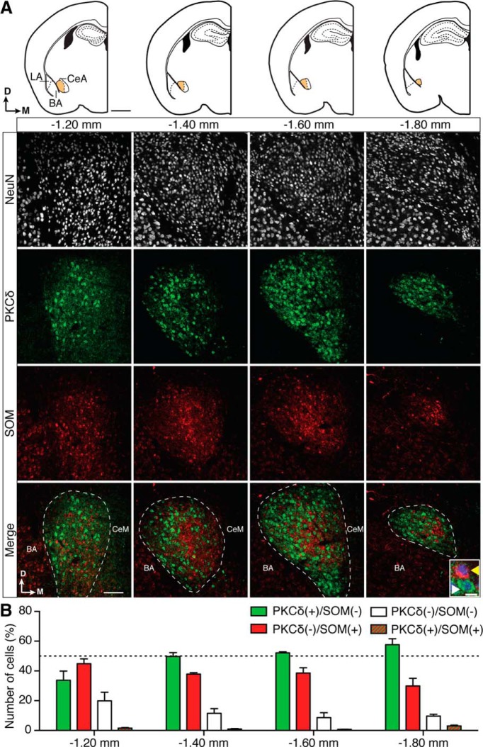
Figure 1.
PKCδ and SOM label distinct populations of neurons in the CeL of wild-type C57BL/6J mice. A, Top, Diagrams of coronal CeL slices of C57BL/6J mouse at −1.20, −1.40, −1.60, and −1.80 mm from bregma. LA, Lateral amygdala; BA, basal amygdala; CeA, central amygdala, which is divided into the CeL (orange) and the central medial amygdala (CeM, in white). Arrows show dorsal and medial orientation. Scale bar, 1 mm. Bottom panels show closeups of the CeL in 50 μm sections that were stained for NeuN (to stain somas of neurons, white fluorescence), PKCδ (green fluorescence), and SOM (red fluorescence). Scale bar in bottom left square, 100 μm. For clarity, the merged panels represent the merging of PKCδ and SOM only. The CeL is outlined in the bottom panel, and this outline was defined both by landmarks visible in bright field (data not shown), and the presence of PKCδ(+) somas. PKCδ(+) fibers can typically be seen in the CeM. The locations of both the BA and the CeM are also labeled in the merged panels, and note that by 1.80 mm the CeM is no longer present. The inset in the lower right corner of the far right merged panel shows a closeup of the most common cells types: PKCδ(+)/SOM(−) (white arrowhead) and SOM(+)/PKCδ(−) neurons (yellow arrowhead; scale bar, 10 μm; PKCδ green fluorescence, SOM red fluorescence, NeuN blue fluorescence). B, Only NeuN(+) neurons were counted to ensure that only mature neuronal cells were taken into account. Of these, 48 ± 5% were PKCδ(+)/SOM(−) (mean n = 83 ± 19 neurons/1.0 × 10−3 mm3), and 38 ± 3% were SOM(+)/PKCδ(−) (mean n = 66 ± 14 neurons/1.0 × 10−3 mm3). These two populations were largely distinct as only 1 ± 0.5% of neurons were PKCδ(+)/SOM(+) (mean n = 2 ± 0.3 neurons/1.0 × 10−3 mm3), and 12 ± 2% NeuN(+) cells were PKCδ(−)/SOM(−) (mean n = 20 ± 4 neurons/1.0 × 10−3 mm3). The dotted line on the graph indicates 50% and bregma-specific percentages were as follows: PKCδ(+)/SOM(−), 34 ± 6% (−1.20 mm), 50 ± 2% (−1.40 mm), 52 ± 1% (−1.60 mm), and 57 ± 4% (−1.80 mm). PKCδ(−)/SOM(+), 45 ± 3% (−1.20 mm), 38 ± 1% (−1.40 mm), 39 ± 3% (−1.60 mm), and 30 ± 5% (−1.80 mm); PKCδ(−)/SOM(−), 20 ± 6% (−1.20 mm), 11 ± 3% (−1.40 mm), 8 ± 3% (−1.60 mm), and 10 ± 1% (−1.80 mm); and PKCδ(+)/SOM(+), 1 ± 0.3% (−1.20 mm), 1 ± 0.2% (−1.40 mm), 1 ± 0.05% (−1.60 mm), and 3 ± 0.5% (−1.80 mm).

