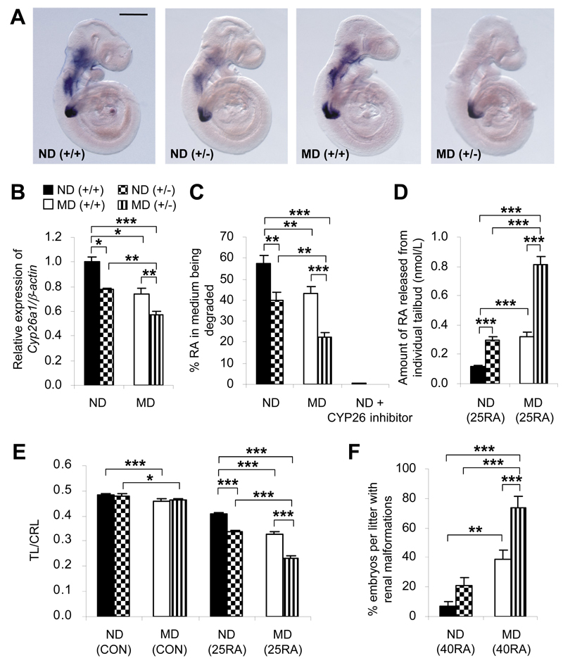
Figure 5.
Cyp26a1 loss-of-function genotype interacts with diabetic maternal environment to influence RA-degrading efficiency and susceptibility to RA teratogenesis. A: Whole-mount in situ hybridization patterns of Cyp26a1 expression in Cyp26a1 heterozygous null (+/-) embryos and their wild-type (+/+) littermates from ND and MD mice at E9. At least 30 embryos from six to eight litters were examined for each group. Scale bar = 0.25 mm. B: Quantification of Cyp26a1 mRNA, normalized to β-actin and expressed relative to ND (+/+), which was set as 1, in tailbuds of embryos from different genotype-maternal environment combinations at E9 (n = 5 from five litters). C: In vitro RA-degrading efficiency, presented as the percentage of RA in the medium being degraded by the tailbud lysate in the absence or presence of 100 nmol/L R115866 (a CYP26 inhibitor) (n = 8-13 from 10 to 14 litters). D: Quantification of RA released from individual tailbuds, using a RA reporter cell line, 3 h after in vivo challenge with 25 mg/kg RA (25RA) at E9 (n = 22-28 from six to seven litters). E: Maternal injection of 25RA or vehicle as the control (CON) at E9. Caudal truncation, measured in terms of the ratio of TL to CRL (TL/CRL), was examined in E13 embryos (n = 41-62 from 10 litters). F: Maternal injection of 40 mg/kg RA (40RA) or CON at E9. Near-term E18 fetuses were examined for renal malformations (n = 11 litters). *P < 0.05; **P < 0.01; ***P < 0.001, one-way ANOVA followed by the Bonferroni test. Error bars represent the mean ± SEM.

