Abstract
A number of lysine-specific methyltransferases (KMTs) are responsible for the post-translational modification of cellular proteins on lysine residues. Most KMTs typically recognize specific motifs in unstructured, short peptide sequences. However, we have recently discovered a novel KMT that appeared to have a more relaxed sequence specificity, namely, valosin-containing protein (VCP)-KMT, which trimethylates Lys-315 in the molecular chaperone VCP. On the basis of this, here, we explored the possibility of using the VCP-KMT/VCP system to obtain specific lysine methylation of desired sequences grafted onto a VCP-derived scaffold. We generated VCP-derived proteins in which three amino acid residues on each side of Lys-315 had been replaced by various sequences representing lysine methylation sites in histone H3. We found that all of these chimeric proteins were subject to efficient VCP-KMT-mediated methylation in vitro, and methylation was also observed in mammalian cells. Thus, we here describe a versatile system for introducing lysine methylation into a desired peptide sequence, and the approach should be readily expandable for generating combinatorial libraries of methylated sequences.
Introduction
Cellular proteins are frequently post-translationally modified by methylation, mainly on the side chain of arginine and lysine residues.1,2 A lysine residue can accept up to three methyl groups, leading to four possible states: mono-, di-, tri- (me1, me2, me3), and unmethylated (me0) forms. Lysine methylation has been most extensively studied in the case of histone proteins, and histone lysine methylation has been established as an important regulator of gene expression and chromatin state.3 In histone proteins, the methylated lysines are mainly found in the unstructured N-terminal tails and are read by specific reader domains, which typically constitute parts of multidomain proteins that can modify and/or remodel chromatin.4 In general, some of the histone methylations represent signals for gene activation and loosening of the chromatin structure, whereas others signal gene repression and heterochromatin formation. In histone H3, for example, trimethylated Lys-9 acts like a repressive mark, whereas trimethylation at Lys-4 is associated with gene activation.3 Importantly, methylated lysines are also found in a number of nonhistone proteins; however, for the majority of these, their functional significance as well as the enzyme introducing the methylation remains elusive.2,5
Protein lysine methylation is catalyzed by a number of S-adenosylmethionine (AdoMet)-dependent lysine (K)-specific methyltransferases (KMTs), which belong to two structurally distinct methyltransferase (MTase) classes, namely, the seven-β-strand (7BS) MTases and the SET domain MTases.6−8 The SET domain MTase family, named after its three founding members (Su(var)-39, E(z), and Trithorax), appears to consist exclusively of KMTs, and these enzymes mainly recognize specific motifs in unstructured, short peptide sequences, such as the flexible N-terminal tails of histone proteins.7 In contrast, the 7BS MTases act on a wide range of substrates, including small metabolites, nucleic acids, and proteins; however, recently, a number of novel human KMTs belonging to this group have been discovered.6 The 7BS KMTs appear to mainly target nonhistone proteins and bind their substrates not primarily through the linear sequence surrounding the methylation site, but rather in a manner involving multiple interactions with the folded substrate protein. This has been studied in detail for the 7BS KMT VCP-KMT (METTL21D), which specifically trimethylates the essential chaperone valosin-containing protein (VCP) on Lys-315.9,10 It was found that VCP-KMT interacts strongly with its substrate and a substantial portion of VCP (amino acids 282–364) was required for the interaction to occur.10 Moreover, simultaneous mutation of the amino acids surrounding the methylation site, Lys-315, in VCP did not negatively affect the efficiency of the VCP-KMT-mediated methylation.10 VCP-KMT appears to be a highly specific enzyme; two independent studies, using different approaches, identified VCP as an interaction partner and substrate of VCP-KMT, and no other substrates have been identified.9,10 Moreover, a comparison of VCP-KMT-mediated methylation in extracts from wild-type and vcpkmt–/– mice also revealed only a single substrate, namely, VCP.11
VCP, also known as p97, is a type II AAA (ATPases associated with diverse cellular activities) ATPase consisting of an N-terminal domain, two ATPase domains referred to as D1 and D2, and a C-terminal tail.12 VCP forms a homohexameric structure, where the D1 and D2 domains constitute two stacked rings surrounding a central channel.12 The site of VCP-KMT-mediated methylation is found within this channel, and accordingly, VCP is not susceptible to methylation by VCP-KMT when present in the hexameric state, suggesting that methylation occurs before hexamer assembly.10 VCP is a highly abundant protein present in both eukaryotes and archaea, where it functions as a ubiquitin-specific protein chaperone mediating protein unfolding and disassembly of protein complexes, and is involved in a wide range of cellular processes, including protein degradation, membrane dynamics, and cell-cycle regulation.12
On the basis of the observation that VCP-KMT-mediated methylation of VCP is apparently rather insensitive to alterations in the sequence surrounding the methylation site, we sought to explore the possibility that short, lysine-containing sequences of choice may be amenable to VCP-KMT-mediated methylation when grafted onto a VCP-derived scaffold. We reasoned that, if successful, such a strategy may allow VCP-KMT-mediated methylation of desired peptide sequences both in vitro and in vivo and may also be utilized for the generation of combinatorial libraries of lysine-methylated proteins. We have therefore, in the present study, generated VCP-derived proteins, where three amino acid residues on each side of the original methylation site (Lys-315) have been replaced by the corresponding sequence from various lysine methylation sites in human histone H3, such as Lys-4, Lys-9, Lys-27, and Lys-36. We demonstrate that the resulting VCP/H3 chimeras are efficiently methylated by VCP-KMT in vitro and also subject to methylation by endogenous cellular VCP-KMT when expressed in mammalian cells.
Results
Determining the Minimal (Lys-315-Encompassing) Part of VCP Required for Efficient Expression and Purification
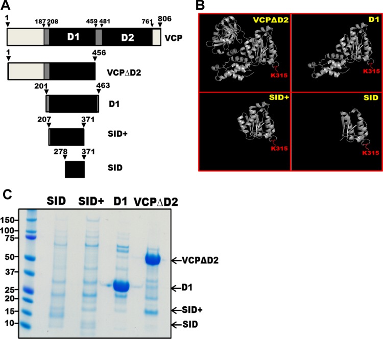
We previously found that VCP was not amenable to VCP-KMT-mediated methylation when present in its naturally occurring homohexameric state. However, a truncated version of VCP, which lacked the D2 domain (VCPΔD2) (Figure 1A,B) and was unable to form a stable VCP homohexamer, was efficiently methylated.10 Moreover, results from a yeast two-hybrid screen defined a relatively short segment (encompassing residues 282–364), the so-called selected interaction domain (SID) (Figure 1A,B), as the minimal part of VCP necessary for interaction with VCP-KMT.10 On the basis of this and for the purpose of generating VCP/peptide chimeras, we set out to define a smallest possible VCP-derived segment that could be efficiently methylated by VCP-KMT and readily be expressed and purified as a recombinant protein. Thus, we generated several constructs encoding N-terminally hexahistidine (6xHis)-tagged, VCP-derived proteins for expression in Escherichia coli. The smallest of these encompassed only the SID (slightly expanded to encompass residues 278–371, thereby avoiding the disruption of secondary structure elements). We also designed a somewhat larger protein, denoted SID+, which encompassed the entire structural subdomain in which the SID resides (residues 207–371). A third construct encompassed the entire D1 ATPase domain of VCP (residues 201–463). The three constructs as well as the previously investigated VCPΔD2 protein were expressed in the BL21-CodonPlus (DE3)-RIPL strain of E. coli and purified using Ni-NTA affinity chromatography. The D1 and VCPΔD2 proteins were expressed at high levels and gave good yields after purification by affinity chromatography, whereas the SID+ and SID proteins were poorly expressed, gave low yield and purity, and were not detected by Coomassie gel staining after purification (Figure 1C).
Figure 1.
Establishing the D1 domain as the minimal (Lys-315-encompassing) part of VCP required for efficient expression and purification. (A) Schematic diagram of VCP and truncated variants thereof (SID = selective interaction domain, that is, portion of VCP found to interact with VCP-KMT in a yeast two-hybrid screen10). (B) Three-dimensional structures of the deletion mutants of VCP. Lys-315 is indicated in red (illustration generated from Protein Data Bank (PDB) entry 1S3S). (C) Expression and purification of truncated variants of VCP. Arrows show the expected sizes of the indicated proteins.
VCP-KMT-Mediated Methylation of VCP Deletion Mutants
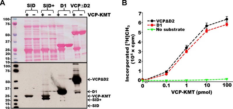
Next, the purified recombinant proteins were tested as substrates for VCP-KMT in an in vitro methylation reaction in the presence of [3H]AdoMet, and we observed by fluorography that D1 and VCPΔD2 were strongly methylated (Figure 2A). Despite our inability to visually detect the corresponding recombinant proteins after purification, we tested the preparations of SID and SID+ proteins as substrates for methylation. Interestingly, the preparation of the SID+ protein showed methylation of a band corresponding to its predicted molecular weight, whereas no methylation was detected in the preparation of the SID protein (Figure 2A). To better quantify the extent of methylation, we performed an experiment where the D1 and VCPΔD2 proteins were subjected to in vitro methylation by varying the concentrations of VCP-KMT in the presence of [3H]AdoMet, followed by scintillation counting of radioactivity incorporated into the trichloroacetic acid (TCA)-precipitable material. The results showed virtually identical VCP-KMT titration curves for D1 and VCPΔD2 (Figure 2B). In conclusion, the above experiments indicate that the SID+ protein can be methylated by VCP-KMT, but suggested D1, which was readily purified and efficiently methylated as the best starting point for generating VCP-derived peptide chimeras.
Figure 2.
In vitro methylation of VCP deletion mutants. (A) VCP-KMT-mediated methylation of VCP deletion mutants in the presence of [3H]AdoMet in vitro. Upper panel: Ponceau S stain. Lower panel: Detection of radioactivity by fluorography. Some automethylation of VCP-KMT in the absence of an optimal substrate was detected, similar to our previous observations.10 (B) Titration of VCP-KMT activity on the VCP deletion mutants, D1 and VCPΔD2 (log-scale x axis). Methylation was assessed by scintillation counting of radioactivity incorporated into the TCA-insoluble material.
VCP-KMT-Mediated Methylation of VCP-D1-Derived Chimeras
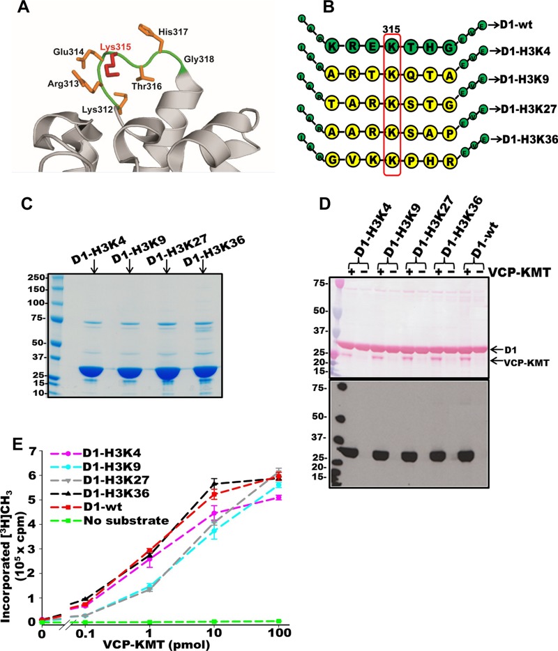
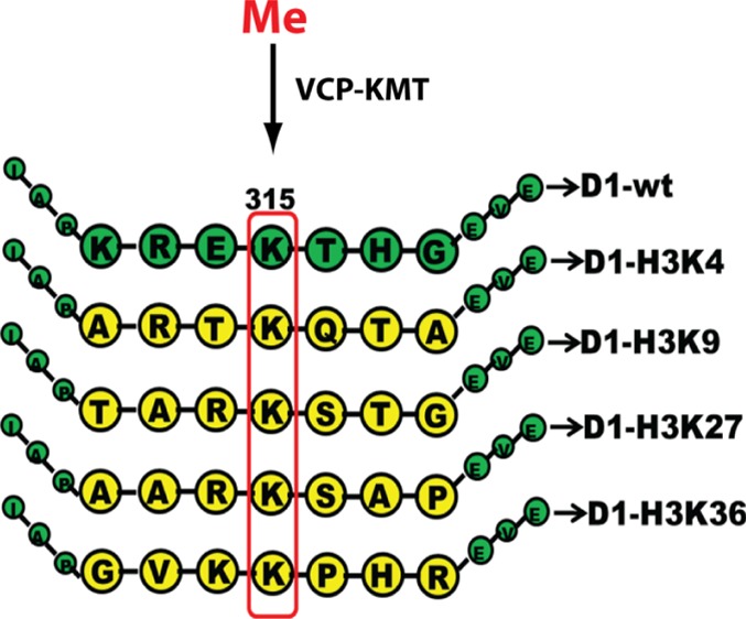
We previously found that VCP-KMT efficiently methylated Lys-315 in VCP even after individual or simultaneous mutation to alanine of the six residues surrounding the methylation site (amino acids 312–314 and 316–318),10 and we therefore reasoned that these neighboring residues are not crucial for the interaction of VCP with VCP-KMT. Moreover, the methylation site and the surrounding residues are localized within a loop in the VCP structure (Figure 3A), indicating that the replacement of these surrounding residues is unlikely to disrupt secondary structure elements and may generally be tolerated with respect to VCP-KMT-mediated methylation. To test this, we replaced these amino acids in the D1 domain with the corresponding sequence derived from several lysine methylation sites in histone H3, thereby generating D1/H3 chimeras where the seven amino acid segment containing Lys-315 and surrounding residues had been replaced by the H3-derived sequence. We generated chimeric proteins representing the methylation sites Lys-4, Lys-9, Lys-27, and Lys-36 in H3, denoted D1-H3K4, D1-H3K9, D1-H3K27, and D1-H3K36, respectively (Figure 3B), and these proteins were efficiently expressed and purified from E. coli (Figure 3C). Next, we investigated whether the generated recombinant D1/H3 chimeras could be methylated by VCP-KMT in vitro, as assessed by the incorporation of radioactivity from [3H]AdoMet. Clearly, all four chimeras (D1-H3K4, D1-H3K9, D1-H3K27, and D1-H3K36) showed strong methylation, as analyzed by fluorography (Figure 3D). To obtain a more quantitative measure of methylation efficiency, the chimeras were methylated in vitro in the presence of varying amounts of VCP-KMT, and the methylation was measured by scintillation counting of the TCA-precipitable material. In these experiments, the D1-H3K4 and D1-H3K36 proteins as well as wild-type D1 gave similar titration curves, reaching an apparent plateau at high enzyme concentrations (Figure 3E). This indicated, on the basis of our previous work with VCP-KMT,10 that close to complete trimethylation was achieved. In contrast, the D1-H3K9 and D1-H3K27 chimeras were less efficiently methylated, and a substantially higher amount of enzyme (approximately 3-fold) was required to obtain a similar level of methylation (Figure 3E). Thus, these experiments demonstrated that all of the four D1/H3 chimeras could be methylated in vitro by VCP-KMT, but that the D1-H3K9 and D1-H3K27 proteins were somewhat poorer substrates than the wild-type D1 protein.
Figure 3.
VCP-KMT-mediated methylation of VCP-D1-derived chimeras containing histone peptides. (A) Localization of Lys-315 and surrounding residues in the VCP structure (illustration generated from PDB entry 5FTK). (B) Schematic representation of D1/H3 chimeras, where amino acids 312–318 in VCP (green) have been replaced by heptameric H3-derived sequences (yellow). Lys-315 is indicated by the red box. (C) Recombinant D1/H3 chimeras purified from E. coli. (D) VCP-KMT-mediated methylation of D1/H3 chimeras in the presence of [3H]AdoMet in vitro. Upper panel: Ponceau S stain. Lower panel: Detection of radioactivity by fluorography. (E) Titration of VCP-KMT activity on D1/ H3 chimeras (log-scale x axis). Methylation was assessed by scintillation counting of radioactivity incorporated into the TCA-insoluble material. Error bars represent the standard deviation of duplicate samples.
Cellular Methylation of D1/H3 Chimeras
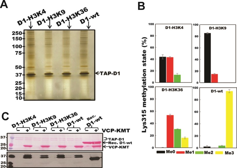
Next, we set out to investigate whether the constructed chimeric proteins could undergo VCP-KMT-mediated methylation also inside cells. To this end, we generated stably transfected, HEK293-derived cell lines that expressed, from a doxycycline (Dox)-inducible promoter, D1 or the D1/H3 chimeras with an added tag for tandem affinity purification (TAP). Utilizing the streptavidin-binding protein (SBP) portion of the TAP-tag, we purified these proteins from the corresponding doxycycline-induced cells by affinity chromatography using streptavidin-coated beads. Unfortunately, we were unable to generate a cell line for the expression of the D1-H3K27 chimera. However, the remaining three chimeras (D1-H3K4, D1-H3K9, and D1-H3K36) as well as the wild-type D1 protein could be successfully purified from the corresponding cell lines (Figure 4A). To assess the methylation status of the purified chimeric proteins, peptidase chymotrypsin was used to produce peptides encompassing the methylation site, and these peptides were subjected to liquid chromatography–mass spectrometry (LC−MS/MS) analysis. Chymotrypsin preferentially cleaves C-terminal to the bulky hydrophobic residues, Phe, Trp, Tyr, Leu, and Met; however, the cleavage varies greatly depending on the sequence context, usually resulting in missed cleavage sites.13 For the wild-type D1 protein as well as the D1-H3K4 and D1-H3K9 chimeras, we primarily detected a Lys-315-encompassing peptide of 27 amino acids containing two missed Leu cleavage sites, whereas for the D1-H3K36 chimera, we primarily detected a 26-mer peptide containing one missed cleavage site. Peptide sequencing by MS/MS confirmed the presence of a methylated lysine at the position corresponding to Lys-315 in all three chimeras as well as in the wild-type D1 protein (Figures S1–S4). To semiquantitatively assess the methylation statuses of the chimeras, extracted-ion chromatograms corresponding to the atomic mass of the 26-mer (D1-H3K36) or 27-mer (remaining proteins) chymotryptic peptides in the four possible methylation states (me0, me1, me2, and me3) were generated (Figure S5), and the area under the relevant peaks was used to determine the methylation status (Figure 4B). Whereas the wild-type D1 protein was found primarily in the trimethylated state, the chimeras showed lower methylation levels, ranging from very little methylation in the case of D1-H3K9 (∼15% monomethylation) to an average of ∼0.9 and ∼1.7 methyl groups per molecule in the case of D1-H3K4 and D1-H3K36, respectively. These results agree reasonably well with those from the experiments where recombinant, E. coli-expressed chimeras were subjected to VCP-KMT-mediated methylation in vitro (Figure 3D,E) and where the D1-H3K9 protein was shown to be less efficiently methylated than D1-H3K4 and D1-H3K36. To assess the purified TAP-tagged chimeras for their susceptibility to VCP-KMT-mediated methylation, they were incubated with VCP-KMT in the presence of [3H]AdoMet, and the methylation was analyzed by fluorography. The results were basically in agreement with those obtained in previous experiments, that is, D1-H3K9 was less efficiently methylated than the two other chimeras (D1-H3K4 and D1-H3K36) (Figure 4C). Notably, the TAP-tagged wild-type D1 protein (expressed and purified from mammalian cells) was a relatively poor substrate for VCP-KMT-mediated methylation in vitro likely because it was in an almost completely trimethylated state. Taken together, the above results demonstrate that the D1/H3 chimeras, when expressed in mammalian cells, become methylated on Lys-315, although the methylation level was lower than that observed for the wild-type D1 domain. On another note, we considered the possibility that chromatin reader proteins that bind to specific methylated histone sequences may also bind to their cognate target sequences when these are present within a D1/H3 chimera. However, by visual inspection of a silver-stained electrophoresis gel containing the TAP-tagged chimera and co-purifying proteins (Figure 4A), we were unable to identify any protein that is bound preferentially to a specific chimeric protein.
Figure 4.
Cellular methylation of D1/H3 chimeras. (A) TAP-tagged D1/H3 chimeras purified from mammalian cells. (B) Methylation status of Lys-315 in TAP-tagged D1/H3 chimeras isolated from cells. Methylation was assessed by LC–MS/MS through quantitation of relevant peaks in extracted-ion chromatograms (examples are shown in Figure S5) corresponding to mass-to-charge ratios of 26-mer (D1-H3K36) or 27-mer (D1-H3K4, D1-H3K9, D1-wt) chymotryptic peptides representing the four possible methylation states (me0, me1, me2, and me3). Representative MS/MS spectra corresponding to these peptides are shown in Figures S1–S4. Error bars indicate the range of duplicate values obtained from independent protein purifications (and following MS analysis). (C) VCP-KMT-mediated in vitro methylation of TAP-tagged D1/H3 chimeras purified from mammalian cells. Upper panel: Ponceau S stain. Lower panel: Detection of radioactivity by fluorography.
Discussion
In this study, we generated four chimeric proteins on the basis of the D1 domain of VCP, where residues surrounding the methylation site, Lys-315, have been replaced by the corresponding sequence from various lysine methylation sites found in histone H3. All four chimeras could be methylated to almost full trimethylation at Lys-315 by VCP-KMT in vitro, and the methylation of chimeras expressed in cells was also observed, albeit at lower efficiency. These results further demonstrate that VCP-KMT-mediated methylation of VCP is rather insensitive to alterations of the sequence surrounding the methylation site and indicate that VCP-KMT and the VCP-D1 domain together constitute a system that can be used for introducing lysine methylation into a sequence of choice, both in vitro or in vivo, and generating combinatorial libraries of lysine-methylated proteins.
VCP-KMT belongs to a recently discovered family of related 7BS KMTs, denoted MTF16. Several other human family members, for example, METTL21A (HSPA-KMT), METTL22 (KIN-KMT), and calmodulin (CaM) KMT, also methylate lysines that, similarly to Lys-315 in VCP, are localized to loops in proteins.9,14,15 We may therefore envision that some of these other enzymes may also show a high degree of tolerance for the replacement of residues in the vicinity of the methylation site and thus may be used to methylate the desired peptide sequences, as has been described here. However, only in the case of CaM-KMT has the effect of systematically mutating residues in the vicinity of the methylation site been investigated, and the methylation was actually abrogated by individual mutation of several residues near the methylation site, Lys-115.16 Another human MTF16 family member, METTL20, targets two neighboring lysine residues (Lys-200 and Lys-203) in the β-subunit of electron transfer flavoprotein (ETFβ), localized in the lysine-rich sequence, Lys200-Ala-Lys-Lys-Lys-Lys205, found as part of an α-helix.17,18 It was shown that the lysines in this motif that are not sites of methylation (Lys-202, Lys-204, Lys-205) could be simultaneously mutated to arginine without affecting methylation, and, in addition, the methylation targets Lys-200 and Lys-203 could be individually replaced by arginine without abolishing methylation at the other site (Lys-203 and Lys-200, respectively).17 This shows that also METTL20 is somewhat tolerant to substitutions of residues in the vicinity of the methylation sites, and it will be interesting to investigate whether this is also the case for other 7BS KMTs belonging to MTF16.
Actually, an approach slightly similar to that described here has been reported for the related enzyme, CaM-KMT, and its substrate, calmodulin.19 However, mutation of residues surrounding the methylation site in calmodulin abrogates CaM-KMT-mediated methylation (as mentioned above), and, in the relevant study, it was merely tested whether the CaM-KMT-mediated methylation would still occur after the replacement of sequence segments found in a flexible linker region of calmodulin (residues 68–92), localized distant to the actual methylation site, Lys-115.19 Methylation was still observed after relative conservative replacements (of polar residues with polar residues and nonpolar residues with nonpolar residues) of sequence in the linker region, indicating that the CaM/CaM-KMT system may be used to generate libraries of methylated proteins, but where the actual methylation site is localized distant to the sequence that is varied.19 Compared with the CaM-KMT/CaM system, the VCP-KMT/D1 system described here has a major and obvious advantage that the varied sequence actually surrounds the methylation site.
We achieved a rather efficient methylation of the D1/H3 chimeras by VCP-KMT in vitro, that is, the level of methylation obtained at the highest enzyme concentration was similar to that obtained with the wild-type domain, reflecting a close to full trimethylation. In contrast, the chimeras were methylated to a much lower occupancy inside cells. This may partly be explained by the chimeras being poorer VCP-KMT substrates per se, as observed in the in vitro experiments; however, it could also be that the chimeras are more prone to aggregation and/or have higher turnover rates or that they interact with endogenous full-length VCP in manners that interfere with methylation. Possibly, overexpression of VCP-KMT, which is normally expressed at very low levels, may increase the methylation of the D1/H3 chimeras in cells.
Several other methods for the site-specific introduction of methylated lysine residues into recombinant proteins have been reported. Methylated recombinant histone proteins have been generated by native chemical ligation of synthetic methylated peptide thioesters corresponding to histone N-termini to histone C-terminal globular domains containing an engineered N-terminal cysteine residue.20,21 Also, genetically modified E. coli strains have been constructed, which allow site-specific incorporation of noncanonical amino acids, such as pyrrolysine or phosphoserine, into recombinant proteins, and methylated lysines can subsequently be formed by specific chemical derivatization of the unnatural amino acid.22,23 Compared with most of the other methods, the current method has the advantage that no chemical transformation is involved and can, therefore, be used for lysine methylation also inside cells (not only in vitro). However, one obvious drawback of the current method is that the peptide sequence to be methylated must be grafted onto the D1 scaffold. It will be an important task for future studies to investigate how the placement of the relevant peptide sequences onto the D1 scaffold will affect their ability to engage in biologically relevant interactions and whether the system described in the present study can be expanded to sequences longer than seven amino acid residues.
The phage-display technology has been extensively used to generate vast libraries of variants of certain proteins, such as the variable domains of antibodies.24 Possibly, the approach described in the present work could be transferred to a phage-display system, where the D1 domain and variants are expressed on a phage surface, and lysine methylation could be mediated either by exogenously added VCP-KMT or by expressing VCP-KMT inside the phage-producing bacteria. Such a system may have a large potential for defining the sequence specificity of methyllysine-specific reader proteins.
Materials and Methods
Cloning and Mutagenesis
Phusion DNA polymerase (Thermo Fisher Scientific) was used to amplify a VCP-derived sequence (corresponding to SID, SID+, and D1) from plasmid pQE9-His-p97deltaD225 using primers that generate PCR products with flanking restriction sites for cloning into pET-28a for expression as 6xHis-tagged protein. DNA fragments encoding VCP/H3 chimeras were generated using PCR SOEing (splicing by overlap extension) and cloned into pET-28a. The genes encoding the D1 and H3/D1 proteins were PCR-amplified and cloned into the pcDNA5/FRT/TO (Thermo Fisher Scientific) plasmid for mammalian expression of fusion proteins with the calmodulin-binding peptide (CBP) and the SBP. The sequences encoding the CBP and SBP tags as well as appropriate restriction enzyme cleavage sites were included as extensions to the relevant PCR primers. All of the sequences were verified by DNA sequencing.
Expression and Purification of His-Tagged Recombinant Proteins
pET-28a-derived plasmids harboring the genes of interest were transformed into the E. coli strain BL21-CodonPlus (DE3)-RIPL (Agilent Technologies). The transformed cells were confirmed by colony PCR and then cultured at 37 °C with agitation at 250 rpm until the optical density at 600 nm reached 0.6. Then, the incubation temperature was reduced to 16 °C, protein expression was induced by 150 μM isopropyl-d-thiogalactopyranoside, and the cultures were incubated with shaking overnight. The cells were harvested by centrifugation and lysed with a lysis buffer [50 mM Tris–HCl (pH 8.0), 500 mM NaCl, 10% glycerol (w/v), 10 mM imidazole, 1 mM dithiothreitol (DTT), 0.5 mg/mL lysozyme (Sigma-Aldrich)], a complete protease inhibitor mixture (Roche Applied Sciences), and 25 units/mL Benzonase (Sigma-Aldrich)). Cell lysates were centrifuged, and the supernatant was collected and allowed to bind to Ni-NTA matrix (QIAGEN) at 4 °C overnight. Thereafter, the Ni-NTA matrix was washed three times with a wash buffer [50 mM Tris–HCl (pH 8.0), 500 mM NaCl, 10% glycerol (w/v), 30 mM imidazole, and 1 mM DTT], and the 6xHis-tagged proteins were eluted with an elution buffer [50 mM Tris–HCl (pH 8.0), 500 mM NaCl, 10% glycerol (w/v), 500 mM imidazole, and 1 mM DTT). The eluate was concentrated and buffer-exchanged with a storage buffer (50 mM Tris–HCl (pH 8.0), 300 mM NaCl, 10% glycerol (w/v), and 1 mM DTT) using Vivaspin 20 ultrafiltration columns with a 10 kDa molecular mass cutoff (SartoriusAG). The protein samples were aliquoted and stored at −80 °C.
MTase Assay
MTase assays were carried out in 50 μL volumes at 37 °C for 1 h in MTase buffer [50 mM Tris (pH 7.8), 50 mM KCl, 5 mM MgCl2, and 1 mM ATP] with 10 μg of protein substrate (17.3 μg of VCPΔD2, corresponding to the same molar amount as that for D1), 100 pmol VCP-KMT, and 13 μM [3H] AdoMet (PerkinElmer; specific activity, ≈80 Ci/mmol). The reaction was stopped either by denaturation in NuPAGE sample buffer for separation on sodium dodecyl sulfate-polyacrylamide gel electrophoresis (SDS-PAGE) gel, followed by fluorography, or by precipitating the protein with 10% TCA for scintillation counting of radioactivity. For fluorography, MTase assay reactions were separated on 4–12% SDS-PAGE gels (Invitrogen) and transferred to polyvinylidene difluoride membranes. Membranes were stained with Ponceau S, dried, sprayed with scintillation enhancer EN3HANCE (PerkinElmer), and then exposed to Kodak Biomax MS film (Sigma-Aldrich) at −80 °C.
For liquid scintillation counting, the precipitated reactions were filtered through Whatman filters and washed three times with 10% TCA, followed by an ethanol wash. The filters containing acid-insoluble protein precipitates were then transferred to a vial with scintillation fluid (Ultima Gold TMXR, PerkinElmer). After a few minutes of incubation, the incorporated radioactivity was measured in a scintillation counter.
Generation of Stable Cell Lines
Inducible overexpressing cell lines for D1 and D1/H3 chimeras were generated using the Flp-In T-REx-293 system (Invitrogen). The pcDNA5/FRT/TO-CBP-SBP-D1/H3 and pOG44 (Invitrogen) plasmids were co-transfected using the FuGENE6 transfection reagent (Roche) into FlpIn-TReX HEK293 cells, according to manufacturer’s instructions. Cells that successfully incorporated CBP-SBP-D1/H3 into the genome were selected by the addition of 200 μg/mL Hygromycin b (Invitrogen) to the medium and further cultured in tetracycline-reduced Dulbecco’s modified Eagle’s medium with 10% fetal bovine serum, 1% glutamine, 1% penicillin, and streptomycin. The expression of constructs in doxycycline-induced cells was confirmed by Western blot using the anti-SBP antibodies (Millipore).
Purification of CBP-SBP-Tagged Proteins
FlpIn-TReX HEK293 CBP-SBP-H3/D1 stable cell lines were cultured in T175 flasks, induced with doxycycline (1 μg/mL) (Sigma-Aldrich) to induce the expression of the incorporated gene and then further incubated at 37 °C for 24 h. Then, the cells were harvested, and CBP–SBP-tagged proteins were purified using the streptavidin resin from Interplay Mammalian TAP system (Agilent) according to the manufacturer’s instructions. The purified proteins were supplemented with 1 mM phenylmethylsulfonyl fluoride (Sigma-Aldrich) and protease inhibitor mixture (Sigma-Aldrich) to promote protein stability.
Protein MS
Proteins were resolved by SDS-PAGE and stained with Coomassie, and the portion of the gel containing the protein of interest was excised and subjected to in-gel chymotrypsin digestion, followed by analysis of the resulting proteolytic fragments by LC coupled to MS. Reversed phase (C18) LC was performed using the Dionex Ultimate 3000 UHPLC systems (Thermo Fisher Scientific). The peptide solution (5 μL) was injected into the extraction μ-Precolumn (Acclaim PepMap100, C18, 5 μm resin, 100 Å, 300 μm i.d. × 5 mm) (Thermo Fisher Scientific), and the peptides were eluted in back-flush mode onto the analytical column (Acclaim PepMap100, C18, 3 μm resin, 100 Å, 75 μm i.d. × 5 cm, nanoViper) (Thermo Fisher Scientific). The mobile phase consisted of acetonitrile and MS-grade water, both containing 0.1% formic acid. Chromatographic separation was achieved by using a binary gradient from 3 to 50% of acetonitrile in water for 60 min, with a flow rate of 0.3 μL/min. The LC system was coupled via a nanoelectrospray ion source to a Q Exactive Hybrid Quadrupole-Orbitrap Mass Spectrometer (Thermo Fisher Scientific). Peptide samples were analyzed with a high-energy collisional dissociation fragmentation method with normalized collision energy set at 28, acquiring one Orbitrap survey scan in the mass-to-charge ratio range (m/z) of 300–2000, followed by MS/MS of the 10 most intense ions in the Orbitrap. MS data were analyzed with in-house-maintained human sequence databases using SEQUEST and Proteome Discoverer (Thermo Fisher Scientific). The mass tolerances of a fragment ion and a parent ion were set at 0.5 Da and 10 ppm, respectively. Methionine oxidation and cysteine carbamidomethylation were selected as variable modifications. MS/MS spectra of peptides corresponding to methylated D1/H3 chimeras were manually searched by Qual Browser (v2.0.7). Ion chromatograms corresponding to the different methylated forms of the D1 protein and the corresponding D1/H3 chimeras were generated and analyzed using Xcalibur Qual Browser (v2.0.7) by gating for relevant mass-to-charge ratios of the 26-mer or 27-mer chymotrypsine-generated peptide, corresponding to Ile303–Leu328 or Ile303–Leu329, respectively, in VCP. The relative abundance of the different methylated species of each chimera was approximated as the ratio of the area under the relevant chromatographic peak (e.g., MeO, Me1, Me2, or Me3) to the sum of the areas under all peaks (MeO, Me1, Me2, and Me3).
Acknowledgments
This work was supported by the Research Council of Norway [grant number FRIMEDBIO-240009] and the University of Oslo.
Glossary
Abbreviations
- AdoMet
S-adenosylmethionine
- KMT
lysine-specific methyltransferase
- MTase
methyltransferase
- SBP
streptavidin-binding protein
- 7BS
seven-β-strand
Supporting Information Available
The Supporting Information is available free of charge on the ACS Publications website at DOI: 10.1021/acsomega.6b00486.
Examples of MS/MS spectra and extracted-ion chromatograms (PDF)
The authors declare no competing financial interest.
Supplementary Material
References
- Bedford M. T. Arginine methylation at a glance. J. Cell Sci. 2007, 120, 4243–4246. 10.1242/jcs.019885. [DOI] [PubMed] [Google Scholar]
- Lanouette S.; Mongeon V.; Figeys D.; Couture J. F. The functional diversity of protein lysine methylation. Mol. Syst. Biol. 2014, 10, 724. 10.1002/msb.134974. [DOI] [PMC free article] [PubMed] [Google Scholar]
- Greer E. L.; Shi Y. Histone methylation: a dynamic mark in health, disease and inheritance. Nat. Rev. Genet. 2012, 13, 343–357. 10.1038/nrg3173. [DOI] [PMC free article] [PubMed] [Google Scholar]
- Taverna S. D.; Li H.; Ruthenburg A. J.; Allis C. D.; Patel D. J. How chromatin-binding modules interpret histone modifications: lessons from professional pocket pickers. Nat. Struct. Mol. Biol. 2007, 14, 1025–1040. 10.1038/nsmb1338. [DOI] [PMC free article] [PubMed] [Google Scholar]
- Biggar K. K.; Li S. S. Non-histone protein methylation as a regulator of cellular signalling and function. Nat. Rev. Mol. Cell Biol. 2015, 16, 5–17. 10.1038/nrm3915. [DOI] [PubMed] [Google Scholar]
- Falnes P. O.; Jakobsson M. E.; Davydova E.; Ho A. Y.; Malecki J. Protein lysine methylation by seven-β-strand methyltransferases. Biochem. J. 2016, 473, 1995–2009. 10.1042/BCJ20160117. [DOI] [PubMed] [Google Scholar]
- Herz H. M.; Garruss A.; Shilatifard A. SET for life: biochemical activities and biological functions of SET domain-containing proteins. Trends Biochem. Sci. 2013, 38, 621–639. 10.1016/j.tibs.2013.09.004. [DOI] [PMC free article] [PubMed] [Google Scholar]
- Petrossian T. C.; Clarke S. G. Uncovering the human methyltransferasome. Mol. Cell Proteomics. 2011, 10, M110.000976 10.1074/mcp.M110.000976. [DOI] [PMC free article] [PubMed] [Google Scholar]
- Cloutier P.; Lavallee-Adam M.; Faubert D.; Blanchette M.; Coulombe B. A newly uncovered group of distantly related lysine methyltransferases preferentially interact with molecular chaperones to regulate their activity. PLoS Genet. 2013, 9, e1003210 10.1371/journal.pgen.1003210. [DOI] [PMC free article] [PubMed] [Google Scholar]
- Kernstock S.; Davydova E.; Jakobsson M.; Moen A.; Pettersen S.; Maelandsmo G. M.; Egge-Jacobsen W.; Falnes P. O. Lysine methylation of VCP by a member of a novel human protein methyltransferase family. Nat. Commun. 2012, 3, 1038 10.1038/ncomms2041. [DOI] [PubMed] [Google Scholar]
- Fusser M.; Kernstock S.; Aileni V. K.; Egge-Jacobsen W.; Falnes P. O.; Klungland A. Lysine Methylation of the Valosin-Containing Protein (VCP) Is Dispensable for Development and Survival of Mice. PLoS One 2015, 10, e0141472 10.1371/journal.pone.0141472. [DOI] [PMC free article] [PubMed] [Google Scholar]
- Wang Q.; Song C.; Li C. C. Molecular perspectives on p97-VCP: progress in understanding its structure and diverse biological functions. J. Struct. Biol. 2004, 146, 44–57. 10.1016/j.jsb.2003.11.014. [DOI] [PubMed] [Google Scholar]
- Giansanti P.; Tsiatsiani L.; Low T. Y.; Heck A. J. Six alternative proteases for mass spectrometry-based proteomics beyond trypsin. Nat. Protoc. 2016, 11, 993–1006. 10.1038/nprot.2016.057. [DOI] [PubMed] [Google Scholar]
- Jakobsson M. E.; Moen A.; Bousset L.; Egge-Jacobsen W.; Kernstock S.; Melki R.; Falnes P. O. Identification and characterization of a novel human methyltransferase modulating Hsp70 function through lysine methylation. J. Biol. Chem. 2013, 288, 27752–27763. 10.1074/jbc.M113.483248. [DOI] [PMC free article] [PubMed] [Google Scholar]
- Magnani R.; Dirk L. M.; Trievel R. C.; Houtz R. L. Calmodulin methyltransferase is an evolutionarily conserved enzyme that trimethylates Lys-115 in calmodulin. Nat. Commun. 2010, 1, 43 10.1038/ncomms1044. [DOI] [PubMed] [Google Scholar]
- Cobb J. A.; Roberts D. M. Structural requirements for N-trimethylation of lysine 115 of calmodulin. J. Biol. Chem. 2000, 275, 18969–18975. 10.1074/jbc.M002332200. [DOI] [PubMed] [Google Scholar]
- Małecki J.; Ho A. Y.; Moen A.; Dahl H. A.; Falnes P. O. Human METTL20 is a Mitochondrial Lysine Methyltransferase that Targets the Beta Subunit of Electron Transfer Flavoprotein (ETFbeta) and Modulates Its Activity. J. Biol. Chem. 2015, 290, 423–434. 10.1074/jbc.M114.614115. [DOI] [PMC free article] [PubMed] [Google Scholar]
- Rhein V. F.; Carroll J.; He J.; Ding S.; Fearnley I. M.; Walker J. E. Human METTL20 methylates lysine residues adjacent to the recognition loop of the electron transfer flavoprotein in mitochondria. J. Biol. Chem. 2014, 289, 24640–24651. 10.1074/jbc.M114.580464. [DOI] [PMC free article] [PubMed] [Google Scholar]
- Magnani R.; Chaffin B.; Dick E.; Bricken M. L.; Houtz R. L.; Bradley L. H. Utilization of a calmodulin lysine methyltransferase co-expression system for the generation of a combinatorial library of post-translationally modified proteins. Protein Expression Purif. 2012, 86, 83–88. 10.1016/j.pep.2012.09.012. [DOI] [PMC free article] [PubMed] [Google Scholar]
- Ruthenburg A. J.; Li H.; Milne T. A.; Dewell S.; McGinty R. K.; Yuen M.; Ueberheide B.; Dou Y.; Muir T. W.; Patel D. J.; Allis C. D. Recognition of a mononucleosomal histone modification pattern by BPTF via multivalent interactions. Cell 2011, 145, 692–706. 10.1016/j.cell.2011.03.053. [DOI] [PMC free article] [PubMed] [Google Scholar]
- He S.; Bauman D.; Davis J. S.; Loyola A.; Nishioka K.; Gronlund J. L.; Reinberg D.; Meng F.; Kelleher N.; McCafferty D. G. Facile synthesis of site-specifically acetylated and methylated histone proteins: reagents for evaluation of the histone code hypothesis. Proc. Natl. Acad. Sci. U.S.A. 2003, 100, 12033–12038. 10.1073/pnas.2035256100. [DOI] [PMC free article] [PubMed] [Google Scholar]
- Nguyen D. P.; Garcia Alai M. M.; Kapadnis P. B.; Neumann H.; Chin J. W. Genetically encoding N(epsilon)-methyl-L-lysine in recombinant histones. J Am. Chem. Soc. 2009, 131, 14194–14195. 10.1021/ja906603s. [DOI] [PubMed] [Google Scholar]
- Yang A.; Ha S.; Ahn J.; Kim R.; Kim S.; Lee Y.; Kim J.; Soll D.; Lee H. Y.; Park H. S. A chemical biology route to site-specific authentic protein modifications. Science 2016, 354, 623–626. 10.1126/science.aah4428. [DOI] [PMC free article] [PubMed] [Google Scholar]
- Carmen S.; Jermutus L. Concepts in antibody phage display. Briefings Funct. Genomics Proteomics 2002, 1, 189–203. 10.1093/bfgp/1.2.189. [DOI] [PubMed] [Google Scholar]
- Ramadan K.; Bruderer R.; Spiga F. M.; Popp O.; Baur T.; Gotta M.; Meyer H. H. Cdc48/p97 promotes reformation of the nucleus by extracting the kinase Aurora B from chromatin. Nature 2007, 450, 1258–1262. 10.1038/nature06388. [DOI] [PubMed] [Google Scholar]
Associated Data
This section collects any data citations, data availability statements, or supplementary materials included in this article.


