Abstract
In recent years, the health-promoting effects of the citrus flavanone naringenin have been examined. The results have provided evidence for the modulation of some key mechanisms involved in cellular damage by this compound. In particular, naringenin has been revealed to have protective properties such as an antioxidant effect in cardiometabolic disorders. Very recently, beneficial effects of naringenin have been demonstrated in old rats. Because aging has been demonstrated to be directly related to the occurrence of cardiac disorders, in the present study, the ability of naringenin to prevent cardiac cell senescence was investigated. For this purpose, a cellular model of senescent myocardial cells was set up and evaluated using colorimetric, fluorimetric, and immunometric techniques. Relevant cellular senescence markers, such as X-gal staining, cell cycle regulator levels, and the percentage of cell cycle-arrested cells, were found to be reduced in the presence of naringenin. In addition, cardiac markers of aging-induced damage, including radical oxidative species levels, mitochondrial metabolic activity, mitochondrial calcium buffer capacity, and estrogenic signaling functions, were also modulated by the compound. These results suggested that naringenin has antiaging effects on myocardial cells.
1. Introduction
Naringenin (Nar), a bitter flavanone mainly present in citrus fruits and tomatoes, is a common component of the human diet. Recently, this compound has received considerable attention for its health-promoting and disease-preventing effects, and interest in its pharmaceutical and nutritional effects has also increased [1]. Among the many biological targets and molecular mechanisms that underlie its beneficial activities, its antioxidant actions are the best characterized. Nar is a free radical scavenger, a metal ion chelator, and an activator of the antioxidant enzyme defense [1, 2]. In addition, Nar stimulates the mitochondrial calcium-dependent potassium channel (mitoBKCa), which causes an influx of potassium ions, a mild depolarization, and a decrease in the mitochondrial matrix calcium uptake, all of which contribute to stabilizing the mitochondria during cellular damage [3, 4]. Furthermore, Nar has been demonstrated to bind to estrogen receptors [5–8] and shows bidirectional adjusting effects [9]. On the basis of these activities, it has been suggested that this compound could be useful as a dietary component during cardiometabolic disorders, also associated with estrogen deficiency [10, 11]. The cardioprotective effects of Nar are well documented in the healthy state, in myocardial infarction, and in daunorubicin-, doxorubicin-, and high-glucose-induced cardiotoxicity [2, 12–17]. A very recent publication has also provided evidence that Nar has beneficial effects in the livers of old rats [18]. Because aging has been demonstrated to be directly related to the occurrence of cardiac disorders, together, the data have prompted us to investigate the effects of Nar in a cellular model of aged myocardial cells. To the best of our knowledge, the effect of Nar in cardiac cell senescence has not yet been studied.
An in vitro model of premature myocardial senescence was established as previously reported [19]. The studies were carried out in the absence and in the presence of Nar. Various cellular senescence hallmarks (the percentage of X-gal staining cells, the mRNA levels of the p16 and p21 cell cycle regulators, and the percentage of cell cycle-arrested cells) were investigated. In addition, some pathways that are typically altered during cardiac aging-induced damage, including the generation of radical oxidative species, the mitochondrial metabolic activity, the modulation of the mitochondrial calcium buffering capacity, and the regulation of estradiol and estrogen-regulated gene expression, were investigated [20–22].
The results demonstrated that Nar exerts effective antiaging properties in myocardial cells.
2. Materials and Methods
2.1. Chemicals
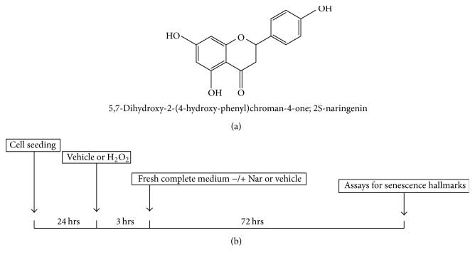
Nar (5,7-dihydroxy-2-(4-hydroxy-phenyl)chroman-4-one) (Figure 1(a)) and paxilline were purchased from Sigma-Aldrich (Milan, Italy). They were dissolved (10−2 M) in DMSO and further diluted in cell culture medium. All other reagents were purchased from standard sources.
Figure 1.
(a) The structure of Nar. (b) Schematic depiction of the cell culture treatment protocol.
2.2. Cell Culture
H9c2 cells (ATTC, Manassas, VA, USA), a subclonal cell line that was derived from embryonic rat hearts [23], were cultured in DMEM (Sigma-Aldrich, Milan, Italy), supplemented with 10% fetal bovine serum (FBS, Sigma-Aldrich, Milan, Italy), 100 units/mL penicillin, and 100 mg/mL streptomycin in tissue culture flasks at 37°C in a humidified atmosphere of 5% CO2.
2.3. H9c2 Cell Senescence Model
H9c2 cell senescence was induced by the exogenous oxidative insult H2O2 as previously reported [19]. Briefly, the cells were seeded at a density of 10 × 103 cells/cm2. After 24 h to allow cell attachment, the cells were treated with vehicle (water) or various concentrations of hydrogen peroxide (H2O2) (5–100 μM) for 3 hours and subsequently cultured for 3 days, after which the senescence hallmarks were determined (Figure 1(b)). In parallel, low-micromolar concentrations (4 and 40 μM) of Nar or vehicle (0.1% DMSO) were added to the cells immediately after the senescence insult and maintained in the medium for 3 days.
2.4. Senescence-Associated β-Galactosidase (sa-β-Gal) Staining
To evaluate the percentage of senescent cells 3 days after the H2O2 insult, staining for the senescence marker sa-β-gal was performed as previously reported [25]. Briefly, the treated cells were fixed in p-formaldehyde and incubated in staining solution for 16 hours (37°C, dry incubator). The cells were then washed in PBS (1x), and images of randomly selected light microscopic fields were captured (5 fields per well, 100x). Both blue (senescent) and uncolored (nonsenescent) cells were counted using ImageJ (ImageJ Software, version 1.41, USA).
2.5. RNA Extraction and Real-Time PCR Analysis
H2O2-injured H9c2 cells were collected, and total RNA was extracted using the RNeasy® Mini Kit (Qiagen, Hilden, Germany) according to the manufacturer's instructions. cDNA synthesis was performed (500 ng of RNA) using the i-Script cDNA synthesis kit (BioRad, Hercules, USA). Real-time RT-PCR reactions (25 μL, Fluocycle® II SYBR® [Euroclone, Milan, Italy], 1.5 μL of 10 μM forward and reverse primers for the p16 and p21 cell cycle regulators (Sigma-Aldrich, Milan, Italy), 3 μL cDNA, and 19 μL H2O) were performed for 38 cycles using the following temperature profiles: 94°C for 1 minute (initial denaturation); 55–59°C for 30 seconds (annealing); and 72°C for 1 second (extension).
Furthermore, the same real-time RT-PCR reactions were performed to assess the expression of two estrogen-regulated genes; estrogen receptor β (ER β) and vitamin D receptor (VDR) were studied as indicators of estrogenic activity [26]. The list of primers used in the study was shown in Table 1.
Table 1.
Nucleotide sequences, annealing temperature, and product size of the primers utilized in real-time RT-PCR experiments.
| Gene | Primer nucleotide sequences | Annealing temperature (°C) | Product size (base pairs) |
|---|---|---|---|
| p16 | FOR 5′- CCGAGAGGAAGGCGAACTC -3′ | 66.3 | 76 |
| REV 5′- GCTGCCCTGGCTAGTCTATCTG -3′ | 66.2 | ||
|
| |||
| p21 | FOR 5′- GAGCAAAGTATGCCGTCGTC -3′ | 64.7 | 127 |
| REV 5′- CTCAGTGGCGAAGTCAAAGTTC -3′ | 65.0 | ||
|
| |||
| ER β | FOR 5′- CTACAGAGAGATGGTCAAAAGTGGA -3′ | 64.4 | 218 |
| REV 5′- GGGCAAGGAGACAGAAAGTAAGT -3′ | 63.6 | ||
|
| |||
| VDR | FOR 5′- GTGACTTTGACCGGAACGTG -3′ | 65.6 | 280 |
| REV 5′- ATCATCTCCCTCTTACGCTG -3′ | 60.8 | ||
2.6. Cell Cycle Analysis
The percentages of H2O2-injured cells in the various cell phases were determined using the Muse™ Cell Cycle reagent (Merck KGaA, Darmstadt, Germany). Briefly, senescent adherent cells were collected and centrifuged at 300 ×g for 5 minutes. The pellet was washed with PBS and suspended in 100 μL of PBS and then slowly added to 1 mL of ice-cold 70% ethanol and maintained overnight at −20°C. Then, an aliquot of the cell suspension (2 × 10⁵ cells) was centrifuged, washed with PBS, suspended in the nuclear DNA stain, propidium iodide, and analyzed [27].
2.7. ROS Production
The ROS generation was assessed using the fluorogenic probe DCFH2-DA (Molecular Probes, Invitrogen) [19, 28]. Briefly, injured H9c2 cells, treated with Nar or vehicle, were washed in PBS/10 mM glucose (loading buffer) and loaded with 8 μM DCFH2-DA for 30 min in the dark (37°C). The cells were then washed and incubated in loading buffer. FDA fluorescence was estimated using a plate reader with wavelengths of 485 nm (excitation) and 520 nm (emission) (Wallac, Victor 2, 1420 multilabel counter, PerkinElmer) 30 minutes later. Then, after washing with PBS/10 mM glucose, the cells were incubated with crystal violet for 30 min at room temperature. After extensive washing, a solution of 1% SDS was added to each well, the plates were mechanically shaken for 1 h, and the absorbance at 595 nm was determined. The DCFH2-DA fluorescence values were normalized to the cell content of each well as indicated by the crystal violet assay.
2.8. Comet Assay
The DNA damage was assessed using the Comet assay as previously reported [29]. Briefly, microscope slides were coated with 0.5% normal-melting-point-agarose (NMA) in calcium- and magnesium-free PBS, covered with a coverslip, and kept at 4°C in a humid box until use. H9c2 cell suspensions (5 × 10³ cells) were added to 1% low-melting-point agarose (LMPA, 35°C). A volume of the LMPA-embedded sample was layered over the NMA layer. After the agarose solidified, a final layer of 1% LMPA was added. Following the solidification of this layer of agarose, the coverslips were removed and the slides were treated with lysing solution (10% DMSO, 1% TRITON X-100, 2.5 M NaCl, 100 mM EDTA, 10 mM TRIS, and 1% sodium sarcosinate, pH 10) for 1 h at 4°C in the dark. The slides were rinsed in distilled water and placed in a horizontal gel electrophoresis apparatus containing 75 mM NaOH and 1 mM EDTA, pH 12, for 20 min. Electrophoresis was carried out at 25 V and 300 mA for 10 min. The slides were then removed and incubated in neutralizing solution (0.4 M TRIS, pH 7.5, 5 min) three times. The slides were drained, stained with ethidium bromide, and observed using a fluorescence microscope (400x). The amount of damaged DNA (migrated in the tail, Comet) was expressed as percent of total fluorescence for each nucleus. To calculate the distribution of DNA damage, the comets were classified into five categories according to the percent of the DNA in the tail: class I: 0–6% (no damage); class II: 6.1–17% (low damage); class III: 17.1–35% (moderate damage); class IV: 35.1–60% (high damage); and class V: 60.1–100% (extreme damage) [30].
2.9. Mitochondrial Metabolic Activity
Briefly, the mitochondrial metabolic activity of senescent H9c2 cells that had been treated with Nar or vehicle was determined using the [3-(4,5-dimethylthiazol-2-yl)-5-(3-carboxymethoxyphenol)-2-(4-sulfophenyl)-2H-tetrazolium, inner salt] (MTS) assay according to manufacturer's instructions (Promega, Milano, Italy) [31]. This tetrazolium dye can be reduced by the metabolic reducing agents NADH and NADPH to a water-soluble formazan salt; the amount of formazan produced is considered to be a marker of the oxidative metabolic activity index [32]. MTS reagent was added to the PIGA ligand-treated cells, and the colorimetric MTS conversion was quantified after 2 h by measuring the absorbance at 490 nm using a microplate reader (WallacVictor 2, 1420 Multilabel Counter, Perkin Elmer, USA).
2.10. Calcium Retention Capacity Measurement
The calcium retention capacity (CRC) was determined as previously described [33] using Calcium Green-5N (λex = 505 nm, λem = 535 nm), a low affinity nonmembrane-permeant probe that increases its fluorescence emission upon binding Ca2+. Senescent H9c2 cells (2 × 10⁶ cells) were suspended in CRC buffer (250 mM sucrose, 1 mM Pi-Tris, and 10 mM MOPS-Tris, pH 7.4), permeabilized with digitonin (40 μM, for 5 min, 0°C), and incubated with the respiratory substrate 5 mM succinate and 0.25 μM Calcium Green-5 N (25°C). Ca2+ pulses (10 nmoles) were added at 3-minute intervals until the onset of the permeability transition, in the presence or absence of Nar. To evaluate the involvement of mitoBKCa channels in the CRC modulation, the selective mitoBKCa blocker paxilline was applied. The fluorescence was measured using a microplate fluorimeter equipped with thermostatic control (Victor Wallac 2, Perkin Elmer, CA, USA). The results are presented as the sample CRC normalized to the CRC of the control (CRC0).
2.11. Estradiol Levels in the Conditioned Cell Medium
The amounts of estradiol in the conditioned medium of senescent H9c2 cells that had been treated with Nar or vehicle were measured using a competitive EIA assay according to the manufacturer's instructions (Estradiol EIA Kit, Cayman Chemical, Ann Arbor, MI, USA). The absorbance data obtained from the standard curve and the cell supernatants were plotted as logit B/B0 versus log concentration and fit using a linear model. To monitor the possible interference of Nar in the assay, standard curves in the presence of Nar concentrations were also performed.
2.12. Statistical Analyses
The nonlinear multipurpose curve-fitting program GraphPad Prism (GraphPad Software Inc., San Diego, CA) was used for the data analysis and graphic presentations. All results are presented as the means ± standard errors of the means (SEM) of data for triplicate samples and are representative of three different experiments. The statistical analyses were performed using a t-test or one-way analysis of variance (ANOVA) with Bonferroni's corrected t-test for post hoc pair-wise comparisons. p < 0.05 was considered statistically significant.
3. Results and Discussion
Several epidemiological studies have suggested an inverse correlation between aging-associated disorders, in particular cardiovascular diseases, and the intake of citrus fruits [34, 35]. Grapefruit, bergamot, and sour oranges contain high amounts of the flavanone-7-O-glycoside naringin, which is metabolized by the human gut microflora to the aglycone Nar [36] (Figure 1(a)). It has been estimated that the mean peak of the plasma levels of Nar reaches micromolar concentrations following consumption of a serving of citrus fruit [37–39]. Various pharmacological effects including antimutagenic, antiatherogenic, antihypertensive, and anti-inflammatory activities have been reported for Nar [40–44]. Recently, the prevention of cell death by flavanone has been studied in H9c2 myocardial cells that were injured by exposure to a high concentration of glucose or to daunorubicin. The results have suggested that Nar significantly ameliorates the drug-induced toxicity by inhibiting apoptosis [1, 2]. A very recent publication has demonstrated that Nar has the ability to improve the antioxidant status and membrane phospholipid composition in old rats [18]. These data prompted us to investigate the effects of Nar in a cellular model of aged myocardial cells.
3.1. Senescence-Associated β-Galactosidase Staining and Cell Cycle Investigations
In the present study, the cellular senescence model that was used to assess the cardioprotective action of Nar was established with H9c2 cells that were exposed to H2O2 to induce senescence. Despite their embryonic origin, H9c2 cells are similar to normal primary cardiomyocytes with respect to their energy metabolism and have been successfully used as an in vitro model for studies of myocardial pathophysiology, including the aging processes [19, 45–49].
Myocardial cellular senescence was triggered by exposing the cells to H2O2 as previously reported [19], and the presence of sa-β-gal product was monitored as a marker of cellular senescence. To establish the effective dose of H2O2, H9c2 cells were exposed to a range of concentrations of H2O2 (from 5 μM to 100 μM) for 3 hours and subsequently cultured with fresh complete medium for 72 h (Figure 1(b)).
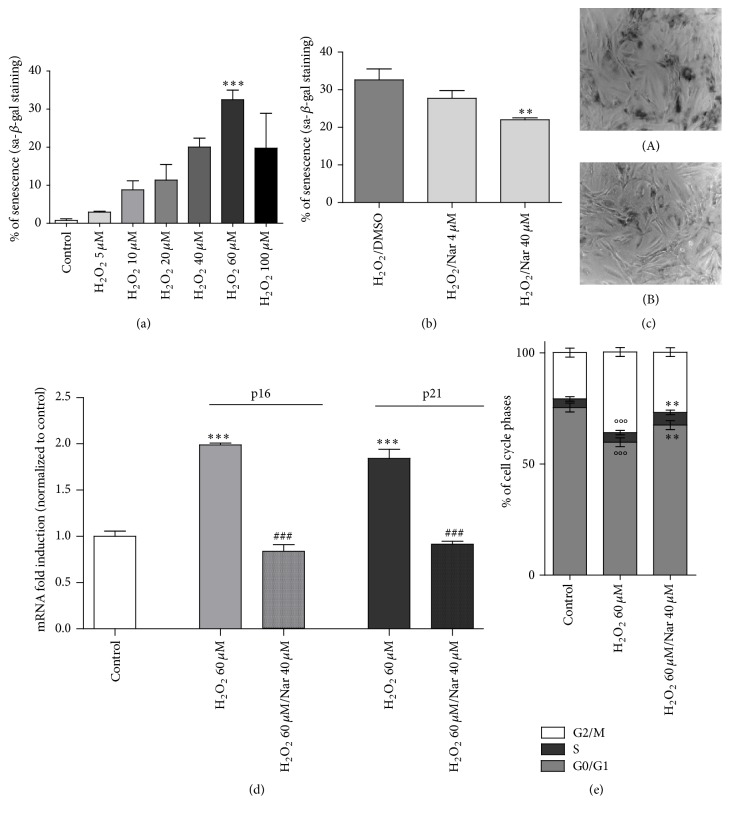
The presence of sa-β-gal product after H2O2 incubations was examined (Figure 2). A concentration-dependent increase in the sa-β-gal activity was observed in the H2O2-exposed cells; the 60 μM H2O2 concentration effectively induced significant staining for sa-β-gal (p < 0.01, Figure 2(a)). A higher dose of H2O2 (100 μM) did not further increase the senescence marker, probably due to its cytotoxic effect (data not shown). Thus, the 60 μM concentration of H2O2 was established as the effective drug concentration (Emax) for use in the cell treatment protocol as shown in Figure 1.
Figure 2.
Nar effects on H9c2 cell senescence hallmarks. (a) Senescence-associated b-galactosidase staining. The data are shown as the percentages of β-galactosidase-positive cells. Each bar represents the mean ± SEM of three replicates from three independent experiments. ∗∗∗p < 0.01 versus the control group; (b) the data are shown as percentage of β-galactosidase-positive cells. Each bar represents the mean ± SEM of three replicates from three independent experiments. ∗∗p < 0.01 versus the H2O2-treated cells. (c) Representative phase contrast photomicrographs of treated cells; (A) H2O2-treated cells; (B) 40 μM Nar-cotreated cells. (d, e) Cell cycle arrest machinery. (d) p16 and p21 mRNA fold induction. Each bar represents the mean ± SEM of three replicates from three independent experiments. ∗∗∗p < 0.001 versus the control group; ###p < 0.001 versus the H2O2-treated group. (e) Cell cycle phases. Each bar represents the mean ± SEM of three replicates from three independent experiments. °°°p < 0.001 versus the control group; ∗∗p < 0.01 versus the H2O2-treated group.
The cell cycle regulators p16 and p21, which are mainly activated during the senescence process, and the percentage of cells in the various cell cycle phases were then evaluated. The real-time RT-PCR results showed that the H2O2 cell treatments significantly increased the levels of p16 and p21 mRNA (p < 0.01 and p < 0.001, Figure 2(d)). Consistent with these results, the cell cycle analysis demonstrated that the cell cycle was arrested in G2/M phase following H2O2 treatment (p < 0.001, Figure 2(e)). All of these results are consistent with the literature [19, 50–54]. Specifically, previous studies have demonstrated that the number of senescent cells as indicated by X-gal staining and the p21 cell cycle regulator are increased in a dose-dependent manner by treatment with H2O2 and that the cell cycle is arrested at the G1 and G2/M phases [19, 53]. Furthermore, it has been demonstrated that challenge of H9c2 cells with H2O2 induces a significant increase in the intracellular ROS level and in the olive tail moment, which indicates an increase in DNA damage [19].
Therefore, H9c2 cells were challenged with 60 μM H2O2 for 3 hours and then cultured with fresh complete medium containing various concentrations of Nar. Senescence hallmarks were assessed 72 h later (Figure 2(b)).
Treatment with 40 μM Nar significantly protected the cells from the senescence-inducing insult; specifically, it decreased the number of sa-β-gal positive cells (p < 0.01, Figures 2(b) and 2(c)). In contrast, treatment with 4 μM Nar failed to prevent the development of cellular senescence, although a trend for a reduction in the number of senescent cells was evident (Figure 2(b)). Thus, the 40 μM concentration was selected for subsequent assays. Notably, previous human pharmacokinetic analyses have shown that Nar, following a single oral administration of 135 mg (equal to 1 or 2 glasses of grapefruit juice), is rapidly absorbed and its concentration in plasma reaches a peak of approximately 10 μM in 3.5 hours [27, 55]. Furthermore, Nar cotreatment abolished the increase in p16 and p21 mRNA due to H2O2 (p < 0.01 and p < 0.001, Figure 2(d)) and reduced the cell cycle arrest in G2/M (p < 0.01, Figure 2(e)).
Collectively, the present data showed for the first time that Nar treatment counteracts the manifestation of the senescent hallmarks in H2O2-treated cells, which suggests that it has the ability to prevent the prematurely induced senescence in H9c2 cells. In a very recent review, Nar was identified as an effective antiaging ingredient among the traditional Chinese medicine natural products [56]. This hypothesis has been proposed based on the activity of Nar to increase glucose uptake in skeletal muscle cells through activation of AMPK [57]. In addition, the recent data obtained using old rats have confirmed this assumption. Indeed, flavanone administration to old male rats was able to modulate the activity of antioxidant enzymes including catalase, superoxide dismutase 1, and glutathione reductase. All of these enzymes significantly decrease during aging, and their activities were found to be increased in flavanone-fed old rats compared to vehicle-treated animals. Furthermore, thiobarbituric acid reactive substances (TBARS) were significantly decreased by Nar administration, and the membrane phospholipid composition was improved in favor of ω-3 PUFA and the ω-6/ω-3 PUFA ratio [18].
3.2. Nar Reduced ROS Generation and DNA Damage
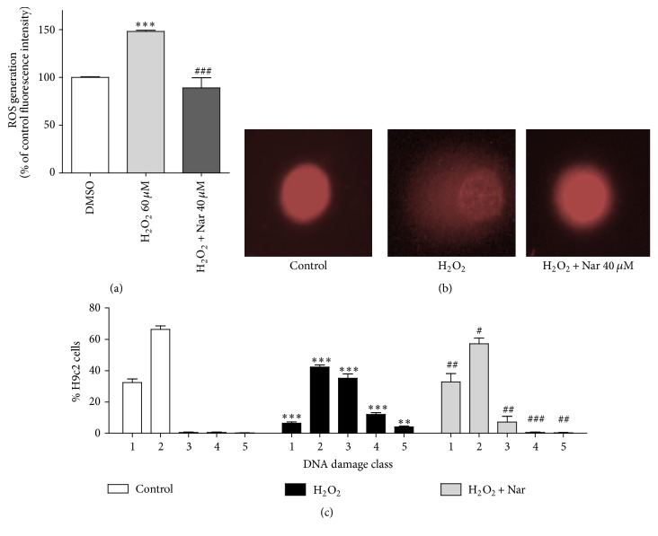
In this context, among the pathways typically impaired during cardiac aging-associated damage, the cellular mechanisms associated with the generation of radical oxidative species (ROS) generation and alterations in DNA integrity are frequently altered [58, 59]. These parameters were therefore assessed in our model. As shown in Figure 3, H2O2 treatment caused a significant increase in ROS generation (p < 0.001, Figure 3(a)), and cotreatment of the cells with Nar resulted in maintenance of the ROS levels as those of the control (p < 0.001 versus cells treated with H2O2 alone).
Figure 3.
ROS production and DNA damage. (a) ROS generation in H9c2 cells. Each bar represents the mean ± SEM of three replicates from three independent experiments. ∗∗∗p < 0.001 versus the control cells, and ###p < 0.001 versus the H2O2-treated cells. (b) Representative photomicrographs of cells assessed using the Comet assay. (c) DNA damage was calculated as classes of DNA damage, as described in Section 2.8. ∗∗∗p < 0.001 and ∗∗p < 0.01 versus control; ###p < 0.001, ##p < 0.01, and #p < 0.05 versus the H2O2-treated cells.
Consistent with the ROS results, the DNA damage assessed using the Comet assay was increased in the H2O2-exposed H9c2 cells (Figures 3(b) and 3(c)). Specifically, senescent cells demonstrated moderate/high DNA damage (corresponding to classes 3, 4, and 5) compared to control cells (p < 0.5, p < 0.1, and p < 0.001, Figure 3(c)). Notably, in the Nar-treated senescent cells, the reduction in the high numbers of cells with moderate/high DNA damage (classes 3, 4, and 5, p < 0.1, p < 0.001, and p < 0.1 versus the H2O2-treated cells, resp.) suggested a protective activity of Nar against ROS-induced genotoxic damage (Figure 3(c)).
It was indirectly shown that the reduction of both ROS and DNA damage is a result of Nar effect on antioxidative enzyme activity or by scavenger function. These results are consistent with previous studies that were carried out using the skin of hairless mice in which UVB irradiation was used to induce oxidative stress and premature aging [60]. Nar has been shown to mitigate UVB irradiation-induced oxidative stress by reducing superoxide anion production and the mRNA expression of a subunit of NADPH oxidase [60].
3.3. Nar Modulated the Mitochondrial Metabolic Activity and Increased the Calcium Retention Capacity
Additional cellular mechanisms that are often impaired during aging processes include those related to mitochondrial homeostasis [61–63]. Cellular senescence and mitochondrial dysfunction are hallmarks of aging; recent works have shown that mitochondrial defects can even cause a distinct senescence phenotype termed mitochondrial dysfunction-associated senescence [62, 63]. The roles of the mitochondrial electron transport chain, bioenergetic balance, redox state, metabolic signature, and calcium homeostasis have been recently studied in the context of the control of cellular growth arrest that is associated with senescence. These studies indicated that multiple mitochondrial signaling pathways in addition to mitochondrial ROS can induce cellular senescence [61]. Perturbation of mitochondrial homeostasis promotes the establishment and maintenance of cellular senescence, and various strategies to improve mitochondrial function have emerged as therapeutic approaches to aging [64].
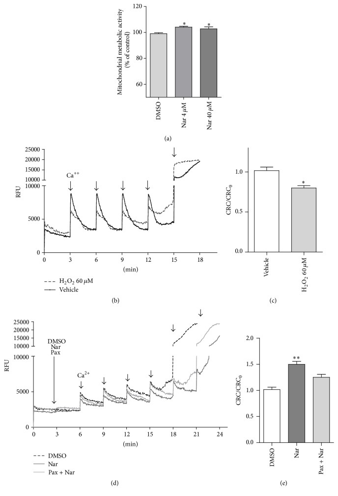
In this context, the mitochondrial metabolic activity was explored in the Nar-treated H9c2 cells using a tetrazolium dye. The results demonstrated that Nar treatment increased the mitochondrial metabolic activity of senescent cells relative to that of the controls (p < 0.05, Figure 4(a)). Interestingly, parallel experiments in naïve H9c2 cells (not senescent) showed that Nar did not modulate mitochondrial metabolism per se, and it did not affect the number of senescent cells relative to the control (data not shown).
Figure 4.
Effects of H2O2 and Nar on the mitochondrial oxidative metabolism activity and CRC. (a) H9c2 cells were treated with H2O2 and DMSO or Nar, and the mitochondrial oxidative metabolism activity was examined. (b–e) The cells were suspended in CRC medium and permeabilized with digitonin. To these cells, 0.25 M Calcium Green-5N and 5 mM succinate were added. This addition was followed by a series of Ca2+ pulses (10 nmoles) at 3-minute intervals until onset of the permeability transition (plateau). The Relative Fluorescence Unit (RFU) was recorded by spectrophotometer technique. ∗p < 0.05 versus DMSO-treated cells. (b) Representative traces of the H9c2 cell treatments with H2O2 and vehicle are shown in dashed black and gray, respectively. (c) The results from the H2O2-treated cells are shown as the CRC normalized to the CRC of control (CRC0). The data shown are the means ± SEM of three independent experiments. ∗p < 0.05 versus vehicle. (d) Representative traces of senescent H9c2 cells cotreated with DMSO (dashed black), Nar (dark gray), and Nar + Pax (light gray). (e) The results from senescent cells treated with Nar and Nar + Pax are shown as the CRC normalized to the CRC of control (CRC0). The data shown are the means ± SEM of three independent experiments. ∗∗p < 0.01 versus DMSO-treated cells.
Next, the effects of Nar on a mitochondrial parameter recognized to be an indicator of damage resistance, the mitochondrial Ca2+-buffering cell capacity, were assessed. In particular, the mitochondrial calcium retention capacity, CRC, was explored; the upper limit of the CRC reflects the opening of the permeability transition pore (MPTP) and the dissipation of mitochondrial potential [65]. As shown in Figure 4, the cells treated with H2O2 (dashed black line) showed a significantly reduced ability to retain Ca2+ as indicated by the decrease in the CRC/CRC0 ratio (p < 0.05) (Figures 4(b) and 4(c)). Cells that had previously been treated with H2O2 were used to investigate the CRC in the presence of Nar. Nar was able to increase the mitochondrial CRC compared to the control cells (Figure 4(d), gray line versus dashed black line, Figure 4(e), p < 0.01).
The present data indicated that senescent H9c2 cells were more susceptible to the opening of the MPTP, in accordance with previous studies on cardiomyocytes isolated from old rats [66]. Notably, treatment of the cells with Nar was able to reduce this altered mitochondrial sensitivity.
It has been previously demonstrated that Nar is able to activate the mitoBKCa channels in mitochondria isolated from young rat hearts. This produces a mild depolarization of the membrane potential. This depolarization is responsible for reducing the uptake of Ca2+ into the mitochondrial matrix and preserving the mitochondria from the Ca2+ overload and the subsequent MPTP opening [4, 14]. To evaluate the involvement of mitoBKCa channels in the Nar-mediated CRC effects, the selective mitoBKCa blocker paxilline was tested in the CRC assessment. Paxilline alone had no significant effect on the CRC (data not shown), which might indicate that mitoBKCa channels were inactive under the resting condition. Interestingly, consistent with the modulation of mitoBKCa channels by Nar [14], the CRC increase exerted by Nar was partially prevented by cotreatment of the cells with paxilline (Figures 4(d), light gray line, and 4(e)). These results indicated that the Nar effects on the CRC might be mediated, at least in part, by the activation of mitoBKCa channels. Interestingly, it has been previously shown that the opening of mitoBKCa channels improves the function and energetic performance of cardiac mitochondria, which is consistent with our results [67]. One possible explanation may be the mild depolarization that occurs during the mitoBKCa channel opening. Indeed, it has been demonstrated that mice produce fewer free radicals and live longer when they have more uncoupled mitochondria, which show a low membrane potential, in muscles [68].
3.4. Nar Modulated the Levels of Estradiol Released by Cells and the Estrogen-Regulated Gene Expression
It is well known that estrogen is strongly implicated in cardiomyocyte survival and that its levels vary widely based on age [69, 70]. In general, premenopausal women are protected from coronary heart disease compared with age-matched men, but this female protection appears to be lost after menopause, which suggests that estrogen has beneficial effects on the cardiovascular system [22]. In addition to adrenal production of estrogen, the synthesis of estrogen by cardiac cells has been proposed, although this process has been very poorly elucidated (for a review, see [71]). Interestingly, some papers have suggested that local estrogen biosynthesis in the heart effectively activates the estrogen receptors α and β and downstream target genes in a gender-based fashion [72]. For these reasons, in the present study, the Nar effects on the estrogenic pathway were studied in senescent H9c2 cells. Specifically, we evaluated the estradiol released by cells and the estrogen-regulated gene expression.
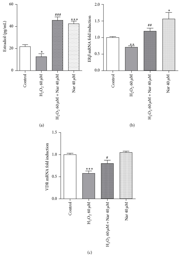
As shown in Figure 5, H2O2 challenge induced a significant decrease in the levels of estradiol released by the cells (p < 0.05). This reduction was significantly counteracted by Nar treatment (p < 0.001 versus the H2O2-treated cells) (Figure 5(a)). Surprisingly, the level of estradiol in Nar-control cells (cells without H2O2 challenge) was higher than in the control cells (p < 0.001 versus control). It is reasonable to presume that such an increase may be due to the activation of cellular response mechanisms during the Nar treatment: one possible explanation may be that the response is related to cellular self-adaptive responses. Although the estrogenic potency of Nar has been controversial [7, 73], some studies have demonstrated the estrogenic effects of Nar and Nar-type flavonoids in mammalian in vitro systems using the estrogen-inducible MVLN luciferase assay [74].
Figure 5.
Effect of Nar on the levels of released estradiol and estrogen-regulated gene mRNA expression. (a) Each bar represents the mean ± SEM of three replicates from three independent experiments. The measured estradiol levels were within the reference range [24]. Specific calibration curves and appropriate positive/negative controls were carried out during the assay. ∗p < 0.05 and ∗∗∗p < 0.001 versus the control; ###p < 0.001 versus H2O2-treated cells. (b) ER β and (c) VDR mRNA expression as indicators of estrogenic activity in the senescent H9c2 cells. Each bar represents the mean ± SEM of three replicates from three independent experiments. ∗p < 0.05, ∗∗p < 0.01, and ∗∗∗p < 0.001 versus the control; #p < 0.05 and ##p < 0.01 versus H2O2-treated cells.
Furthermore, the Nar-mediated expression of some commonly used parameters of drug estrogenic activity, specifically the estrogen-regulated genes ER β and VDR, was investigated. Quantitative real-time PCR experiments demonstrated that the expression of the ERβ and VDR mRNA was significantly reduced in H2O2-treated cells (p < 0.01, Figure 5(b)) and that Nar was able to prevent this decrease (p < 0.01 versus the H2O2-treated cells). Consistent with the estradiol results, mRNA expression of both of these genes was increased by the Nar-only treatment.
Together, these data suggested that the ability of Nar to counterbalance the effects of H2O2 on the estrogenic pathway may contribute to its antisenescence effects.
4. Conclusions
In conclusion, Nar treatment of H9c2 cells prevented the prematurely induced senescence; Nar protection may involve multiple cellular pathways on the basis that it modulated the ROS levels, the mitochondrial potassium channels, and the estrogen-related pathways. In particular, the specific pathways involved seem to be those associated with the mitochondria. These results pave the way to exciting nutraceutical advances.
Acknowledgments
Funding for this study was provided by the University of Pisa (Progetti di Ricerca di Ateneo (PRA)).
Conflicts of Interest
All of the authors declare that they have no competing interests or other interests that might be perceived to influence the results and discussion reported in this paper.
Authors' Contributions
Eleonora Da Pozzo designed and conducted the research, analyzed the data, and drafted the manuscript; Barbara Costa performed the Comet assay and critically revised the manuscript; Chiara Cavallini performed the cell culturing; Lara Testai and Alma Martelli critically revised the manuscript; Vincenzo Calderone and Claudia Martini designed the research and critically revised the manuscript. All authors read and approved the final manuscript.
References
- 1.Rani N., Bharti S., Krishnamurthy B., et al. Pharmacological properties and therapeutic potential of naringenin: a citrus flavonoid of pharmaceutical promise. Current Pharmaceutical Design. 2016;22(28):4341–4359. doi: 10.2174/1381612822666160530150936. [DOI] [PubMed] [Google Scholar]
- 2.Alam M. A., Subhan N., Rahman M. M., Uddin S. J., Reza H. M., Sarker S. D. Effect of citrus flavonoids, naringin and naringenin, on metabolic syndrome and their mechanisms of action. Advances in Nutrition. 2014;5(4):404–417. doi: 10.3945/an.113.005603. [DOI] [PMC free article] [PubMed] [Google Scholar]
- 3.Sung H. K., Won S. P., Kim N., et al. Mitochondrial Ca2+-activated K+ channels more efficiently reduce mitochondrial Ca2+ overload in rat ventricular myocytes. American Journal of Physiology—Heart and Circulatory Physiology. 2007;293(1):H307–H313. doi: 10.1152/ajpheart.00789.2006. [DOI] [PubMed] [Google Scholar]
- 4.Testai L., Rapposelli S., Martelli A., Breschi M. C., Calderone V. Mitochondrial potassium channels as pharmacological target for cardioprotective drugs. Medicinal Research Reviews. 2015;35(3):520–553. doi: 10.1002/med.21332. [DOI] [PubMed] [Google Scholar]
- 5.Kuiper G. G. J. M., Carlsson B., Grandien K., et al. Comparison of the ligand binding specificity and transcript tissue distribution of estrogen receptors α and β. Endocrinology. 1997;138(3):863–870. doi: 10.1210/en.138.3.863. [DOI] [PubMed] [Google Scholar]
- 6.Kuiper G. G., Lemmen J. G., Carlsson B., et al. Interaction of estrogenic chemicals and phytoestrogens with estrogen receptor beta. Endocrinology. 1998;139(10):4252–4263. doi: 10.1210/endo.139.10.6216. [DOI] [PubMed] [Google Scholar]
- 7.Ruh M. F., Zacharewski T., Connor K., Howell J., Chen I., Safe S. Naringenin: a weakly estrogenic bioflavonoid that exhibits antiestrogenic activity. Biochemical Pharmacology. 1995;50(9):1485–1493. doi: 10.1016/0006-2952(95)02061-6. [DOI] [PubMed] [Google Scholar]
- 8.Totta P., Acconcia F., Leone S., Cardillo I., Marino M. Mechanisms of naringenin-induced apoptotic cascade in cancer cells: involvement of estrogen receptor α and β signalling. IUBMB Life. 2004;56(8):491–499. doi: 10.1080/15216540400010792. [DOI] [PubMed] [Google Scholar]
- 9.Guo D., Wang J., Wang X., et al. Double directional adjusting estrogenic effect of naringin from Rhizoma drynariae (Gusuibu) Journal of Ethnopharmacology. 2011;138(2):451–457. doi: 10.1016/j.jep.2011.09.034. [DOI] [PubMed] [Google Scholar]
- 10.Swarnkar G., Sharan K., Siddiqui J. A., et al. A naturally occurring naringenin derivative exerts potent bone anabolic effects by mimicking oestrogen action on osteoblasts. British Journal of Pharmacology. 2012;165(5):1526–1542. doi: 10.1111/j.1476-5381.2011.01637.x. [DOI] [PMC free article] [PubMed] [Google Scholar]
- 11.Mulvihill E. E., Burke A. C., Huff M. W. Citrus flavonoids as regulators of lipoprotein metabolism and atherosclerosis. Annual Review of Nutrition. 2016;36:275–299. doi: 10.1146/annurev-nutr-071715-050718. [DOI] [PubMed] [Google Scholar]
- 12.Rajadurai M., Stanely Mainzen Prince P. Preventive effect of naringin on lipid peroxides and antioxidants in isoproterenol-induced cardiotoxicity in Wistar rats: biochemical and histopathological evidences. Toxicology. 2006;228(2-3):259–268. doi: 10.1016/j.tox.2006.09.005. [DOI] [PubMed] [Google Scholar]
- 13.Rajadurai M., Prince P. S. M. Preventive effect of naringin on cardiac mitochondrial enzymes during isoproterenol-induced myocardial infarction in rats: A Transmission Electron Microscopic Study. Journal of Biochemical and Molecular Toxicology. 2007;21(6):354–361. doi: 10.1002/jbt.20203. [DOI] [PubMed] [Google Scholar]
- 14.Testai L., Martelli A., Marino A., et al. The activation of mitochondrial BK potassium channels contributes to the protective effects of naringenin against myocardial ischemia/reperfusion injury. Biochemical Pharmacology. 2013;85(11):1634–1643. doi: 10.1016/j.bcp.2013.03.018. [DOI] [PubMed] [Google Scholar]
- 15.Mojžišová G., Mirossay L., Kučerová D., Kyselovič J., Miroššay A., Mojžiš J. Protective effect of selected flavonoids on in vitro daunorubicin-induced cardiotoxicity. Phytotherapy Research. 2006;20(2):110–114. doi: 10.1002/ptr.1811. [DOI] [PubMed] [Google Scholar]
- 16.Shiromwar S. S., Chidrawar V. R. Combined effects of p-coumaric acid and naringenin against doxorubicin-induced cardiotoxicity in rats. Pharmacognosy Research. 2011;3(3):214–219. doi: 10.4103/0974-8490.85012. [DOI] [PMC free article] [PubMed] [Google Scholar]
- 17.Reddy T. K., Seshadri P., Reddy K. K. R., Jagetia G. C., Reddy C. D. Effect of Terminalia arjuna extract on adriamycin-induced DNA damage. Phytotherapy Research. 2008;22(9):1188–1194. doi: 10.1002/ptr.2428. [DOI] [PubMed] [Google Scholar]
- 18.Miler M., Živanović J., Ajdžanović V., et al. Citrus flavanones naringenin and hesperetin improve antioxidant status and membrane lipid compositions in the liver of old-aged Wistar rats. Experimental Gerontology. 2016;84:49–60. doi: 10.1016/j.exger.2016.08.014. [DOI] [PubMed] [Google Scholar]
- 19.Dong R., Xu X., Li G., et al. Bradykinin inhibits oxidative stress-induced cardiomyocytes senescence via regulating redox state. PLoS ONE. 2013;8(10) doi: 10.1371/journal.pone.0077034.e77034 [DOI] [PMC free article] [PubMed] [Google Scholar]
- 20.Dai D.-F., Chiao Y. A., Marcinek D. J., Szeto H. H., Rabinovitch P. S. Mitochondrial oxidative stress in aging and healthspan. Longevity & Healthspan. 2014;3(1, article 6) doi: 10.1186/2046-2395-3-6. [DOI] [PMC free article] [PubMed] [Google Scholar]
- 21.Pepe S. Mitochondrial function in ischaemia and reperfusion of the ageing heart. Clinical and Experimental Pharmacology and Physiology. 2000;27(9):745–750. doi: 10.1046/j.1440-1681.2000.03326.x. [DOI] [PubMed] [Google Scholar]
- 22.Gouva L., Tsatsoulis A. The role of estrogens in cardiovascular disease in the aftermath of clinical trials. HORMONES. 2004;3(3):171–183. doi: 10.14310/horm.2002.11124. [DOI] [PubMed] [Google Scholar]
- 23.Hescheler J., Meyer R., Plant S., Krautwurst D., Rosenthal W., Schultz G. Morphological, biochemical, and electrophysiological characterization of a clonal cell (H9c2) line from rat heart. Circulation Research. 1991;69(6):1476–1486. doi: 10.1161/01.RES.69.6.1476. [DOI] [PubMed] [Google Scholar]
- 24.Mitak M., Gojmerac T., Mandić B., Cvetnić Ž. Changes in serum concentration of 17β-estradiol in female rats during estrous cycle after treatment with atrazine and zearalenone. Veterinarni Medicina. 2001;46(5):145–148. [Google Scholar]
- 25.Costa B., Bendinelli S., Gabelloni P., et al. Human glioblastoma multiforme: p53 reactivation by a novel MDM2 inhibitor. PLoS ONE. 2013;8(8) doi: 10.1371/journal.pone.0072281.e72281 [DOI] [PMC free article] [PubMed] [Google Scholar]
- 26.Höfer N., Diel P., Wittsiepe J., Wilhelm M., Kluxen F. M., Degen G. H. Investigations on the estrogenic activity of the metallohormone cadmium in the rat intestine. Archives of Toxicology. 2010;84(7):541–552. doi: 10.1007/s00204-010-0524-x. [DOI] [PubMed] [Google Scholar]
- 27.Daniele S., Taliani S., Da Pozzo E., et al. Apoptosis therapy in cancer: the first single-molecule co-activating p53 and the translocator protein in Glioblastoma. Scientific Reports. 2014;4, article 4749 doi: 10.1038/srep04749. [DOI] [PMC free article] [PubMed] [Google Scholar]
- 28.Barzegar A., Moosavi-Movahedi A. A. Intracellular ROS protection efficiency and free radical-scavenging activity of curcumin. PLoS ONE. 2011;6(10) doi: 10.1371/journal.pone.0026012.e26012 [DOI] [PMC free article] [PubMed] [Google Scholar]
- 29.Singh N. P., McCoy M. T., Tice R. R., Schneider E. L. A simple technique for quantitation of low levels of DNA damage in individual cells. Experimental Cell Research. 1988;175(1):184–191. doi: 10.1016/0014-4827(88)90265-0. [DOI] [PubMed] [Google Scholar]
- 30.Giovannelli L., Cozzi A., Guarnieri I., Dolara P., Moroni F. Comet assay as a novel approach for studying DNA damage in focal cerebral ischemia: differential effects of NMDA receptor antagonists and poly(ADP-ribose) polymerase inhibitors. Journal of Cerebral Blood Flow and Metabolism. 2002;22(6):697–704. doi: 10.1097/00004647-200206000-00008. [DOI] [PubMed] [Google Scholar]
- 31.da Pozzo E., Giacomelli C., Costa B., et al. TSPO PIGA ligands promote neurosteroidogenesis and human astrocyte well-being. International Journal of Molecular Sciences. 2016;17(7, article 1028) doi: 10.3390/ijms17071028. [DOI] [PMC free article] [PubMed] [Google Scholar]
- 32.Dunigan D. D., Waters S. B., Owen T. C. Aqueous soluble tetrazolium/formazan MTS as an indicator of NADH- and NADPH-dependent dehydrogenase activity. BioTechniques. 1995;19(4):640–649. [PubMed] [Google Scholar]
- 33.Costa B., Da Pozzo E., Giacomelli C., et al. TSPO ligand residence time influences human glioblastoma multiforme cell death/life balance. Apoptosis. 2015;20(3):383–398. doi: 10.1007/s10495-014-1063-3. [DOI] [PubMed] [Google Scholar]
- 34.Mulero J., Bernabé J., Cerdá B., et al. Variations on cardiovascular risk factors in metabolic syndrome after consume of a citrus-based juice. Clinical Nutrition. 2012;31(3):372–377. doi: 10.1016/j.clnu.2011.11.014. [DOI] [PubMed] [Google Scholar]
- 35.Buscemi S., Rosafio G., Arcoleo G., et al. Effects of red orange juice intake on endothelial function and inflammatory markers in adult subjects with increased cardiovascular risk. American Journal of Clinical Nutrition. 2012;95(5):1089–1095. doi: 10.3945/ajcn.111.031088. [DOI] [PubMed] [Google Scholar]
- 36.Kawaii S., Tomono Y., Katase E., Ogawa K., Yano M. Quantitation of flavonoid constituents in Citrus fruits. Journal of Agricultural and Food Chemistry. 1999;47(9):3565–3571. doi: 10.1021/jf990153+. [DOI] [PubMed] [Google Scholar]
- 37.Erlund I., Meririnne E., Alfthan G., Aro A. Plasma kinetics and urinary excretion of the flavanones naringenin and hesperetin in humans after ingestion of orange juice and grapefruit juice. Journal of Nutrition. 2001;131(2):235–241. doi: 10.1093/jn/131.2.235. [DOI] [PubMed] [Google Scholar]
- 38.Bugianesi R., Catasta G., Spigno P., D'Uva A., Maiani G. Naringenin from cooked tomato paste is bioavailable in men. Journal of Nutrition. 2002;132(11):3349–3352. doi: 10.1093/jn/132.11.3349. [DOI] [PubMed] [Google Scholar]
- 39.Brett G. M., Hollands W., Needs P. W., et al. Absorption, metabolism and excretion of flavanones from single portions of orange fruit and juice and effects of anthropometric variables and contraceptive pill use on flavanone excretion. British Journal of Nutrition. 2009;101(5):664–675. doi: 10.1017/S000711450803081X. [DOI] [PMC free article] [PubMed] [Google Scholar]
- 40.Choi J. S., Park K. Y., Moon S. H., Rhee S. H., Young H. S. Antimutagenic effect of plant flavonoids in the Salmonella assay system. Archives of Pharmacal Research. 1994;17(2):71–75. doi: 10.1007/BF02974226. [DOI] [PubMed] [Google Scholar]
- 41.Lee C.-H., Jeong T.-S., Choi Y.-K., et al. Anti-atherogenic effect of citrus flavonoids, naringin and naringenin, associated with hepatic ACAT and aortic VCAM-1 and MCP-1 in high cholesterol-fed rabbits. Biochemical and Biophysical Research Communications. 2001;284(3):681–688. doi: 10.1006/bbrc.2001.5001. [DOI] [PubMed] [Google Scholar]
- 42.Ribeiro I. A., Rocha J., Sepodes B., Mota-Filipe H., Ribeiro M. H. Effect of naringin enzymatic hydrolysis towards naringenin on the anti-inflammatory activity of both compounds. Journal of Molecular Catalysis B: Enzymatic. 2008;52-53(1–4):13–18. doi: 10.1016/j.molcatb.2007.10.011. [DOI] [Google Scholar]
- 43.Renugadevi J., Prabu S. M. Cadmium-induced hepatotoxicity in rats and the protective effect of naringenin. Experimental and Toxicologic Pathology. 2010;62(2):171–181. doi: 10.1016/j.etp.2009.03.010. [DOI] [PubMed] [Google Scholar]
- 44.Wang B.-D., Yang Z.-Y., Wang Q., Cai T.-K., Crewdson P. Synthesis, characterization, cytotoxic activities, and DNA-binding properties of the La(III) complex with Naringenin Schiff-base. Bioorganic and Medicinal Chemistry. 2006;14(6):1880–1888. doi: 10.1016/j.bmc.2005.10.031. [DOI] [PubMed] [Google Scholar]
- 45.Kuznetsov A. V., Javadov S., Sickinger S., Frotschnig S., Grimm M. H9c2 and HL-1 cells demonstrate distinct features of energy metabolism, mitochondrial function and sensitivity to hypoxia-reoxygenation. Biochimica et Biophysica Acta—Molecular Cell Research. 2015;1853(2):276–284. doi: 10.1016/j.bbamcr.2014.11.015. [DOI] [PMC free article] [PubMed] [Google Scholar]
- 46.Crawford R. M., Jovanović S., Budas G. R., et al. Chronic mild hypoxia protects heart-derived H9c2 cells against acute hypoxia/reoxygenation by regulating expression of the SUR2A subunit of the ATP-sensitive K+ channel. Journal of Biological Chemistry. 2003;278(33):31444–31455. doi: 10.1074/jbc.M303051200. [DOI] [PMC free article] [PubMed] [Google Scholar]
- 47.Jovanović S., Du Q., Sukhodub A., Jovanović A. A dual mechanism of cytoprotection afforded by M-LDH in embryonic heart H9C2 cells. Biochimica et Biophysica Acta (BBA)—Molecular Cell Research. 2009;1793(8):1379–1386. doi: 10.1016/j.bbamcr.2009.04.007. [DOI] [PMC free article] [PubMed] [Google Scholar]
- 48.Jovanović S., Du Q., Sukhodub A., Jovanović A. M-LDH physically associated with sarcolemmal K ATP channels mediates cytoprotection in heart embryonic H9C2 cells. The International Journal of Biochemistry & Cell Biology. 2009;41(11):2295–2301. doi: 10.1016/j.biocel.2009.05.012. [DOI] [PMC free article] [PubMed] [Google Scholar]
- 49.Zhang Y., Wang D., Cao K., Chen M., Yang X., Tao Y. Rat induced pluripotent stem cells protect H9C2 cells from cellular senescence via a paracrine mechanism. Cardiology (Switzerland) 2014;128(1):43–50. doi: 10.1159/000357423. [DOI] [PubMed] [Google Scholar]
- 50.de Jesus B. B., Blasco M. A. Assessing cell and organ senescence biomarkers. Circulation research. 2012;111(1):97–109. doi: 10.1161/circresaha.111.247866. [DOI] [PMC free article] [PubMed] [Google Scholar]
- 51.Kuilman T., Michaloglou C., Mooi W. J., Peeper D. S. The essence of senescence. Genes and Development. 2010;24(22):2463–2479. doi: 10.1101/gad.1971610. [DOI] [PMC free article] [PubMed] [Google Scholar]
- 52.Gire V., Dulic V. Senescence from G2 arrest, revisited. Cell Cycle. 2015;14(3):297–304. doi: 10.1080/15384101.2014.1000134. [DOI] [PMC free article] [PubMed] [Google Scholar]
- 53.Oyama K., Takahashi K., Sakurai K. Hydrogen peroxide induces cell cycle arrest in cardiomyoblast H9c2 cells, which is related to hypertrophy. Biological and Pharmaceutical Bulletin. 2011;34(4):501–506. doi: 10.1248/bpb.34.501. [DOI] [PubMed] [Google Scholar]
- 54.Lüpertz R., Wätjen W., Kahl R., Chovolou Y. Dose- and time-dependent effects of doxorubicin on cytotoxicity, cell cycle and apoptotic cell death in human colon cancer cells. Toxicology. 2010;271(3):115–121. doi: 10.1016/j.tox.2010.03.012. [DOI] [PubMed] [Google Scholar]
- 55.Kanaze F. I., Bounartzi M. I., Georgarakis M., Niopas I. Pharmacokinetics of the citrus flavanone aglycones hesperetin and naringenin after single oral administration in human subjects. European Journal of Clinical Nutrition. 2007;61(4):472–477. doi: 10.1038/sj.ejcn.1602543. [DOI] [PubMed] [Google Scholar]
- 56.Shen C., Jiang J., Yang L., Wang D., Zhu W. Anti-ageing active ingredients from herbs and nutraceuticals used in traditional Chinese medicine: pharmacological mechanisms and implications for drug discovery. British Journal of Pharmacology. 2016 doi: 10.1111/bph.13631. [DOI] [PMC free article] [PubMed] [Google Scholar]
- 57.Zygmunt K., Faubert B., MacNeil J., Tsiani E. Naringenin, a citrus flavonoid, increases muscle cell glucose uptake via AMPK. Biochemical and Biophysical Research Communications. 2010;398(2):178–183. doi: 10.1016/j.bbrc.2010.06.048. [DOI] [PubMed] [Google Scholar]
- 58.Dai D.-F., Rabinovitch P. S. Cardiac aging in mice and humans: the role of mitochondrial oxidative stress. Trends in Cardiovascular Medicine. 2009;19(7):213–220. doi: 10.1016/j.tcm.2009.12.004. [DOI] [PMC free article] [PubMed] [Google Scholar]
- 59.Wu J., Xia S., Kalionis B., Wan W., Sun T. The role of oxidative stress and inflammation in cardiovascular aging. BioMed Research International. 2014;2014:13. doi: 10.1155/2014/615312.615312 [DOI] [PMC free article] [PubMed] [Google Scholar]
- 60.Martinez R. M., Pinho-Ribeiro F. A., Steffen V. S., et al. Naringenin inhibits UVB irradiation-induced inflammation and oxidative stress in the skin of hairless mice. Journal of Natural Products. 2015;78(7):1647–1655. doi: 10.1021/acs.jnatprod.5b00198. [DOI] [PubMed] [Google Scholar]
- 61.Ziegler D. V., Wiley C. D., Velarde M. C. Mitochondrial effectors of cellular senescence: beyond the free radical theory of aging. Aging Cell. 2015;14(1):1–7. doi: 10.1111/acel.12287. [DOI] [PMC free article] [PubMed] [Google Scholar]
- 62.Wiley C. D., Velarde M. C., Lecot P., et al. Mitochondrial dysfunction induces senescence with a distinct secretory phenotype. Cell Metabolism. 2016;23(2):303–314. doi: 10.1016/j.cmet.2015.11.011. [DOI] [PMC free article] [PubMed] [Google Scholar]
- 63.Gallage S., Gil J. Mitochondrial dysfunction meets senescence. Trends in Biochemical Sciences. 2016;41(3):207–209. doi: 10.1016/j.tibs.2016.01.005. [DOI] [PubMed] [Google Scholar]
- 64.Dai D.-F., Rabinovitch P. S., Ungvari Z. Mitochondria and cardiovascular aging. Circulation Research. 2012;110(8):1109–1124. doi: 10.1161/CIRCRESAHA.111.246140. [DOI] [PMC free article] [PubMed] [Google Scholar]
- 65.Argaud L., Gateau-Roesch O., Augeul L., et al. Increased mitochondrial calcium coexists with decreased reperfusion injury in postconditioned (but not preconditioned) hearts. American Journal of Physiology—Heart and Circulatory Physiology. 2008;294(1):H386–H391. doi: 10.1152/ajpheart.01035.2007. [DOI] [PubMed] [Google Scholar]
- 66.Picard M., Wright K. J., Ritchie D., Thomas M. M., Hepple R. T. Mitochondrial function in permeabilized cardiomyocytes is largely preserved in the senescent rat myocardium. PLoS ONE. 2012;7(8) doi: 10.1371/journal.pone.0043003.e43003 [DOI] [PMC free article] [PubMed] [Google Scholar]
- 67.Aon M. A., Cortassa S., Wei A.-C., Grunnet M., O'Rourke B. Energetic performance is improved by specific activation of K+ fluxes through KCa channels in heart mitochondria. Biochimica et Biophysica Acta. 2010;1797(1):71–80. doi: 10.1016/j.bbabio.2009.08.002. [DOI] [PMC free article] [PubMed] [Google Scholar]
- 68.Speakman J. R., Talbot D. A., Selman C., et al. Uncoupled and surviving: individual mice with high metabolism have greater mitochondrial uncoupling and live longer. Aging Cell. 2004;3(3):87–95. doi: 10.1111/j.1474-9728.2004.00097.x. [DOI] [PubMed] [Google Scholar]
- 69.Murphy E. Estrogen signaling and cardiovascular disease. Circulation Research. 2011;109(6):687–696. doi: 10.1161/CIRCRESAHA.110.236687. [DOI] [PMC free article] [PubMed] [Google Scholar]
- 70.Mendelsohn M. E., Karas R. H. The protective effects of estrogen on the cardiovascular system. New England Journal of Medicine. 1999;340(23):1801–1811. doi: 10.1056/NEJM199906103402306. [DOI] [PubMed] [Google Scholar]
- 71.Luo T., Kim J. K. The role of estrogen and estrogen receptors on cardiomyocytes: an overview. Canadian Journal of Cardiology. 2016;32(8):1017–1025. doi: 10.1016/j.cjca.2015.10.021. [DOI] [PMC free article] [PubMed] [Google Scholar]
- 72.Grohé C., Kahlert S., Löbbert K., Vetter H. Expression of oestrogen receptor α and β in rat heart: role of local oestrogen synthesis. Journal of Endocrinology. 1998;156(2):R1–R7. doi: 10.1677/joe.0.156r001. [DOI] [PubMed] [Google Scholar]
- 73.Rosenberg Zand R. S., Jenkins D. J. A., Diamandis E. P. Steroid hormone activity of flavonoids and related compounds. Breast Cancer Research and Treatment. 2000;62(1):35–49. doi: 10.1023/A:1006422302173. [DOI] [PubMed] [Google Scholar]
- 74.Zierau O., Hamann J., Tischer S., et al. Naringenin-type flavonoids show different estrogenic effects in mammalian and teleost test systems. Biochemical and Biophysical Research Communications. 2005;326(4):909–916. doi: 10.1016/j.bbrc.2004.11.124. [DOI] [PubMed] [Google Scholar]


