Fig. 4.
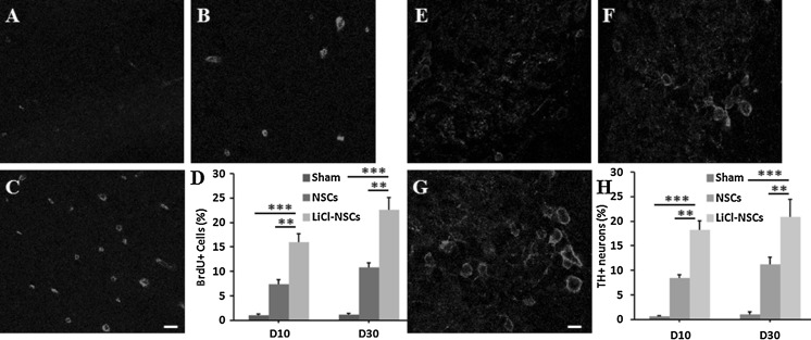
LiCl enhanced the proliferation and dopaminergic differentiation in PD models. BrdU staining in sham group (a), NSCs transplanted group (b) and PD animals transplanted with LiCl-treated NSCs (c). d Statistical analysis showed increasing ratio of BrdU+ cells in LiCl-NSCs transplanted rats compared to that in sham control or NSCs transplanted group. TH staining in sham group (e), NSCs transplanted group (f) and PD animals transplanted with LiCl-treated NSCs (g). h Ratio of TH+ cells was increased in LiCl-NSCs transplanted rats compared to that in sham control or NSCs transplanted group. **p < 0.01; ***p < 0.001. Scale bars in c was 20 μm and in g was 10 μm

