Abstract
Knee osteoarthritis (OA) is a painful and incapacitating disease affecting a large portion of the elderly population, for which no cure exists. There is a critical need to enhance our understanding of OA pathogenesis, as a means to improve therapeutic options.
Knee OA is a complex disease influenced by many factors, including the loading environment. Analysing knee biomechanics during walking - the primary cyclic load-bearing activity - is therefore particularly relevant.
There is evidence of meaningful differences in the knee adduction moment, flexion moment and flexion angle during walking between non-OA individuals and patients with medial knee OA. Furthermore, these kinetic and kinematic gait variables have been associated with OA progression.
Gait analysis provides the critical information needed to understand the role of ambulatory biomechanics in OA development, and to design therapeutic interventions. Multidisciplinary research is necessary to relate the biomechanical alterations to the structural and biological components of OA.
Cite this article: Favre J, Jolles BM. Analysis of gait, knee biomechanics and the physiopathology of knee osteoarthritis in the development of therapeutic interventions. EFORT Open Rev 2016;1:368-374. DOI: 10.1302/2058-5241.1.000051.
Keywords: knee, osteoarthritis, gait analysis, ambulatory mechanics, adduction moment, flexion moment, flexion angle
Introduction
Osteoarthritis (OA), the most common type of arthritis, is a disease causing pain, deformity, and dysfunction of the joints. This condition affects a constantly increasing portion of the population, inducing serious socio-economic concerns worldwide. Costs related to OA in the USA were estimated have already exceeded $330 billion in 2003,1 and numbers are expected to rise with the growth of obesity and the ageing of the population to the point of becoming the fourth leading cause of disability by 2020 and affecting a third of the population by 2030.2,3 The knee is the most frequently affected load-bearing joint, with disease developing more often in the medial than in any other knee compartment. An important element contributing to the burden of knee OA is the absence of a cure. Current therapeutic options consist primarily of medication and rehabilitation to reduce symptoms, and of arthroplasty later in the disease, when the joint becomes too severely damaged. Consequently, there is a critical need to enhance therapeutic options, which requires the improvement of our understanding of OA pathogenesis.
Knee OA is a joint disease involving complex interactions between biomechanical, structural and biological pathways at an in vivo systems level.4 Specifically, multidisciplinary research suggests that, in young healthy joints, the properties of the tissues, such as morphology, biology or mechanics of the tissues (bone, cartilage or ligament, for example) are adapted to each other and conditioned to the unique characteristics of each individual, including genetics or lifestyle. Unfortunately, knee tissues have limited adaptation capacity and a change in individual features such as ageing, an increase in weight or knee injury could modify the multi-parametric joint system to a point where homeostasis is disrupted, thus leading to knee OA. While changes of different types can alter joint homeostasis, changes in the loading environment are among the most important because they could modify the mechanical stress exerted on the tissues, a major component of tissue integrity.5
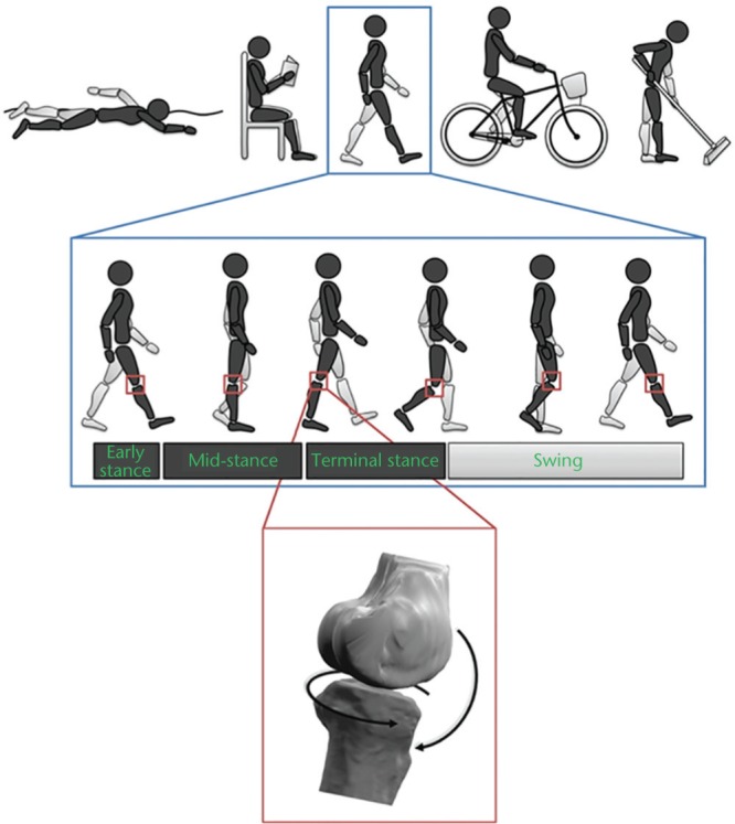
When analysing knee loading in the context of OA, occupation and physical activity are clearly among the first elements to consider as the literature indicates that workers with duties involving kneeling or squatting as well as top-level athletes of certain disciplines are at higher risk of developing knee OA.6,7 However, there are also studies suggesting that intensity of physical activity is not associated with the risks of OA development in the general population.8,9 Together, these observations indicate the biomechanical function of the knee as a critical factor in OA development, more than the frequency or duration of activities. Therefore, apart from some particularly ‘risky’ activities, this situation motivates the analysis of knee motion during walking as a basis for understanding the role of biomechanics in OA (Fig. 1).4,5 In fact, knee tissues are primarily conditioned by cyclic loading and gait is the primary load-bearing activity, particularly with knee and OA.
Fig. 1.

Quantifying three-dimensional ambulatory biomechanics is critical to understand knee osteoarthritis.
Combining the results of prior research indentifying some occupations and sport activities as risk factors for knee osteoarthritis (OA) with the results suggesting that the intensity of physical activity is not associated with the risks of OA development in the general population, indicates the need for gait analysis in order to better understand the mechanical pathway leading to knee OA. While an overall description of the ambulatory function using spatio-temporal parameters is enough to study numerous pathologies, such parameters are not specific enough to detect the subtle biomechanical differences involved in knee OA. Consequently, gait analyses in the framework of knee OA were mostly based on three-dimensional kinetic and kinematic patterns.
This paper aims to review the gait alterations consistently associated with knee OA and discuss their role in OA pathogenesis and treatment. Since OA develops more frequently in the medial compartment, and since past research has primarily analysed medial OA, this review focusses on this sub-type of OA.
Gait alterations
Speed and other spatio-temporal parameters are important outcomes used to characterise gait in a wide range of pathologies. Knee OA is no exception, with numerous studies reporting slower walking speed in OA patients compared with non-OA individuals, and slower walking speed in severe OA compared to moderate OA patients.10,11 While these basic parameters are efficient to describe the overall ambulatory function, they are not specific enough to detect subtle differences in knee biomechanics. It is therefore necessary to go further and analyse the three-dimensional kinetic and kinematic patterns of the knee (Fig. 1). The most frequent method used to measure these patterns is based on non-invasive motion capture technology, generally a combination of cameras and forceplates.12,13 Invasive methods such as instrumented prosthesis or medical imaging are also used as they offer the possibility of directly measuring tibio-femoral contacts.14,15 Although internal measures would appear to be more precise than estimations based on external sensors, selecting the method for a gait study remains a trade-off between this internal/external measurement aspect and other critical considerations such as a testing environment which modifies the natural walking patterns of patients as little as possible, which allows the testing of sufficiently large cohorts to account for inter-patient variability, and which minimises the burden on patients. These considerations have frequently led to the use of non-invasive movement analysis. Today, the non-invasive method is well-established and there is substantial evidence to indicate that it provides a sufficiently valid description of knee biomechanics for the study of OA.5
In general, non-invasive gait data in literature on knee OA were obtained in a gait laboratory equipped with a camera-based motion capture system and forceplates embedded in the floor.12 The procedure consists of attaching reflective markers to the subject using adhesive tape. A calibration procedure is then performed to build a subject-specific anatomical model based on the positions of markers placed on anatomical landmarks and anthropometric data. Next, the subject performs a number of walking trials across the laboratory (Fig. 2a). Marker trajectories (Fig. 2b) are processed using the anatomical model to determine the position and orientation of the pelvis, thigh, shank and foot anatomical frames and hip, knee and ankle joint throughout the trials. The knee angles (flexion/extension, adduction/abduction and internal/external rotation) are derived from the orientation of the thigh and shank anatomical frames (Fig. 2c). In addition to this kinematic description, an inverse dynamic procedure is commonly performed to estimate knee kinetics.13 This calculation results in three moments corresponding to the external forces acting on the joint (muscle forces are not included in this calculation), namely to flex/extend, adduct/abduct and internally/externally rotate the knee. Moments are generally expressed in normalised units (i.e. in percentage of bodyweight and height, %BW*Ht) to allow for comparison between individuals. Finally, the amplitudes of characteristic peaks are measured on the kinetic and kinematic time-curves (Fig. 2d) and these amplitudes are used to compare ambulatory knee biomechanics between individuals.
Fig. 2.
Illustration of a standard gait analysis procedure.
a) A subject with reflective markers(1) walks in a gait laboratory instrumented with a network of cameras(2) and floor-mounted forceplates(3). b) The camera and forceplate signals are processed to determine the three-dimensional trajectories of the markers and the forces exchanged between the foot of the subject and the ground. c) Biomechanical modelling is performed to determine the dynamics of the pelvis, thigh, shank, and foot segments based on marker trajectories and force data. d) Descriptive modelling is carried out to quantify three-dimensional joint kinetics and kinematics according to clinically relevant variables, such as the knee flexion/extension angle. Finally, the continuous data over the entire walking trial are limited to a period of interest, for example a stance phase in the middle of the walkway, and the amplitude of characteristic peaks are measured.
Although most gait data relative to knee OA were obtained following the method described above, actual collection and post-processing protocols differ from one institution to another and these differences can influence the results. Several other confounders, such as participant characteristics (age, BMI or disease location, for example) or the statistical approach could also affect the findings and limit comparison across gait studies. Walking speed is also an important factor to consider when interpreting knee biomechanics because it influences the amplitude of many gait variables. A systematic review by Mills, Hunt and Ferber provided a first reference dataset regarding lower-limb biomechanics in knee OA.11 The present article extends this prior work by weighting the results of gait studies in the literature based on experimental conditions. This procedure highlighted three variables with generally consistent findings regarding non-OA, moderate OA and severe OA individuals. Before discussing these variables, it is important to note that this shortlist of gait variables is the result of a conservative literature analysis, and it is possible that additional variables are important in the disease process but divergences among publications did not allow for their identification. Furthermore, for the sake of consistency and according to the journal guidelines, the following sections only include a selection of representative references.
Knee adduction moment
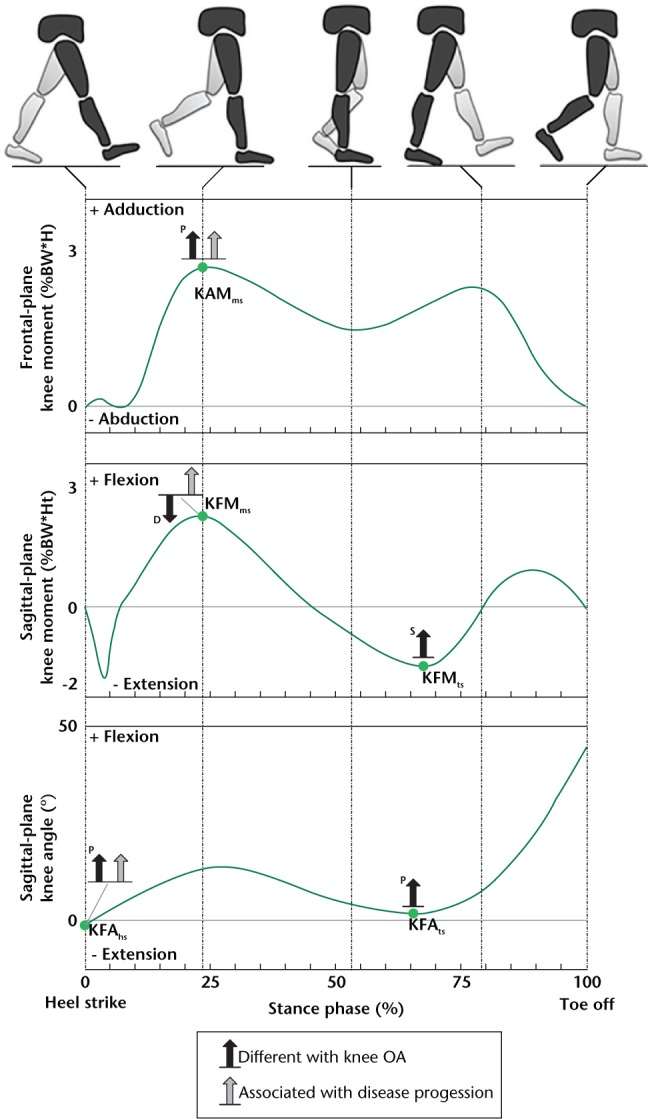
The knee adduction moment (KAM) is undoubtedly the variable which received the most attention. Critically reviewing the literature suggests that the mid-stance KAM peak (KAMms in Fig. 3) is larger in medial knee OA patients when compared with non-OA individuals and larger in severe OA patients than in those with moderate OA.16 -18 Additionally, there are indications for the magnitude of this peak to be positively associated with pain intensity.19 Interestingly, beyond cross-sectional research, a few longitudinal studies have highlighted an association with disease progression, where OA progressed faster in patients with higher mid-stance KAM.20,21 These consistent observations motivate a deeper explanation of the meaning of this gait variable. In fact, the KAM can be interpreted as a surrogate variable for the load distribution between the medial and lateral knee compartments, with a larger KAM indicating that a larger portion of the load is transmitted through the medial compartment. The relationship between this variable and medial knee OA could thus be interpreted as knees with larger KAM being knees with increased relative mechanical stress on the medial compartment. This increased medial loading could disrupt the multiparametric homeostatic joint system and contribute to tissue degradation.4 While the mid-stance peak occurs during a period of high tibio-femoral loading due to large external forces acting on the lower-limbs, an overall characterisation is also interesting to assess the loading during the entire stance phase. Indeed, the KAM impulse, a parameter measuring the area below the KAM curve, is frequently used for this purpose and has also been associated with disease severity and progression.22,23 The consistency of the results obtained with the KAM mid-stance peak and impulse parameters confirms the importance of the frontal-plane deviation (varus alignment) and of gait biomechanics (cyclic loading) in medial knee OA.
Fig. 3.

Overview of gait alterations consistently reported with medial knee OA.
The black arrows indicate differences between groups of individuals at diverse stages of the disease, and the gray arrows indicate gait parameters which have been associated with OA progression in longitudinal studies.
Please note that the gait alterations reported in this figure are the result of a conservative literature analysis and it is possible that additional alterations are important in OA but divergences among publications did not allow their identification.
P, progressive differences from non-OA subjects to moderate and severe OA patients; D, differences between non-OA and OA individuals; S, differences between patients with severe knee OA and both non-OA individuals and moderate OA patients; KAM, knee adduction moment; KFM, knee flexion moment; KFA, knee flexion angle; hs, heel-strike; ms, mid-stance; ts, terminal stance.
Knee flexion moment
Another important kinetic variable used to describe ambulatory biomechanics in the framework of knee OA is the knee flexion moment (KFM). Specifically, cross-checking prior literature suggests that knee OA patients walk with a smaller mid-stance KFM peak (KFMms in Fig. 3) in comparison with non-OA individuals.10,24 Review of the literature further tends to indicate that mid-stance KFM does not differ between groups of patients with moderate and severe OA. In addition, several studies have reported an association between this KFM peak and knee pain. In particular, it has been suggested that OA and non-OA individuals reduce the magnitude of this peak in response to pain.25 A recent study highlighted an association between mid-stance KFM and OA progression, where patients walking with a larger KFM peak at baseline lost more cartilage during the five-year follow-up period.21 Together, these observations suggest that patients reduce their mid-stance KFM as disease develops, certainly to decrease pain. However, the amount of KFM reduction and the moment this reduction develops seem to be patient-specific, therefore possibly explaining the absence of systematic differences between moderate and severe OA patients. Further, the association between the magnitude of this KFM peak and cartilage loss suggests a protective effect of smaller mid-stance KFM against disease progression, in addition to pain reduction.
Past studies with a model to estimate internal knee loading based on gait measures indicates that the mid-stance KFM could be considered as a surrogate marker for the magnitude of load transferred through the knee. Consequently, the implication of the KFM peak in the OA process could be interpreted as knees walking with a larger mid-stance KFM having larger mechanical forces at their tibio-femoral interface. Conditions that change these forces could disrupt the multi-parametric homeostatic joint system and thus lead to tissue degradation. In this sense, the mechanism associating mid-stance KFM and OA seems to be related to the mechanism described for the mid-stance KAM: the KFM and KAM peaks are two parameters characterising different aspects of the loading environment, so it would make sense that their role in disease development is co-dependent. This comment is particularly supported by studies showing that the mechanical load acting on the medial compartment is better estimated by a combination of the KAM and KFM, rather than by any of these moments in isolation.14
The literature also suggests that OA-related differences exist in terminal stance KFM peak (KFMts in Fig. 3), specifically, a reduced peak (i.e. less extension) has been reported in patients with more severe OA compared to both non-OA and moderate OA individuals.10,26 Nevertheless, no association has been reported in longitudinal studies between this KFM peak and disease initiation or progression, limiting the analysis of the role of this parameter in knee OA. It has nonetheless been suggested that OA patients could adopt this walking pattern to increase knee stability and reduce pain.10,27
Knee flexion angle
The particular sagittal plane motion of the knee and the specific properties of the tibio-femoral articular surfaces make the knee flexion angle (KFA) an essential variable in the analysis of ambulatory biomechanics. The smaller ranges of motion over the entire gait cycle consistently reported with increasing disease severity confirm the importance of the KFA in the biomechanical analysis of knee OA.16,26 A critical review of the literature suggests that the KFA at heel-strike (KFAhs in Fig. 3) is larger (i.e. the knee is less extended) in OA compared with non-OA individuals, and that this parameter is larger in severe compared with moderate OA patients.16,26,27 Furthermore, a recent longitudinal study reported an association between this parameter and OA progression, with less extended knees showing more rapid disease progression.28 These consistent observations indicate that knee kinematics is also involved in the OA process, supporting the general hypotheses that a disruption of the multi-parametric homeostatic joint system contributes to knee OA and that a range of alterations can disturb the system. Although this review analysed the KAM, KFM, and KFA successively, these variables are certainly not independent, and their role in knee OA certainly not totally different. Whereas the KAM and KFM could be related to the amount of load transferred through the medial compartment, the KFA could be related to the location where the load is applied. Indeed, the points of tibio-femoral contact vary with the angle of knee flexion. The importance of the KFA is further supported by research showing that cartilage properties vary spatially and that the spatial variations depend on the heel-strike KFA.29 Therefore, a difference in this KFA parameter could alter the local stress on some knee tissues in a manner that the tissues cannot accommodate, thus contributing to homeostasis disruption.4 This interpretation is further supported by the fact that heel-strike is another period of high loading due to the important muscle co-contractions necessary to stabilise the joint.
Cross-checking prior literature also suggests that the KFA peak during terminal stance (KFAts in Fig. 2) is larger (i.e. the knee is less extended) in OA compared with non-OA individuals and is greater in severity compared with moderate OA patients.26,27 However, no longitudinal study has reported an association between this parameter and OA progression. Therefore, similar to the differences in KFM during terminal stance, it is possible that this gait alteration is a compensation to reduce pain and increase joint stability.10,27
Discussion
The data reviewed in the previous sections highlight a clear and meaningful relationship between ambulatory biomechanics and medial knee OA. Specifically, disease progression was associated with larger heel-strike KFA and larger mid-stance KAM and KFM. In parallel, the literature also suggested that heel-strike KFA and mid-stance KAM increase with disease severity. Together the observations summarised in this review suggested a sort of amplification mechanism, where the disease can produce specific gait alterations and, in the reverse direction, the same gait alterations can contribute to disease development. It is worth noting that this amplification mechanism fits well with the progressive development of OA. Observations regarding the mid-stance KFM were different, as patients seem to reduce this kinetic parameter, which is opposite to the association of mid-stance KFM and OA progression. The difference with the two other parameters could be explained by the relationship between mid-stance KFM and pain and the possibility of walking with reduced mid-stance KFM. Nevertheless, this sagittal plane kinetic parameter was associated with OA progression, so even if OA-related differences seem protective, it remains a parameter of primary interest.
In addition to the descriptive value, better understanding the differences in ambulatory biomechanics between non-OA, moderate OA and severe OA individuals offers a unique basis to study the disease and improve prevention and treatment options. For example, it is still unclear why a portion of the population, apparently without predisposition for knee OA, develops this disease when they become older. In this regard, analysing the differences in gait which naturally occur with increasing age, and contrasting them with those listed in this review could help in the understanding of the possible mechanical pathway leading to idiopathic knee OA. Specifically, the literature has reported that older individuals walk with a larger heel-strike KFA (i.e. a less-extended knee) in comparison with younger individuals,26 which is a similar alteration to that observed in OA. It is therefore possible that this age-related modification plays a role in idiopathic OA initiation. Similar analyses could be performed for other risk factors such as obesity or trauma.5
The design of therapeutic options is an area which is strongly dependent on the understanding of the disease process. Knowledge regarding ambulatory biomechanics in knee OA is particularly valuable, as illustrated by the range of interventions developed on the basis of the KAM alterations described above. Mechanical interventions notably include braces,29 orthotic insoles,31 and gait re-training.32 These interventions were reported to decrease mid-stance KAM and to improve pain, function and medication intake with small to moderate effect sizes. Interestingly, most mechanical intervention until now has focussed on decreasing the KAM, as this gait alteration was the most documented and the first to be associated with progression of the disease. Recently, the effects of braces, insoles and gait re-training on the KFM has received much attention, and it is expected that in the near future other interventions will be introduced to modify the KFA and KFM in addition to the KAM. In the coming years, one can also expect an increase of individualised interventions, where the type and amplitude of modification will be determined independently for each patient based on a gait test. New options have been introduced recently to measure the function of the knee, and will certainly play a central role in the success of individualised treatment. For example, portable systems based on inertial sensors could offer an alternative to full gait laboratories, which are too complex for routine practice.33 Additionally, external harnesses could contribute to a more precise analysis of knee kinematics during walking.34
Conclusion
There is clear evidence to support the value of gait analysis in understanding the role of biomechanics in knee OA. Compiling findings across studies is not an easy task due to large inter-individual variations, numerous confounders and errors inherent in the method used to quantify gait.35 Nevertheless, today there are enough consistent data to begin identifying a widespread alteration in knee biomechanics associated with OA, including the KAM, KFM and KFA. In this regard, it is important to keep in mind that the data in Figure 3 is a conservative summary, and that other differences could exist notably in relation to specific factors such as obesity or a history of trauma. Additional research is necessary to improve our understanding of the factors influencing gait and OA development. A better understanding of ambulatory biomechanics in OA will also require the analysis of other variables at the knee and at other joints. For example, the anterior displacement of the femur relative to the tibia was recently shown to differ with OA severity and to be associated with disease progression.4,26 Similarly, hip and trunk motions have been reported to differ with knee OA.10,24 Multidisciplinary studies will also be critical in understanding the relationship between the structural and biological aspects of the disease. A framework, assuming that knee health is dependent on a multi-parametric system, provides a unique basis to connect the various components of the disease.4 Nevertheless, many additional years of research will be necessary to refine this framework.
Finally, while our understanding of the role of ambulatory biomechanics in knee OA is increasing, knowledge regarding which gait modification to recommend to which patient and the long-term benefits of gait modifications remains fairly limited.
Acknowledgments
The authors would like to thank Baptiste Ulrich for his assistance with the illustrations in this article.
Footnotes
Conflict of Interest: No interests declared.
Funding Statement
No benefits in any form have been received or will be received from a commercial party related directly or indirectly to the subject of this article.
References
- 1. Yelin E, Murphy L, Cisternas MG, et al. Medical care expenditures and earnings losses among persons with arthritis and other rheumatic conditions in 2003, and comparisons with 1997. Arthritis Rheum 2007;56:1397-1407. [DOI] [PMC free article] [PubMed] [Google Scholar]
- 2. Helmick CG, Felson DT, Lawrence RC, et al. ; National Arthritis Data Workgroup. Estimates of the prevalence of arthritis and other rheumatic conditions in the United States. Part I. Arthritis Rheum 2008;58:15-25. [DOI] [PubMed] [Google Scholar]
- 3. Lawrence RC, Felson DT, Helmick CG, et al. ; National Arthritis Data Workgroup. Estimates of the prevalence of arthritis and other rheumatic conditions in the United States. Part II. Arthritis Rheum 2008;58:26-35. [DOI] [PMC free article] [PubMed] [Google Scholar]
- 4. Andriacchi TP, Favre J, Erhart-Hledik JC, Chu CR. A systems analysis of knee osteoarthritis reveals new insights into the pathogenesis of the disease. Ann Biomed Eng 2015;43:376-387. [DOI] [PMC free article] [PubMed] [Google Scholar]
- 5. Andriacchi TP, Favre J. The nature of in vivo mechanical signals that influence cartilage health and progression to knee osteoarthritis. Curr Rheumatol Rep 2014;16:463. [DOI] [PubMed] [Google Scholar]
- 6. Coggon D, Croft P, Kellingray S, et al. Occupational physical activities and osteoarthritis of the knee. Arthritis Rheum 2000;43:1443-1449. [DOI] [PubMed] [Google Scholar]
- 7. Kujala UM, Kettunen J, Paananen H, et al. Knee osteoarthritis in former runners, soccer players, weight lifters, and shooters. Arthritis Rheum 1995;38:539-546. [DOI] [PubMed] [Google Scholar]
- 8. Felson DT, Niu J, Yang T, et al. ; MOST and OAI investigators. Physical activity, alignment and knee osteoarthritis: data from MOST and the OAI. Osteoarthritis Cartilage 2013;21:789-795. [DOI] [PMC free article] [PubMed] [Google Scholar]
- 9. Barbour KE, Hootman JM, Helmick CG, et al. Meeting physical activity guidelines and the risk of incident knee osteoarthritis: a population-based prospective cohort study. Arthritis Care Res (Hoboken) 2014;66:139-146. [DOI] [PMC free article] [PubMed] [Google Scholar]
- 10. Astephen JL, Deluzio KJ, Caldwell GE, Dunbar MJ. Biomechanical changes at the hip, knee, and ankle joints during gait are associated with knee osteoarthritis severity. J Orthop Res 2008;26:332-341. [DOI] [PubMed] [Google Scholar]
- 11. Mills K, Hunt MA, Ferber R. Biomechanical deviations during level walking associated with knee osteoarthritis: a systematic review and meta-analysis. Arthritis Care Res (Hoboken) 2013;65:1643-1665. [DOI] [PubMed] [Google Scholar]
- 12. Cappozzo A, Della Croce U, Leardini A, Chiari L. Human movement analysis using stereophotogrammetry. Part 1: theoretical background. Gait Posture 2005;21:186-196. [DOI] [PubMed] [Google Scholar]
- 13. Andriacchi TP, Johnson TS, Hurwitz DE, Natarajan RN. Musculoskeletal dynamics, locomotion, and clinical applications. In Mow VC, Huiskes R. ed. Basic orthopaedic biomechanics and mechano-biology. Third ed. Philadephia: Lippincott Williams & Wilkins, 2005. [Google Scholar]
- 14. Walter JP, D’Lima DD, Colwell CW, Jr, Fregly BJ. Decreased knee adduction moment does not guarantee decreased medial contact force during gait. J Orthop Res 2010;28:1348-1354. [DOI] [PMC free article] [PubMed] [Google Scholar]
- 15. DeFrate LE, Sun H, Gill TJ, Rubash HE, Li G. In vivo tibiofemoral contact analysis using 3D MRI-based knee models. J Biomech 2004;37:1499-1504. [DOI] [PubMed] [Google Scholar]
- 16. Baliunas AJ, Hurwitz DE, Ryals AB, et al. Increased knee joint loads during walking are present in subjects with knee osteoarthritis. Osteoarthritis Cartilage 2002;10:573-579. [DOI] [PubMed] [Google Scholar]
- 17. Mündermann A, Dyrby CO, Hurwitz DE, Sharma L, Andriacchi TP. Potential strategies to reduce medial compartment loading in patients with knee osteoarthritis of varying severity: reduced walking speed. Arthritis Rheum 2004;50:1172-1178. [DOI] [PubMed] [Google Scholar]
- 18. Erhart-Hledik JC, Favre J, Andriacchi TP. New insight in the relationship between regional patterns of knee cartilage thickness, osteoarthritis disease severity, and gait mechanics. J Biomech 2015;48:3868-3875. [DOI] [PubMed] [Google Scholar]
- 19. Thorp LE, Sumner DR, Wimmer MA, Block JA. Relationship between pain and medial knee joint loading in mild radiographic knee osteoarthritis. Arthritis Rheum 2007;57:1254-1260. [DOI] [PubMed] [Google Scholar]
- 20. Miyazaki T, Wada M, Kawahara H, et al. Dynamic load at baseline can predict radiographic disease progression in medial compartment knee osteoarthritis. Ann Rheum Dis 2002;61:617-622. [DOI] [PMC free article] [PubMed] [Google Scholar]
- 21. Chehab EF, Favre J, Erhart-Hledik JC, Andriacchi TP. Baseline knee adduction and flexion moments during walking are both associated with 5 year cartilage changes in patients with medial knee osteoarthritis. Osteoarthritis Cartilage 2014;22:1833-1839. [DOI] [PMC free article] [PubMed] [Google Scholar]
- 22. Bennell KL, Bowles KA, Wang Y, et al. Higher dynamic medial knee load predicts greater cartilage loss over 12 months in medial knee osteoarthritis. Ann Rheum Dis 2011;70:1770-1774. [DOI] [PubMed] [Google Scholar]
- 23. Creaby MW, Wang Y, Bennell KL, et al. Dynamic knee loading is related to cartilage defects and tibial plateau bone area in medial knee osteoarthritis. Osteoarthritis Cartilage 2010;18:1380-1385. [DOI] [PubMed] [Google Scholar]
- 24. Huang SC, Wei IP, Chien HL, et al. Effects of severity of degeneration on gait patterns in patients with medial knee osteoarthritis. Med Eng Phys 2008;30:997-1003. [DOI] [PubMed] [Google Scholar]
- 25. Henriksen M, Graven-Nielsen T, Aaboe J, Andriacchi TP, Bliddal H. Gait changes in patients with knee osteoarthritis are replicated by experimental knee pain. Arthritis Care Res (Hoboken) 2010;62:501-509. [DOI] [PubMed] [Google Scholar]
- 26. Favre J, Erhart-Hledik JC, Andriacchi TP. Age-related differences in sagittal-plane knee function at heel-strike of walking are increased in osteoarthritic patients. Osteoarthritis Cartilage 2014;22:464-471. [DOI] [PMC free article] [PubMed] [Google Scholar]
- 27. Heiden TL, Lloyd DG, Ackland TR. Knee joint kinematics, kinetics and muscle co-contraction in knee osteoarthritis patient gait. Clin Biomech (Bristol, Avon) 2009;24:833-841. [DOI] [PubMed] [Google Scholar]
- 28. Favre J, Erhart-Hledik JC, Chehab EF, Andriacchi TP. Baseline ambulatory knee kinematics are associated with changes in cartilage thickness in osteoarthritic patients over 5 years. J Biomech 2016;49:1859-1864. [DOI] [PubMed] [Google Scholar]
- 29. Scanlan SF, Favre J, Andriacchi TP. The relationship between peak knee extension at heel-strike of walking and the location of thickest femoral cartilage in ACL reconstructed and healthy contralateral knees. J Biomech 2013;46:849-854. [DOI] [PubMed] [Google Scholar]
- 30. Moyer RF, Birmingham TB, Bryant DM, et al. Biomechanical effects of valgus knee bracing: a systematic review and meta-analysis. Osteoarthritis Cartilage 2015;23:178-188. [DOI] [PubMed] [Google Scholar]
- 31. Penny P, Geere J, Smith TO. A systematic review investigating the efficacy of laterally wedged insoles for medial knee osteoarthritis. Rheumatol Int 2013;33:2529-2538. [DOI] [PubMed] [Google Scholar]
- 32. Shull PB, Silder A, Shultz R, et al. Six-week gait retraining program reduces knee adduction moment, reduces pain, and improves function for individuals with medial compartment knee osteoarthritis. J Orthop Res 2013;31:1020-1025. [DOI] [PubMed] [Google Scholar]
- 33. Favre J, Aissaoui R, Jolles BM, de Guise JA, Aminian K. Functional calibration procedure for 3D knee joint angle description using inertial sensors. J Biomech 2009;42:2330-2335. [DOI] [PubMed] [Google Scholar]
- 34. Lustig S, Magnussen RA, Cheze L, Neyret P. The KneeKG system: a review of the literature. Knee Surg Sports Traumatol Arthrosc 2012;20:633-638. [DOI] [PubMed] [Google Scholar]
- 35. McGinley JL, Baker R, Wolfe R, Morris ME. The reliability of three-dimensional kinematic gait measurements: a systematic review. Gait Posture 2009;29:360-369. [DOI] [PubMed] [Google Scholar]


