

Abstract
Cancer has become a global problem. On all continents, a great number of people are diagnosed with this disease. In spite of the progress in medical care, cancer still ends fatal for a great number of the ill, either as a result of a late diagnosis or due to inefficiency of therapies. The majority of the tumors are resistant to drugs. Thus, the search for new, more effective therapy methods continues. Recently, nanotechnology has been attributed with big expectations in respect of the cancer fight. That interdisciplinary field of science creates nanomaterials (NMs) and nanoparticles (NPs) that can be applied, e.g., in nanomedicine. NMs and NPs are perceived as very promising in cancer therapy since they can perform as drug carriers, as well as photo- or sonosensitizers (compounds that generate the formation of reactive oxygen species as a result of either electromagnetic radiation excitation with an adequate wavelength or ultrasound activation, respectively). Consequently, two new treatment modalities, the photodynamic therapy (PDT) and the sonodynamic therapy (SDT) have been created. The attachment of ligands or antibodies to NMs or to NPs improve their selective distribution into the targeted organ or cell; hence, the therapy effectiveness can be improved. An important advantage of the targeted tumor treatment is lowering the cyto- and genotoxicity of active substance towards healthy cells. Therefore, both PDT and SDT constitute a valuable alternative to chemo- or radiotherapy. The vital role in cancer eradication is attributed to two inorganic sensitizers in their nanosized scale: titanium dioxide and zinc oxide.
Keywords: Nanotechnology, Nanoparticles, Titanium dioxide, Zinc oxide, Cancer, Photodynamic therapy, Sonodynamic therapy
Review
Introduction
During the congress of the American Physical Society in Caltech, on 26th December 1959, an American physicist R. Feynman [1] held a presentation. He was the first to claim the matter can be altered at the level of atoms and particles. That statement, the main thesis of the speech entitled “There’s plenty of room at the bottom,” earned him the attribute of a forerunner of nanotechnology. In the lecture, R. Feynman suggested that in his day, the only limit to the development of nanotechnology resulted from the lack of sufficiently precise tools and techniques. For the first time, the term “nanotechnology” was applied by a Japanese scientist N. Taniguchi [2] and meant the possibility of material engineering at the nanometers level. A book by K. Drexler [3] “Nanosystems: molecular machinery, manufacturing and computation” contributed to the popularization of the knowledge in this area. At present, nanotechnology is a rapidly developing multidisciplinary field that combines the latest achievements of many disciplines of science, including biology, chemistry, physics, informative technology, and mechanics. It designs nanomaterials (NMs) and nanoparticles (NPs) with at least one dimension ranging in the scale from 1 to 100 nm [4], confers on them the desired properties, and alters them accordingly. For comparison, the diameters of DNA, viruses, and lymphocytes are 2.5, 30–50, and about 7000 nm, respectively [5]. Nanotechnology, besides biotechnology and genetics, has recently become one of the most highlighted fields of science [6]. It gave rise to the creation of nanomedicine [7]. According to Fakruddin et al. [8], thanks to the achievements of nanotechnology, it will soon be possible to increasingly or even fully control the matter structure; hence, to overcome numerous obstacles, the medicine is nowadays struggling against. For example, by using quantum dots, nanotechnology might greatly assist in an early stage cancer diagnosis and therapy [9]. The regeneration and remodeling of damaged neuronal connections is going to be possible, thanks to dendrimers [10]. Carbon nanotubes can deliver drugs to cells in a more targeted manner [11]. Another example might be NM-based video capsule endoscopy providing high quality images of formerly inaccessible spots, e.g., small bowel [12].
The challenge the nanomedicine is now facing is to find the most effective way of the tumor cells eradication [13]. The main area this discipline is trying to explore is designing systems of targeted drug delivery and release into pathologically altered cells, increasing the drug treatment effectiveness, and limiting their side effects [14, 15]. There are several key attributes of NPs in their performance as drug carriers. One of them is their ability to become conjugated with ligands that are specific to cancer cells and that enable the targeted drug delivery [16–18]. NPs can be also conjugated with monoclonal antibodies, a process preventing them from immunological system response [19]. Moreover, thanks to the possibility of modifying the physicochemical properties of NPs, a smooth endocytosis into the cancer cells can occur [20]. NPs have also found their application in nanodiagnostics as biosensors for an early stage tumor markers detection [21] and in gene therapy as carriers delivering into cancer cells foreign nucleic acids that encode proteins killing the tumor cells [22].
The past decade brought about the discovery that many materials used in a variety of industries exhibit formerly undisplayed catalytic properties, after they have been powdered to NPs and subsequently exposed to ultraviolet (UV) radiation or ultrasound (US) wave. Among such materials, there are titanium dioxide (TiO2) and zinc oxide (ZnO) [23–25].
Nanoparticles of Titanium and Zinc Oxides
Size, Surface Area/Volume Ratio, and Surface Chemistry
Nanoparticles of titanium dioxide (TiO2 NPs) and zinc oxide (ZnO NPs) are single elements with a diameter of 1 < φ ≤ 100 nm [4]. Their size is one of their key properties. The smaller the NP diameter, the bigger becomes the surface area/volume ratio. A high value of surface area/volume ratio allows a greater number of constituting atoms to be located around the surface of the NPs, a phenomenon making the NPs highly reactive and attributing them with new, unique physicochemical properties [26, 27]. Moreover, the smaller the NP diameter, the longer becomes the blood circulation time [28]. Metal oxide nanoparticles whose diameter ranges between 10 < φ ≤ 100 nm are considered perfect in respect of their medical applications. The NPs with φ ≤ 10 nm are able to deeper penetrate tumors and better accumulate in tumor cells, but they are also toxic to healthy cells. Bigger NPs, in the range of 10 < φ ≤ 100 nm, can only hardly be uptaken by healthy cells while they can easily penetrate tumor cells [29]. Nowadays, thanks to the nanoengineering, it is possible not only to precisely control the size of TiO2 and ZnO NPs but also to functionalize their surface. The NP functionalization provides for an easier transport within tissues, creates the NP ability to cross plasma membranes and allows them to be delivered to cancer cells in a targeted manner [30]. Thus, NPs of TiO2 and ZnO have become a promising tool in medicine [28–30].
Photo and Sonocatalytic Properties
The electronic structure of TiO2 and ZnO, both belonging to the family of semiconducting metal oxides, determines their catalytic properties [31]. The semiconductors have a valence band (VB), which is filled with electrons, and an electron-free conduction band (CB). The energy difference (ΔE) between those bands is known as band gap, also referred to as energy gap. The band gap is an equivalent of the energy amount necessary to excite an electron from VB to CB. For nanosized titanium dioxide (nano-TiO2, anatase polymorph) and nanosized zinc oxide (nano-ZnO), the width of the energy gap is 3.20 and 3.37 eV, respectively [26, 32]. Both amounts approximate the value of the electromagnetic radiation photon energy with a wavelength of λ < 400 nm. In biological trials, semiconducting metal oxides (e.g., TiO2, ZnO) are excited by the UV radiation (photoexcitation), mainly in the near ultraviolet range (UV-A, 315 ≤ λ < 400 nm) [33–38]. Less common is the photoexcitation with a wavelength in the medium ultraviolet range (UV-B, 280 ≤ λ < 315 nm) [33, 34, 36]. The use of far ultraviolet range (UV-C, 100 < λ < 280 nm) as an agent responsible for advanced oxidation processes may be hazardous to human health [37]. Another way to excite semiconductor NPs in aqueous solutions is the application of the US wave (sonoexcitation). The US wave (20 kHz < f ≤ 1 GHz), while propagating in the liquid, causes a twofold effect: a temporary punctual dilution (result of the loss of pressure), facilitating the formation of cavitation bubbles; as well as liquid’s thickening (result of the rise of pressure) that facilitates the bubbles’ collapse. This phenomenon is referred to as acoustic cavitation. It is accompanied by a number of secondary effects, among them sonoluminescence, an emission of sonoluminescent light (mainly UV light) during the implosion of the cavitation bubbles suspended in a liquid [39, 40].
The consequence of the semiconductor photo- or sonoexcitation is that electron (e−) transfers from VB to CB, leaving behind a positive hole (h+). Therefore, a specific “electron-hole” pair (e− + h+) is created [40–42]. This bound state, called exciton, lacks stability and exhibits redox properties. Charge carriers (e−, h+) can migrate to the surface of the catalyst (e.g., TiO2 or ZnO) NPs where they can be trapped by adsorbate molecules, initiating thereby the formation of reactive oxygen species (ROS). Excited negative electrons (e−) reduce molecular oxygen (O2), forming superoxide radical anion (•O2 −). Generated positive holes (h+) oxidize water molecules (H2O) and hydroxide ions (OH−), producing hydroxyl radicals (•OH) and hydrogen peroxide molecules (H2O2) [40–43] (Fig. 1).
Fig. 1.
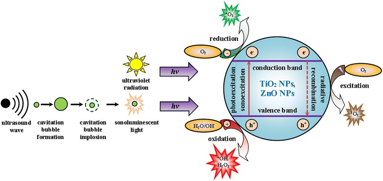
Mechanisms of generating reactive oxygen species (ROS). ROS are generated on the surface of titanium dioxide or zinc oxide nanoparticles (TiO2 NPs, ZnO NPs), exposed to ultraviolet (UV) radiation or ultrasound (US) wave in aqueous solutions. Semiconductor NPs can be excited by UV (photoexcitation) or US (sonoexcitation). The acoustic wave, while propagating in the liquid, induces the formation and the subsequent collapse of cavitation bubbles. During their implosion, the emission of the sonoluminescent light takes place. As a result of the TiO2 NPs or ZnO NPs photo- or sonoexcitation, electron (e−) transfers from valence band to conduction band, leaving behind hole (h+). The follow-up process can be twofold. The electrons (e−) together with holes (h+) react with adjacent molecules (O2, H2O, OH−), generating the formation of ROS (•O2 −, •OH, H2O2). Alternatively, radiative recombination of electron-hole pair (e− + h+) results in the emission of photon that transforms the oxygen molecule in the ground state (O2) to the oxygen molecule’s excited state, singlet oxygen (1O2)
The inverse process to the generation of charge carriers in semiconductor (e.g., TiO2 or ZnO) NPs is the decay of exciton. The recombination of the electron-hole pair (e− + h+) is accompanied by the emission of a photon (radiative recombination). In this process, the excitation of ground state oxygen (O2) by photon occurs and singlet oxygen (1O2) is formed [40, 43] (Fig. 1).
Oxidative Stress and its Anticarcinogenic Effects
The oxidative stress is an underlying factor for adverse biological effects caused by NPs of TiO2 and ZnO. In the cells, it is reflected by the increase of the level of ROS and oxidative products, as well as by the depletion of cellular antioxidants [44]. ROS forming is an inherent element of the aerobic cellular metabolism. In their physiological concentrations, ROS perform a series of important functions. They mediate in the intracellular signal transduction [45], regulate protein phosphorylation [46], and control intracellular Ca2+ homeostasis [47]. Balance disruptions between the ROS formation and the efficiency of antioxidative systems lead to oxidative stress that poses a serious danger to the integrity of cells and their correct functioning [48].
Eukaryotic cells, similarly to prokaryotic cells, are equipped with mechanisms protecting them from ROS. The first defense line, preventing ROS from forming and from reactions with cell compounds, are proteins (e.g., lactoferrin) that sequestrate metal ions (e.g., Cu, Fe). Interruption of free radical chain reactions is the task of the second defense line that consists of a system of three enzymes: superoxide dismutase (SOD; EC 1.15.1.1), catalase (CAT; EC 1.11.1.6), and glutathione peroxidase (GPX; EC 1.11.1.9), supported by low molecular weight antioxidants, such as glutathione (GSH), thioredoxin (Trx), or coenzyme Q10 (CoQ10). The third defense line present repair enzymes responsible for nucleic acids damage overhaul, e.g., DNA ligase (EC 6.5.1.1) [48].
ROS can inflict cell injury as an effect of reaction with lipids in cellular membranes [49], nucleotides in DNA [50], or sulfhydryl groups in proteins [51]. Depending on the intensity of the oxidative stress, the cell can undergo carcinogenesis or enter one of the cell death pathways [52]. Antunes and Cadenas [53] proved that as the concentration of certain ROS (e.g., H2O2) increases, the cell’s viability decreases. In physiological conditions, the concentration of H2O2 is low and ranges within 5–50 nM. The gradual increase of its quantity in a cell up to the level of 0.7 μM triggers mechanisms that result in a cell proliferation and tumor formation. In case the H2O2 concentration reaches the value of 1–3 μM, the apoptosis or autophagy (programmed cell death pathways) induction occurs. The concentration in excess of the level of 3 μM becomes toxic for a cell hence leads to its necrosis (unprogrammed cell death pathway). Therefore, the phenomenon of the oxidative stress in tumor cells has recently been used to design new tumor therapies [54, 55].
Increasingly frequent are the opinions that in near future, the NPs of semiconductor metal oxides (e.g., TiO2 and ZnO) are going to play the crucial role in medicine as photo- or sonosensitizers applied in ROS-based cancer therapy [56, 57], a finding that naturally corroborates the necessity to study their cyto- and genotoxicity.
Toxicity of Titanium Dioxide and Zinc Oxide Nanoparticles
Despite the assumption that the microsized titanium dioxide (micro-TiO2) is harmless to people and animals [58, 59], the issue of TiO2 toxicity has been a research subject in many scientific centers. Recent years have witnessed a dynamic development of nanotechnology. Along with it, there has been an increased concern, though that this compound might be toxic in its NPs form. There are studies proving that NPs (1 < φ ≤ 100 nm) of any compound are more harmful to people and animals than microparticles (MPs) (0.1 < φ ≤ 100 μm) of the same compound [60, 61].
Studies In Vivo
Wang and Fan [62] conducted studies showing that the level of the rats’ pulmonary cell impairment, inflicted by the intratracheally instilled TiO2, clearly depended upon the diameter of its particles. Kobayashi et al. [63] reported the pulmonary toxicity of TiO2 NPs in rats, evaluated on the basis of the short-term post-installation lung injury (up to 7 days after the installation), increased proportionally to the fragmentation of the TiO2 particles. Sycheva et al. [64] pointed out the genotoxicity of TiO2 NPs (φ = 30 nm), as measured by the number of DNA double-strand breaks and the number of micronuclei, was significantly higher in brain, liver, or bone marrow cells in mice, compared to the genotoxicity of TiO2 MPs (φ = 160 nm). Danio rerio, when exposed to TiO2 (50 mg dm−3) in their NPs form, exhibits more serious oxidative injuries of liver and gill cells, in comparison to the exposition to the MPs form of those compounds [65]. The studies by Ma-Hock et al. [66] showed the rats’ exposition to the aerosol of TiO2 NPs (50 mg m−3) caused lung inflammation, accompanied by the increase of total neutrophil count in bronchoalveolar lavage (BAL) fluid. Bermudez et al. [67] examined pulmonary responses in rats, mice, and hamsters after their exposition to the aerosol of TiO2 NPs (10 mg m−3). They concluded there was a clear increase of cytotoxicity markers, i.e., an increase in the total macrophage and neutrophil count in BAL fluid in rats and mice, while in hamsters, there were no distinct changes in the cellular response. Ze et al. [68] proved the TiO2 NPs, administered to mice through the nose for 90 days, moved up and built up in the brain, leading to the oxidative stress, excessive glial cells proliferation, and tissue necrosis, as well as inflicting hippocampal cells apoptosis. Lindberg et al. [69] reported that the intraperitoneal administration of TiO2 NPs to mice resulted in an increase of DNA damages in leukocytes and in higher micronuclei count in peripheral blood polychromatic erythrocytes. The intratracheal instillation of TiO2 NPs showed the same effects in BAL cells. The cyto- and genotoxicity of TiO2 NPs were confirmed by many other authors [70–73]. Ferin and Oberdörster [74] showed the dust of TiO2 NPs, introduced into the trachea of a rat in the amount of 5 mg per animal, caused respiratory tract cancer within 4 months. Therefore, TiO2 NPs in the dust form, contrary to the TiO2 MPs in the same form, were classified in the 2B group by the International Agency for Research on Cancer (IARC) [75]. The 2B group assembles compounds that might be carcinogenic for humans.
Studies In Vitro
One of the main differences between TiO2 MPs and TiO2 NPs is a much bigger active surface area of TiO2 NPs, a feature resulting in a higher absorption rate of UV radiation and a greater photocatalytic activity [41]. Gurr et al. [76] prove there is a strong correlation between the TiO2 NPs genotoxicity and their diameter. TiO2 NPs (φ < 50 nm) caused the formation of 8-hydroxy-2-deoxyguanosine (8-OHdG) adducts and the increase of the micronuclei number in human bronchial epithelial cells, while TiO2 MPs (φ > 200 nm) in the same concentration were practically harmless. Kongseng et al. [77] report TiO2 NPs (φ < 30 nm) were responsible for the rise of the pro-inflammatory cytokine (IL-1β and TNFα) level in human peripheral blood mononuclear cells (PBMCs) in serum-free medium. The in vitro studies of Long et al. [78, 79], conducted on mouse brain microglia cells (BV2 line), proved that TiO2 particles with 10 ≤ φ ≤ 20 nm stimulated the generation of ROS and, as a consequence, induced damages of neurons, whereas UV radiation was not a precondition for generating ROS. There are, however, numerous contrary opinions. Fujita et al. [80], drawing on DNA microarray analysis of human keratinocytes (HaCaT line), proved that the natural (with no UV irradiation) toxicity of TiO2 NPs was very low. Similar conclusions were presented by Verves and Jha [81] who examined in vitro the toxicity of TiO2 NPs (50 mg mL−1) to rainbow trout (Oncorhynchus mykiss) gonadal tissue cells (RTG-2 line). They reported cyto- and genotoxic effects of TiO2 NPs in irradiated cells, only. Saquib et al. [82], while examining the toxicity of TiO2 NPs (10 μg mL−1) to UV-A irradiated human amniotic epithelial cells (WISH line), noted a decrease in the CAT activity, lowering the GSH level, as well as the arrest of the cellular cycle in the G2/M phase. In similar studies on human epidermoid carcinoma cells (A431 line), Shukla et al. [83] observed an increase in lipid hydroperoxide (LPO) concentration, numerous oxidative DNA damages, and the formation of many micronuclei. Vamanu et al. [84] exposed human leukemic monocyte lymphoma cells (U937 line) in vitro to 0.1% colloidal nano-TiO2 for 120 min and, subsequently, to UV-A irradiation. After 30 min of UV-A irradiation, significant plasma membrane damages and DNA fragmentation were noticed. Other studies show that TiO2 powdered to NPs exhibited ROS-dependent cyto- and genotoxicity towards human HaCaT cells [85], protozoan (Cryptosporidium parvum) oocysts [86], as well as towards cysts of Giardia lamblia [87] and Acanthamoeba castellanii [88], only if the parasites were exposed to UV radiation. The cytotoxicity was determined on the basis of the lysosomal membrane stability, using neutral red retention time (NRRT) assay whereas the genotoxicity was measured by the number of DNA double-strand breaks, using the single-cell gel electrophoresis (SCGE).
ZnO toxicity is also dependent on the scale of its fragmentation [65]. Microsized zinc oxide (micro-ZnO), similarly to micro-TiO2, is practically harmless to people and animals [75], contrary to nano-ZnO. Wang et al. [89] studied the toxicity of ZnO NPs to human HaCaT cells and discovered the viability of those cells was clearly reduced, the integrity of their membranes was disrupted, and numerous oxidative DNA damages occurred. Those effects significantly increased when the keratinocytes were exposed to the UV radiation. Similar cyto- and genotoxicity effects of ZnO NPs were observed in human monocytes (THP-1 line) [90, 91] and human lung adenocarcinoma epithelial cells (A549 line) [92], as well as in fish embryos, e.g., Danio rerio [93].
Cyto- and genotoxicity of TiO2 NPs, as well as of ZnO NPs have been proved in vitro also with many other cells, both healthy, e.g., human dermal fibroblasts [94, 95], human periodontal ligament fibroblasts [96], human alveolar macrophages [97], human gastric epithelial cells [98]; and malignant, e.g., human colonic mucinous adenocarcinoma cells (Ls-174-t line) [99], human colon adenocarcinoma cells (LoVo line) [100, 101], human cervical carcinoma cells (HeLa line) [102–105], human breast adenocarcinoma cells (MDA-MB-468 line) [106], human hepatocellular carcinoma cells (Bel 7402 line) [107], human brain glioblastoma cells (T98G line) [108, 109], or human bladder carcinoma cells (T-24 line) [110].
Future of Cancer Therapy in View of the Application of Titanium Dioxide and Zinc Oxide Nanoparticles
In recent years, there has been an increase in cancer disease prevalence, an issue inducing an acute need to invent new, effective, and safe therapy methods. In the past decade, nanotechnology and nanomedicine gained quite an importance in anticancer therapy. Those two fields of science joined forces not only to seek substances that would effectively eradicate cancer cells but also to find ways of delivering chemotherapeutics to tumor cells, in a targeted manner [111, 112]. The introduction of nanosized photo- and sonosensitizers to the medicine gave rise to develop new therapeutic strategies, photodynamic therapy (PDT) [113] and sonodynamic therapy (SDT) [114], respectively. Designing of new ways of NM-based targeted drug delivery into cancer cells (targeted therapy) lowered side effects inflicted by many marketed drugs, such as doxorubicin and daunorubicin [115–117]. Among the scientists, great hopes to fight cancer more effectively are attributed to the application of NPs of metal oxides (e.g., TiO2, ZnO) in PDT [113, 118, 119], a treatment strategy presenting an alternative to chemo- and radiotherapy.
Photodynamic Therapy
PDT is a treatment in which the administration of photosensitizer to the tumor tissue is followed by the excitation of a photosensitizer by electromagnetic radiation with a specific wavelength, e.g., UV-A or UV-B. Consequently, on the surface of NPs of photoexcited metal oxides, ROS are formed which then damage cancer cells [120] (Fig. 2).
Fig. 2.
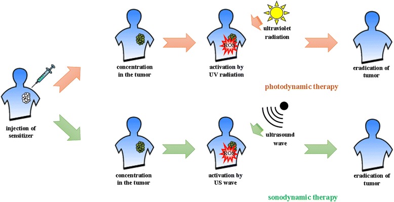
Milestones of photodynamic therapy (PDT) and sonodynamic therapy (SDT) in tumor treatment. Cancer therapies, such as PDT or SDT, build on the administration of sensitive agents into tumor, followed by their activation by UV radiation or US wave, respectively. Activated photo- or sonosensitizers generate ROS that lead to cancer cell eradication
Photosensitizers, such as TiO2 or ZnO NPs, can be delivered to the tumor cells as the result of passive or active processes [121]. The passive process takes advantage of the enhanced permeability and retention (EPR) effect. Leaky vasculature in the cancer tissue allows the NPs to be freely uptaken into the tumor cells, a phenomenon leading to the cell death. The uptake takes place also in the inflamed tissue; hence, the passive process does not provide for the targeted NPs delivery to the tumor cells [122]. In the active process, the functionalization of NPs occurs; therefore, the NPs can be delivered to tumor cells in a targeted manner. The functionalization of NPs means their conjugation with specific antibodies or ligands. PDT clearly lowers the cancer therapy side effects, compared to chemo- or radiotherapy [123]. The way the cellular uptake of nanoparticles takes place, their subcellular position and the ability to cause toxic effects depend mainly upon the NPs diameter, their surface area, surface chemistry, and charge [124]. There are two major ways of the NPs uptake into the cell: active uptake by endocytosis and passive uptake by diffusion. Due to their strong hydrophilic properties, both TiO2 and ZnO NPs are uptaken into the cells by phagocytosis [125].
It is assumed that TiO2 NPs, if exposed to the UV radiation, induce, depending on their subcellular position, one of the cell death pathways: apoptosis [105, 126], autophagy [127, 128], or necrosis [129, 130], whereas the mechanism of those processes is still insufficiently examined (Fig. 3).
Fig. 3.
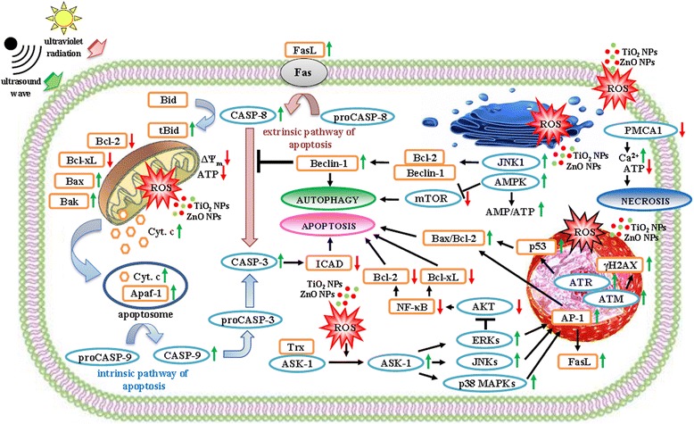
Molecular mechanisms of reactive oxygen species (ROS)-mediated cell death pathways. Activated by ultraviolet (UV) radiation or ultrasound (US) wave, nanoparticles of titanium dioxide (TiO2 NPs) and zinc oxide (ZnO NPs) induce ROS-mediated cell apoptosis, autophagy or necrosis. The increase of ROS level in cytoplasm results in thioredoxin (Trx) oxidation and apoptosis signal-regulating kinase 1 (ASK1) activation. ASK1 phosphorylates mitogen-activated protein kinases (ERKs, JNKs, p38 MAPKs) that control the AP-1-mediated synthesis of pro- (Bak, Bax) and anti-apoptotic (Bcl-2, Bcl-xL) proteins, as well as death ligands (FasL), thus promoting the cell apoptosis. Likewise, ERKs promote the cell apoptosis trough attenuation of activity of AKT kinase that controls the NF-κB-mediated synthesis of Bcl-2 and Bcl-xL proteins. The oxidative damages of mitochondrion result in dissipation of mitochondrial membrane potential (ΔΨm), decrease of ATP level and release of cytochrome c (Cyt. c) to the cytosol. Cyt. c, binding with apoptotic protease activating factor 1 (Apaf-1), induces the caspase-dependent apoptosis. The increase of ROS level in nucleus results in the activation of ataxia telangiectasia mutated (ATM), as well as ataxia telangiectasia and Rad 3-related (ATR) kinases that induce the p53-mediated apoptosis and account for the increase of DNA double-strand breaks marker (γH2AX) level. The ROS-induced endoplasmic reticulum stress results in the JNK1-mediated increase of pro-autophagic protein (Beclin-1) level and the AMPK-mediated inhibition of autophagic inhibitor, mammalian target of rapamycin (mTOR) kinase. Both processes lead to the cell autophagy. The oxidative injuries of plasma membrane result in the downregulation of plasma membrane calcium ATPase 1 (PMCA1), in the decrease of ATP level and the increase of Ca2+ level. As a consequence, the cell necrosis follows
Zhang et al. [131], having examined UV-excited TiO2 NPs toxicity towards human hepatocarcinoma cells (SMMC-7721 line), reported an increase and a decrease of protein levels that are, correspondingly, pro- (e.g., Bax, Bak, tBid) and anti-apoptotic (e.g., Bcl-2, Bcl-xL) molecules. Zhao et al. [132] point out there is an overexpression of the tBid in the TiO2 NPs-treated mouse epidermal cells (JB6 line), this protein playing a crucial role in the involvement of mitochondria in oxidative stress-dependent cell death. PDT-induced mitochondria damages result in a selective Bcl-2 degradation, with no effect on the Bax. Therefore, the Bax/Bcl-2 ratio is increased, and the intrinsic pathway of apoptosis is promoted [133–135]. Those effects were noticed in PDT-treated human breast epithelial cells (MCF10A line) [136], human brain glioblastoma cells (U87MG line) [137] or human gastric adenocarcinoma cells (MGC803 line) [138]. Nearly identical changes in levels of proteins regulating apoptosis were observed by Ahamed et al. [139] and by Wahab et al. [140] whereby the purpose of their experiments was to examine the ZnO NPs toxicity towards human A549 cells, as well as human breast adenocarcinoma cells (MCF-7 line) and human hepatocellular liver carcinoma cells (HepG2 line). The level of Bax and Bcl-2 proteins in ZnO NPs-treated (50 μg mL−1, 24 h) human HepG2 cells was twofold higher and threefold lower, respectively, compared to control cells [140]. The principle function of Bax is the formation, with the assistance of Bak, of pores in the outer mitochondrial membrane that enhance its permeability. Effectively, the release of cytochrome c (Cyt. c) from the mitochondrial intermembrane space into the cytosol follows. Cyt. c, along with the apoptotic protease-activating factor 1 (Apaf-1), contributes to the formation of the structure called apoptosome. This structure activates the initiator caspase-9 (CASP-9; EC 3.4.22.62) that subsequently activates the executioner caspase-3 (CASP-3; EC 3.4.22.56). Bcl-2 is an inhibitor of apoptosis. It associates with the outer mitochondrial membrane, keeping therefore its integrity [141, 142] (Fig. 3).
According to many authors [139, 140, 143, 144], the key role in the cell death induced by the UV-excited NPs of TiO2 or ZnO is played by ROS. They are strong oxidants but also important agents modulating ROS-sensitive signaling pathways that lead to the cell death. Biola-Clier et al. [145] report the ROS-mediated downregulation of DNA repair processes such as base excision repair and nucleotide excision repair. Toyooka et al. [146], when examining toxicity of TiO2 NPs (15 μg mL−1) to human A549 cells, concluded there was an increased H2AX histone phosphorylation that followed the oxidative stress-induced DNA double-strand breaks. Similar results obtained Premanathan et al. [147], having examined human HL-60 cells exposed to ZnO NPs (10 μg mL−1). Phosphorylated H2AX (γH2AX) histone is considered to be a marker of DNA damages [148]. Its phosphorylation is caused by ataxia telangiectasia mutated kinase (ATM; EC 2.7.11.1), as well as by ataxia telangiectasia and Rad 3-related kinase (ATR; EC 2.7.11.1), enzymes participating in the DNA damage response (DDR) pathway. The key effector protein on this pathway is p53. The importance of the ROS-induced activation of p53-mediated DNA damage check point signals in apoptosis of TiO2 NP-treated human peripheral blood lymphocytes (PBL) was stressed by Kang et al. [143]. Responding to the big scale of the DNA damages, p53 induces the pro-apoptotic protein gene expression, followed by the cell death [143] (Fig. 3).
Yoo et al. [144] studied the ROS mediation in the induction of both intrinsic and extrinsic apoptotic pathways since ROS stimulate the increase of pro-apoptotic protein (Bax) and death ligand (FasL) levels, respectively. ROS induce apoptosis in tumor cells also by modification of the activity of mitogen-activated protein kinases (MAPKs; EC 2.7.11.24) [149] and of the nuclear factor kappa B (NF-κB) [150]. Cheng et al. [151] report that PDT-generated ROS modify the ability of thioredoxin (Trx), the intracellular sensor of oxidative stress, to inhibit the apoptosis signal-regulating kinase 1 (ASK1; EC 2.7.11.25). ASK1 forms an inactive structure with reduced Trx. In the presence of ROS, the oxidation of Trx takes place and the structure degrades. The free form of ASK1 phosphorylates MAPKs such as c-Jun N-terminal protein kinase isoform 1 and 2 (JNK1/2), p38 mitogen-activated protein kinases (p38 MAPKs), and extracellular signal-regulated kinase isoform 1 and 2 (ERK1/2) [152]. Phosphorylated MAPKs translocate into the nucleus, where they induce the formation of the activator protein 1 (AP-1), a transcription factor regulating the expression of the AP-1-dependant pro- and anti-apoptotic genes [153]. JNK1/2 and p38 MAPKs lead to AP-1-mediated inactivation of anti-apoptotic proteins (Bcl-2 and Bcl-xL), the result of which is the formation of apoptosome and the activation of caspases cascade, both subsequently leading to the proteolysis of inhibitor of caspase-activated DNase (ICAD) and then, as a consequence, to the DNA fragmentation [154, 155]. JNK1/2 and p38 MAPKs promote apoptosis in case the ERK1/2 are inhibited or remain inactive [156, 157]. This conclusion was confirmed by the studies on the cytotoxicity of ZnO NPs to human HepG2 cells performed by Yuan et al. [158]. Nevertheless, in oxidative stress conditions, ERK1/2 might adopt a pro-apoptotic role since they attenuate the activity of protein kinase B (PKB, EC 2.7.11.1), called also AKT1 [159]. This phenomenon was observed in renal proximal tubule epithelial cells (LLC-PK1 line) [160]. AKT1 activates NF-κB through IκB kinase (IKK; EC 2.7.11.10) phosphorylation [161]. Coupienne et al. [162] suggest that ROS induce the AKT-mediated inactivation of NF-κB, an inhibitor of apoptosis. The NF-κB activation occurs in the cytoplasm and follows the detachment of the κB inhibitor (IκB). The active form of NF-κB translocates into the nucleus, where it initiates expression of genes that encode anti-apoptotic proteins (Fig. 3). The studies by Granville et al. [163] revealed PDT-treated human promyelocytic leukemia cells (HL-60 line) showed no detectable IκB level in the cytoplasm. It might constitute an explanation of typical morphological symptoms of the apoptotic cell death, as noticed using the confocal laser scanning microscope (CLSM), such as nucleus fragmentation, cytoplasm shrinkage, apoptotic bodies formation, or lack of inflammation reaction.
PDT-induced damages of the endoplasmic reticulum habitually result in the cell death by autophagy [164] (Fig. 3). In the oxidative stress, the activation of JNK1 that phosphorylates Bcl-2 occurs. Consequently, the dissociation of Beclin 1/Bcl-2 complex, hence the activation of Beclin 1, a pro-autophagic protein, takes place [165–167]. Such effects were noticed in PDT-treated human prostate adenocarcinoma cells (DU-145 and PC3 lines) [168]. Beclin 1 inhibits apoptosis by the inactivation of caspase-8 (CASP-8; EC 3.4.22.61), an enzyme transforming Bid to tBid, a pro-apoptotic protein. Beclin 1 also contributes, along with the phosphatidylinositol 3-kinase class III (PI3K3C; EC 2.7.1.137), towards the formation of the autophagosome that eliminates cytoplasmic “waste” material [166]. Autophagosome fuses with lisosome, leading therefore to the formation of autophagolisosome, in which lysosomal hydrolases complete the process of the definite destruction of its content [169–171]. Among the morphological symptoms of the autophagic cell death, there are increased number of autophagic vesicles, the degradation of Golgi apparatus, and endoplasmic reticulum, as well as no inflammation reaction [164]. Those symptoms were observed by Yu et el. [128] in TiO2 NP-treated human bronchial epithelial cells (16HBE14o-line). High level of ROS in a cell results in the increase of the AMP/ATP ratio, an effect stimulating the AMP-activated protein kinase (AMPK; EC, 2.7.11.31) [172, 173]. AMPK may efficiently inhibit the mammalian target of rapamycin kinase (mTOR; EC. 2.7.11.1). mTOR is a key autophagy inhibitor and is frequently overexpressed in miscellaneous tumor cells [174, 175]. Its PDT-induced inhibition, leading up to the autophagy, was observed in human breast adenocarcinoma cells (MDA-MB-231 line) [176].
High level of the PDT-generated ROS, inflicting pro-apoptotic (Bax, Bak) and pro-autophagic (Beclin 1) protein damages, as well as plasma membrane disruption, results in a cell death by necrosis [177, 178] (Fig. 3). Cell membrane oxidative damages cause the increase of the cytosolic Ca2+ level (>1 μM) that activates Ca2+-dependent proteases. Other effects are dissipation of mitochondrial membrane potential (Δψm) and a dramatic decrease of the intracellular ATP level [178]. Guo et al. [179, 180] exposed rat retinal ganglion cells (RGC-5 line) to ZnO NPs and concluded there was a remarkable increase in Ca2+ level in cytosol that resulted from the downregulation of plasma membrane calcium ATPase isoform 1 (PMCA1; EC 3.6.3.8) activity. Disrupted homeostasis of Ca2+ was also observed in human lens epithelial cells (HLE-B3 line) incubated with ZnO NPs and, subsequently, exposed to UV-B radiation [181]. Typical morphological symptoms of the necrotic cell death encompass: cell swelling, destruction of nucleus and of other cell organelles, plasma membrane disruption–all of them resulting in a cell disintegration, followed by the release of the cell content into the intercellular space hence inflammation reaction [182]. Xu et al. [108] described the necrosis of human lung adenocarcinoma cells (APC-A1 line), induced by photoexcited TiO2 NPs.
PDT-treatment of tumor cells, using the metal oxide nanoparticles, results in the cell death, presenting the consequence of irreversible DNA damage [146], plasma membrane disintegration [183], intracellular Ca2+ homeostasis disruption [179, 180], strong reduction or complete exhaustion of the redox compound pool, essential in ATP synthesis [184]. The main factor of reported damages is PDT-induced oxidative stress [184–187].
Nanocomposites, e.g., gold-doped TiO2 (Au/TiO2) [101], silver-doped TiO2 (Ag/TiO2) [102], platinum-doped TiO2 (Pt/TiO2) [188], or silver-doped ZnO (Ag/ZnO) [189] are compounds increasingly often applied in anticancer therapy. The conjugation of NPs of a precious metal with TiO2 NPs increases the catalytic activity of this oxide, a phenomenon confirmed in the studies of Abdulla-al-Mamun et al. [102]. They reported that Ag/TiO2 nanocomposite showed an 80% higher effectiveness in eradication of human HeLa cells than TiO2 NPs. In other studies, TiO2 NPs were conjugated with folic acid (FA) molecules [190]. Such conjugation enhanced the ability of human HeLa cells to uptake FA/TiO2 nanocomposite by FA receptors in plasma membrane and, therefore, to leverage the efficiency of cancer cells eradication. Feng et al. [191] confirmed high biocompatibility of FA/TiO2 nanocomposite and its incorporation into human nasopharyngeal carcinoma (KB) cells. Rozhkova et al. [109] designed TiO2-based nanocomposites that target and eradicate brain carcinoma cells in vivo with no harm to adjacent healthy cells. IL13α2R antibodies were conjugated with TiO2 NPs through 3,4-dihydroxyphenylacetic acid (DOPAC). The antibodies recognize cancer cells, bind to them and are eventually uptaken by means of phagocytosis. In the process of tumor cells exposition to UV-A radiation, TiO2-based nanoconstructs generate the formation of ROS that subsequently damage the cancer cells and induce their apoptosis. The selection of suitable antibodies can help design nanocomposites able to precisely target and eradicate many types of cancer cells [82, 83]. Xu et al. [100] advocate a simultaneous application of conjugates of TiO2 NPs with monoclonal antibodies, along with reversible plasma membrane injury by means of electric field, so called electroporation. As the authors suggest, such combination may increase the accuracy of cancer cell recognition by TiO2-based nanocomposites and, in consequence, enhance the TiO2-based nanoconstructs penetration into cytosol. Using that method, it was possible to eradicate in vitro all human LoVo cells that were subject to experiment, within merely 90 min.
One of the main disadvantages of PDT is the limited penetration depth of the tissues by the UV radiation. Therefore, this method is suitable to eradicate tumor cells located in superficial depths [192]. This shortcoming might be rectified, and deeper located tumor cells can still be eradicated using PDT, if NPs of TiO2 and ZnO are conjugated with upconverting nanoparticles (UCNPs) [193]. Ytterbium and erbium doped sodium yttrium fluoride (NaYF4:Yb,Er) is frequently applied as transducer [194]. UCNPs absorb near infrared (NIR, 700 ≤ λ < 850 nm) radiation and emit UV (λ < 400 nm) radiation. NIR radiation, compared to UV radiation, penetrates tissues in bigger depths but is also less harmful to the organism. Therefore, UCNP-based PDT gains enhanced effectiveness in the eradication of deeper depths tumor cells [195]. This finding has been confirmed both in vitro, in human cancer cells (HeLa and MCF-7 lines) and in vivo, in female Balb/c nude mice [196, 197].
Thevenot et al. [198] withdrew from TiO2 NPs excitation by UV radiation in favor of the chemical functionalization of their surfaces by binding to them –OH, –NH2, or –COOH groups. Then, they examined TiO2 NPs, as modified on their surface, in respect of their influence on the viability of a number of cancer cell lines (e.g., B16F10 mouse melanoma cell line, 3T3 mouse embryonic fibroblast cell line, or PC-3M human prostate carcinoma cell line) in vitro. According to the authors, the viability of the examined cancer cell lines was dependent upon both the concentration of TiO2 NPs in the examined cells and the type of functional group altering the surface properties of TiO2 NPs. The highest cyto- and genotoxicity exhibited NPs functionalized by –OH groups. The design of surface properties of NMs and NPs presents a valuable tool in the targeted cancer therapy.
Sonodynamic Therapy
Limited possibilities to apply PDT in the cancer therapy gave rise to seek different cancer treatment modalities. An example of an alternative tumor eradication method is SDT. This therapy is based on a synergistic effect on a cancer cell by sonosensitizer and US [199]. US can activate particles of compounds called sonosensitizers. In comparison with UV or NIR radiation, US penetrates tissues much more effectively as it reaches deeper depths tumor cells. The introduction of sonosensitizers into cancer cells and their subsequent exposure to US in order to generate ROS is the underlying factor of SDT [199, 200] (Fig. 2).
Harada et al. [114] showed in their studies in vitro that there was a remarkable reduction (in excess of 50%) in viability of human melanoma cells (C32 line) upon exposure to the TiO2 NP-based SDT (1 MHz, 30 s). Moosavi Nejda et al. [200] examined in vitro the influence of the US-excited TiO2 NPs (100 μg mL−1) on the viability of human oral squamous carcinoma cells (HSC-2 line). The authors proved the increased intensity of US (20, 32, 55, 73 W cm−2) and the prolonged sonication period (0.1, 1.0, 3.0 s) enhanced the damage ratio in the human HSC-2 cells, finally leading to their necrosis at the conditions of 73 W cm−2 and 3 s. Yamaguchi et al. [183] believe that TiO2 NP-based cancer therapies will soon become a valuable tool in fighting malignant gliomas. The authors compared PDT to SDT in respect of their effectiveness in eradication of human glioblastoma cells (U251 line). With PDT, the toxicity effect of TiO2 NPs (20 μg mL−1) was noticeable after 2-h exposure of single cancer cell layer to UV-A radiation (5.0 mW cm−2). Moreover, the effect was fairly restricted by free radical scavengers. Cytotoxicity of TiO2 NPs (20 μg mL−1) in case of SDT was proportional to the sonication period, and it was not dependent on antioxidants concentration, an observation indicating there must be a different mechanism of intracellular structure damages. US, propagating in cell, induce the formation of cavitation bubbles which free energy as they implode. It is assumed that acoustic cavitation can not only lead to sonosensitizer excitation, the result of which is 1O2 formation, but it can also cause—with no ROS involved—a disruption in plasma membrane integrity. Saito et al. [201] report that the necrotic death of human corneal endothelial cells after the US exposure can take place rapidly and could be attributed to mechanically induced damage to the plasma membrane. Thanks to fluorescent dyes, a greater amount of plasma membrane damages resulting from the TiO2 NP-based SDT could be noticed than in the TiO2 NP-based PDT.
In SDT, similarly to PDT, TiO2 NPs are also conjugated with precious metals or antibodies. The subject of studies conducted by Ogino et al. [202], as well as Ninomiya et al. [203, 204], was the examination of TiO2 NP toxicity to human HepG2 cells. To increase the TiO2 NPs uptake rate by cancer cells, pre-S1/S2, a model antibody recognizing hepatocytes, was conjugated with the TiO2 NPs. The ability of such conjugates to recognize cancer cells was examined using the surface plasmon resonance and immunofluorescence techniques. Apoptosis of some cells of the human HepG2 cells in vitro was firstly observed after 6 h from the termination of the TiO2 NP sonication (1 MHz, 30 s), and after the following 90 h, the number of dividing cells decreased by 45%. The studies conducted by Ninomiya et al. [205] revealed the high efficiency of the TiO2 NP-based SDT as a tool to reduce the proliferation of human MCF-7 cells, too. In this case, avidin was immobilized on the surface of TiO2 NPs in order to enhance its uptake rate by cancer cells. Within 96 h after the termination of the TiO2 NP sonication (1 MHz, 30 s), the number of viable cancer cells was reduced by 30%.
TiO2 and ZnO NPs are excellent sensitizers and oxidizing agents. The feature of the ROS generated on their surface is a higher oxidation potential as well as a short half-life (<0.04 μs) [41]. Therefore, the occurrence of ROS-induced oxidative injuries is limited to a small distance (<0.02 μm) from the subcellular position of the sensitizer NPs [54]. NPs administered into the bloodstream become covered by a plasma proteins layer, called protein corona, that lowers their cytotoxicity and, depending on the type of adsorbed plasma proteins, modulates their immunotoxicity and the targeted delivery into tumor cells [206, 207]. Garvas et al. [208] proved the cytotoxicity of TiO2 NPs in vivo is limited by serum proteins covering their surface that exhibit a trapping ability of photogenerated ROS. The knowledge on the potential application of NPs of TiO2 and ZnO in therapy and diagnostics of neoplastic maladies is continuously growing. Nonetheless, further studies are necessary to improve the tumor targetability of TiO2 and ZnO NPs, as well as to increase the therapeutic window of PDT using metal oxide NPs [209, 210].
Conclusions
Traditional methods of cancer therapy inflict a series of side effects, including the impairment of the immune system. Recent years have witnessed many efforts in creating alternative cancer therapy methods that would be less invasive and would exhibit a site-specific activity. PDT might present a viable solution. Among photosensitizers that have been thoroughly examined in view of their potential application in cancer therapies, there are TiO2 and ZnO in their NPs form. UV-excited NPs of TiO2 and ZnO are a source of ROS that form on their surfaces. ROS are agents with a high oxidative potential that damage primarily DNA and cell membranes of cancer cells. Plasma membrane damages lead to cell necrosis, nucleus and mitochondria damages induce apoptotic cell death, and endoplasmic reticulum damages cause cell death by autophagy. Limited tissue penetration ability of UV radiation presents a serious obstacle in the application of UV-sensitizing NPs of TiO2 and ZnO in cancer therapy. Efforts to apply NIR radiation, since it is less harmful and can penetrate deeper depths tissues, have so far not been successful. Therefore, a new method had to be set up, allowing to generate ROS in deeper depths tissues. Such a method is SDT, using US with low tissue attenuation coefficient. However, SDT exhibits one major disadvantage: insufficient tumor targetability by NPs of TiO2 and ZnO. The difficulty in preparation of a drug formula that would allow the targeted delivery of NPs to cancer cells is an important factor that restricts the application of photo- and sonosensitizers in cancer therapy. Thanks to the development of nanotechnology, various drug delivery systems in organisms have been designed. One of them is compiling conjugates of TiO2 or ZnO in their NPs form with monoclonal antibodies, a method that allows targeting the photo- and sonosensitizers on a selected receptor. The design of modern, more effective cancer fight methods remains an important challenge to contemporary medicine. The scientists are still facing many years of studies on how to optimize cancer therapy using nanotechnology whereas the results have so far been fairly promising.
Acknowledgments
Authors’ Contributions
JB performed the overall review, drafted the manuscript, and designed all figures. JPC assisted in the research work on the latest developments and perspectives of the application of TiO2 and ZnO in anticancer therapy and helped draft the manuscript. JZ modified the first version of the draft, prepared its revision, and assisted in the sequence alignment. All authors read and approved the final manuscript.
Competing Interests
The authors declare that they have no competing interests.
Publisher’s Note
Springer Nature remains neutral with regard to jurisdictional claims in published maps and institutional affiliations.
Abbreviations
- AMPK
AMP-activated protein kinase
- AP-1
Activator protein 1
- Apaf-1
Apoptotic protease-activating factor 1
- ASK
Apoptosis signal-regulating kinase
- ATM
Ataxia telangiectasia mutated kinase
- ATR
Ataxia telangiectasia Rad 3-related kinase
- BAL
Bronchoalveolar lavage
- CASP
Caspase
- CAT
Catalase
- CB
Conduction band
- CLSM
Confocal laser scanning microscope
- DDR
DNA damage response
- DOPAC
3,4-Dihydroxyphenylacetic acid
- ERK
Extracellular signal-regulated kinase
- FA
Folic acid
- GPX
Glutathione peroxidase
- GSH
Glutathione
- ICAD
Inhibitor of caspase-activated DNase
- IKK
κB inhibitor kinase
- IκB
κB inhibitor
- JNK
c-Jun N-terminal protein kinase
- LPOs
Lipid hydroperoxides
- MAPKs
Mitogen-activated protein kinases
- MPs
Microparticles
- mTOR
Mammalian target of rapamycin kinase
- NF-κB
Nuclear factor-kappa B
- NMs
Nanomaterials
- NPs
Nanoparticles
- NRRT
Neutral red retention time
- PDT
Photodynamic therapy
- PI3K3C
Phosphatidylinositol 3-kinase class III
- PKB
Protein kinase B
- PMCA
Plasma membrane calcium ATPase
- ROS
Reactive oxygen species
- SCGE
Single-cell gel electrophoresis
- SDT
Sonodynamic therapy
- SOD
Superoxide dismutase
- Trx
Thioredoxin
- US
Ultrasound
- UV
Ultraviolet
- VB
Valence band
Contributor Information
Janusz Bogdan, Email: janusz_bogdan@sggw.pl.
Joanna Pławińska-Czarnak, Email: joanna_plawinska@sggw.pl.
Joanna Zarzyńska, Email: joanna_zarzynska@sggw.pl.
References
- 1.Feynman RP. There’s plenty of room at the bottom. Eng. Sci. 1960;23(5):22–36. [Google Scholar]
- 2.Taniguchi N. Proc Intl Conf Prod Eng 26-29 August 1974, Tokyo, Part II. Tokyo: Japan Society of Precision Engineering; 1974. On the basic concept of “nano-technology”; pp. 18–23. [Google Scholar]
- 3.Drexler KE. Nanosystems: molecular machinery, manufacturing and computation. New York: John Wiley and Sons, Inc.; 1992. [Google Scholar]
- 4.International Organization for Standardization . Nanotechnologies—vocabulary—part 2. Nano-objects. 2015. [Google Scholar]
- 5.Whitesides GM. The “right” size in nanobiotechnology. Nat Biotechnol. 2003;21(10):1161–1165. doi: 10.1038/nbt872. [DOI] [PubMed] [Google Scholar]
- 6.Dowling AP. Development of nanotechnologies. Mater Today. 2004;7(12):30–35. doi: 10.1016/S1369-7021(04)00628-5. [DOI] [Google Scholar]
- 7.Sweeney AE. Nanomedicine concepts in the general medical curriculum: initiating a discussion. Int J Nanomedicine. 2015;10(1):7319–7331. doi: 10.2147/IJN.S96480. [DOI] [PMC free article] [PubMed] [Google Scholar]
- 8.Fakruddin M, Hossain Z, Afroz H. Prospects and applications of nanobiotechnology: a medical perspective. J Nanobiotechnology. 2012;10:31. doi: 10.1186/1477-3155-10-31. [DOI] [PMC free article] [PubMed] [Google Scholar] [Retracted]
- 9.Kamila S, McEwan C, Costley D, Atchison J, Sheng Y, Hamilton GR, Fowley C, Callan JF. Diagnostic and therapeutic applications of quantum dots in nanomedicine. Top Curr Chem. 2016;370:203–224. doi: 10.1007/978-3-319-22942-3_7. [DOI] [PubMed] [Google Scholar]
- 10.Orive G, Anitua E, Pedraz JL, Emerich DF. Biomaterials for promoting brain protection, repair and regeneration. Nat Rev Neurosci. 2009;10(9):682–692. doi: 10.1038/nrn2685. [DOI] [PubMed] [Google Scholar]
- 11.Iannazzo D, Piperno A, Pistone A, Grassi G, Galvagno S. Recent advances in carbon nanotubes as delivery systems for anticancer drugs. Curr Med Chem. 2013;20(11):1333–1354. doi: 10.2174/0929867311320110001. [DOI] [PubMed] [Google Scholar]
- 12.Ankri R, Peretz D, Motiei M, Sella-Tavor O, Popovtzer R. New optical method for enhanced detection of colon cancer by capsule endoscopy. Nanoscale. 2013;5(20):9806–9811. doi: 10.1039/c3nr02396f. [DOI] [PubMed] [Google Scholar]
- 13.Sanna V, Pala N, Sechi M. Targeted therapy using nanotechnology: focus on cancer. Int J Nanomedicine. 2014;9:467–483. doi: 10.2147/IJN.S36654. [DOI] [PMC free article] [PubMed] [Google Scholar]
- 14.Mukherjee B. Nanosize drug delivery system. Curr Pharm Biotechnol. 2013;14(15):1221. doi: 10.2174/138920101415140804121008. [DOI] [PubMed] [Google Scholar]
- 15.Krishnamurthy S, Ke X, Yang YY. Delivery of therapeutics using nanocarriers for targeting cancer cells and cancer stem cells. Nanomedicine. 2015;10(1):143–160. doi: 10.2217/nnm.14.154. [DOI] [PubMed] [Google Scholar]
- 16.Ediriwickrema A, Saltzman WM. Nanotherapy for cancer: targeting and multifunctionality in the future of cancer therapies. ACS Biomater Sci Eng. 2015;1(2):64–78. doi: 10.1021/ab500084g. [DOI] [PMC free article] [PubMed] [Google Scholar]
- 17.Lee BK, Yun YH, Park K. Smart nanoparticles for drug delivery: boundaries and opportunities. Chem Eng Sci. 2015;125:158–164. doi: 10.1016/j.ces.2014.06.042. [DOI] [PMC free article] [PubMed] [Google Scholar]
- 18.Cui W, Li J, Decher G. Self-assembled smart nanocarriers for targeted drug delivery. Adv Mater. 2016;28(6):1302–1311. doi: 10.1002/adma.201502479. [DOI] [PubMed] [Google Scholar]
- 19.Moyano DF, Liu Y, Peer D, Rotello VM. Modulation of immune response using engineered nanoparticle surfaces. Small. 2016;12(1):76–82. doi: 10.1002/smll.201502273. [DOI] [PMC free article] [PubMed] [Google Scholar]
- 20.Huang C, Ozdemir T, Xu LC, Butler PJ, Siedlecki CA, Brown JL, Zhang S. The role of substrate topography on the cellular uptake of nanoparticles. J Biomed Mater Res B Appl Biomater. 2016;104(3):488–495. doi: 10.1002/jbm.b.33397. [DOI] [PMC free article] [PubMed] [Google Scholar]
- 21.Azzazy HM, Mansour MM, Kazmierczak SC. From diagnostics to therapy: prospects of quantum dots. Clin Biochem. 2007;40(13-14):917–927. doi: 10.1016/j.clinbiochem.2007.05.018. [DOI] [PubMed] [Google Scholar]
- 22.Mokhtarzadeh A, Alibakhshi A, Yaghoobi H, Hashemi M, Hejazi M, Ramezani M. Recent advances on biocompatible and biodegradable nanoparticles as gene carriers. Expert Opin Biol Ther. 2016;16(6):771–784. doi: 10.1517/14712598.2016.1169269. [DOI] [PubMed] [Google Scholar]
- 23.Ahmad M, Ahmed E, Hong ZL, Ahmed W, Elhissi A, Khalid NR. Photocatalytic, sonocatalytic and sonophotocatalytic degradation of Rhodamine B using ZnO/CNTs composites photocatalysts. Ultrason Sonochem. 2014;21(2):761–773. doi: 10.1016/j.ultsonch.2013.08.014. [DOI] [PubMed] [Google Scholar]
- 24.Jassby D, Farner Budarz J, Wiesner M. Impact of aggregate size and structure on the photocatalytic properties of TiO2 and ZnO nanoparticles. Environ Sci Technol. 2012;46(13):6934–6941. doi: 10.1021/es202009h. [DOI] [PubMed] [Google Scholar]
- 25.Soleimani F, Madaah Hosseini HR, Ordikhani F, Mokhtari-Dizaji M. Enhancing sonocatalytic properties of TiO2 nanocatalysts by controlling the surface conditions: effect of particle size and PVA modification. Desalin. Water Treat. 2016;57(58):28378–28385. doi: 10.1080/19443994.2016.1185746. [DOI] [Google Scholar]
- 26.Gupta SM, Tripathi M. A review of TiO2 nanoparticles. Chin Sci Bull. 2011;56(16):1639–1657. doi: 10.1007/s11434-011-4476-1. [DOI] [Google Scholar]
- 27.Salata O. Applications of nanoparticles in biology and medicine. J Nanobiotechnology. 2004;2(1):3. doi: 10.1186/1477-3155-2-3. [DOI] [PMC free article] [PubMed] [Google Scholar]
- 28.Zhang L, Gu FX, Chan JM, Wang AZ, Langer RS, Farokhzad OC. Nanoparticles in medicine: therapeutic applications and developments. Clin Pharmacol Ther. 2008;83(5):761–769. doi: 10.1038/sj.clpt.6100400. [DOI] [PubMed] [Google Scholar]
- 29.Bhattacharyya S, Kudgus RA, Bhattacharya R, Mukherjee P. Inorganic nanoparticles in cancer therapy. Pharm Res. 2011;28(2):237–259. doi: 10.1007/s11095-010-0318-0. [DOI] [PMC free article] [PubMed] [Google Scholar]
- 30.Rasmussen JW, Martinez E, Louka P, Wingett DG. Zinc oxide nanoparticles for selective destruction of tumor cells and potential for drug delivery applications. Expert Opin Drug Deliv. 2010;7(9):1063–1077. doi: 10.1517/17425247.2010.502560. [DOI] [PMC free article] [PubMed] [Google Scholar]
- 31.Khan MM, Adil SF, Al-Mayouf A. Metal oxides as photocatalysts. J Saudi Chem Soc. 2015;19(5):462–464. doi: 10.1016/j.jscs.2015.04.003. [DOI] [Google Scholar]
- 32.Lee KM, Lai CW, Ngai KS, Juan JC. Recent developments of zinc oxide based photocatalyst in water treatment technology: a review. Water Res. 2016;88:428–448. doi: 10.1016/j.watres.2015.09.045. [DOI] [PubMed] [Google Scholar]
- 33.Haidaka H, Horikoshi S, Serpone N, Knowland J. In vitro photochemical damage to DNA, RNA and their bases by an inorganic sunscreen agent on exposure to UVA and UVB radiation. J Photochem Photobiol A Chem. 1997;111(1-3):205–213. doi: 10.1016/S1010-6030(97)00229-3. [DOI] [Google Scholar]
- 34.Kim S, An YJ. Effect of ZnO and TiO2 nanoparticles preilluminated with UVA and UVB light on Escherichia coli and Bacillus subtilis. Appl Microbiol Biotechnol. 2012;95(1):243–253. doi: 10.1007/s00253-012-4153-6. [DOI] [PubMed] [Google Scholar]
- 35.Kühn KP, Chaberny IF, Massholder K, Stickler M, Benz VW, Sonntag HG, Erdinger L. Disinfection of surfaces by photocatalytic oxidation with titanium dioxide and UVA light. Chemosphere. 2003;53(1):71–77. doi: 10.1016/S0045-6535(03)00362-X. [DOI] [PubMed] [Google Scholar]
- 36.Lee JE, Ko G. Norovirus and MS2 inactivation kinetics of UV-A and UV-B with and without TiO2. Water Res. 2013;47(15):5607–5613. doi: 10.1016/j.watres.2013.06.035. [DOI] [PubMed] [Google Scholar]
- 37.Pablos C, Marugán J, van Grieken R, Serrano E. Emerging micropollutant oxidation during disinfection processes using UV-C, UV-C/H2O2, UV-A/TiO2 and UV-A/TiO2/H2O2. Water Res. 2013;47(3):1237–1245. doi: 10.1016/j.watres.2012.11.041. [DOI] [PubMed] [Google Scholar]
- 38.Robertson JMC, Robertson PKJ, Lawton LA. A comparison of the effectiveness of TiO2 photocatalysis and UVA photolysis for the destruction of three pathogenic microorganisms. J Photochem Photobiol A Chem. 2005;175(1):51–56. doi: 10.1016/j.jphotochem.2005.04.033. [DOI] [Google Scholar]
- 39.Anju SG, Jyothi KP, Joseph S, Suguna Y, Yesodharan EP. Ultrasound assisted semiconductor mediated catalytic degradation of organic pollutants in water: comparative efficacy of ZnO, TiO2 and ZnO-TiO2. Res J Recent Sci. 2012;1:191–201. [Google Scholar]
- 40.Grčić I, Vujević D, Žižek K, Koprivanac N. Treatment of organic pollutants in water using TiO2 powders: photocatalysis versus sonocatalysis. React Kinet Mech Cat. 2013;109(2):335–354. doi: 10.1007/s11144-013-0562-5. [DOI] [Google Scholar]
- 41.Fujishima A, Rao TN, Tryk DA. Titanium dioxide photocatalysis. J. Photochem. Photobiol. C Photchem. Rev. 2000;1(1):1–21. doi: 10.1016/S1389-5567(00)00002-2. [DOI] [Google Scholar]
- 42.Mills A, le Hunte S. An overview of semiconductor photocatalysis. J Photochem Photobiol A Chem. 1997;108(1):1–35. doi: 10.1016/S1010-6030(97)00118-4. [DOI] [Google Scholar]
- 43.Linsebigler AL, Lu G, Yates JY. Photocatalysis on TiO2 surfaces: principles, mechanisms, and selected results. Chem Rev. 1995;95(3):735–758. doi: 10.1021/cr00035a013. [DOI] [Google Scholar]
- 44.Nel A, Xia T, Mädler L, Li N. Toxic potential of materials at the nanolevel. Science. 2006;311(5761):622–627. doi: 10.1126/science.1114397. [DOI] [PubMed] [Google Scholar]
- 45.Reczek CR, Chandel NS. ROS-dependent signal transduction. Curr Opin Cell Biol. 2015;33:8–13. doi: 10.1016/j.ceb.2014.09.010. [DOI] [PMC free article] [PubMed] [Google Scholar]
- 46.Quast SA, Berger A, Eberle J. ROS-dependent phosphorylation of Bax by wortmannin sensitizes melanoma cells for TRAIL-induced apoptosis. Cell Death Dis. 2013;4:e839. doi: 10.1038/cddis.2013.344. [DOI] [PMC free article] [PubMed] [Google Scholar]
- 47.Görlach A, Bertram K, Hudecova S, Krizanova O. Calcium and ROS: a mutual interplay. Redox Biol. 2015;6:260–271. doi: 10.1016/j.redox.2015.08.010. [DOI] [PMC free article] [PubMed] [Google Scholar]
- 48.Sun Y. Free radicals, antioxidant enzymes, and carcinogenesis. Free Radic Biol Med. 1990;8(6):583–599. doi: 10.1016/0891-5849(90)90156-D. [DOI] [PubMed] [Google Scholar]
- 49.Porter NA, Caldwell SE, Mills KA. Mechanisms of free radical oxidation of unsaturated lipids. Lipids. 1995;30(4):277–290. doi: 10.1007/BF02536034. [DOI] [PubMed] [Google Scholar]
- 50.Aust AE, Eveleigh JF. Mechanisms of DNA oxidation. Proc Soc Exp Biol Med. 1999;222(3):246–252. doi: 10.1046/j.1525-1373.1999.d01-141.x. [DOI] [PubMed] [Google Scholar]
- 51.Sohal RS. Role of oxidative stress and protein oxidation in the aging process. Free Radic Biol Med. 2002;33(1):37–44. doi: 10.1016/S0891-5849(02)00856-0. [DOI] [PubMed] [Google Scholar]
- 52.Martindale JL, Holbrook NJ. Cellular response to oxidative stress: signaling for suicide and survival. J Cell Physiol. 2002;192(1):1–15. doi: 10.1002/jcp.10119. [DOI] [PubMed] [Google Scholar]
- 53.Antunes F, Cadenas E. Cellular titration of apoptosis with steady state concentrations of H2O2: submicromolar levels of H2O2 induce apoptosis through Fenton chemistry independent of the cellular thiol state. Free Radic Biol Med. 2001;30(9):1008–1018. doi: 10.1016/S0891-5849(01)00493-2. [DOI] [PubMed] [Google Scholar]
- 54.Liu J, Wang Z. Increased oxidative stress as a selective anticancer therapy. Oxid Med Cell Longev. 2015;2015:294303. doi: 10.1155/2015/294303. [DOI] [PMC free article] [PubMed] [Google Scholar]
- 55.Mileo AM, Miccadei S. Polyphenols as modulator of oxidative stress in cancer disease: new therapeutic strategies. Oxid Med Cell Longev. 2016;2016:6475624. doi: 10.1155/2016/6475624. [DOI] [PMC free article] [PubMed] [Google Scholar]
- 56.Ahmad J, Akhter S, Rizwanullah M, Amin S, Rahman M, Ahmad MZ, Rizvi MA, Kamal MA, Ahmad FJ. Nanotechnology-based inhalation treatments for lung cancer: state of the art. Nanotechnol. Sci. Appl. 2015;8:55–66. doi: 10.2147/NSA.S49052. [DOI] [PMC free article] [PubMed] [Google Scholar]
- 57.Vinardell MP, Mitjans M. Antitumor activities of metal oxide nanoparticles. Nanomaterials. 2015;5:1004–1021. doi: 10.3390/nano5021004. [DOI] [PMC free article] [PubMed] [Google Scholar]
- 58.Bystrzejewska B, Golimowski J, Urban PL. Nanoparticles: their potential toxicity, waste and environmental management. Waste Manage. 2009;29(9):2587–2595. doi: 10.1016/j.wasman.2009.04.001. [DOI] [PubMed] [Google Scholar]
- 59.Menard A, Drobne D, Jemec A. Ecotoxicity of nanosized TiO2. Review of in vivo data. Environ Pollut. 2011;159(3):677–684. doi: 10.1016/j.envpol.2010.11.027. [DOI] [PubMed] [Google Scholar]
- 60.Chihara Y, Fujimoto K, Kondo H, Moriwaka Y, Sasahira T, Hirao Y, Kuniyasu H. Anti-tumor effects of liposome-encapsluated titanium dioxide in nude mice. Pathobiology. 2007;74(6):353–358. doi: 10.1159/000110029. [DOI] [PubMed] [Google Scholar]
- 61.Landsiedel R, Kapp MD, Schulz M, Wiench K, Oesch F. Genotoxicity investigations on nanomaterials: methods, preparation and characterization of test material, potential artifacts and limitations-many questions, some answers. Mutat Res. 2009;681(2-3):241–258. doi: 10.1016/j.mrrev.2008.10.002. [DOI] [PubMed] [Google Scholar]
- 62.Wang J, Fan Y. Lung injury induced by TiO2 nanoparticles depends on their structural features: size, shape, crystal phases, and surface coating. Int J Mol Sci. 2014;15(12):22258–22278. doi: 10.3390/ijms151222258. [DOI] [PMC free article] [PubMed] [Google Scholar]
- 63.Kobayashi N, Naya M, Endoh S, Maru J, Yamamoto K, Nakanishi J. Comparative pulmonary toxicity study of nano-TiO2 particles of different sizes and agglomerations in rats: different hort- and long-term post-instillation results. Toxicology. 2009;264(1-2):110–118. doi: 10.1016/j.tox.2009.08.002. [DOI] [PubMed] [Google Scholar]
- 64.Sycheva LP, Zhurkov VS, Iurchenko VV, Daugel-Dauge NO, Kovalenko MA, Krivtsova EK, Durnev AD. Investigation of genotoxic and cytotoxic effects of micro- and nanosized titanium dioxide in six organs of mice in vivo. Mutat Res. 2011;726(1):8–14. doi: 10.1016/j.mrgentox.2011.07.010. [DOI] [PubMed] [Google Scholar]
- 65.Xiong D, Fang T, Yu L, Sima X, Zhu W. Effects of nano-scale TiO2, ZnO and their bulk counterparts on zebrafish: acute toxicity, oxidative stress and oxidative damage. Sci Total Environ. 2011;409(8):1444–1452. doi: 10.1016/j.scitotenv.2011.01.015. [DOI] [PubMed] [Google Scholar]
- 66.Ma-Hock L, Burkhardt S, Strauss V, Gamer AO, Wiench K, van Ravenzwaay B, Landsiedel R. Development of a short-term inhalation test in the rat using nano-titanium dioxide as a model substance. Inhal Toxicol. 2009;21(2):102–118. doi: 10.1080/08958370802361057. [DOI] [PubMed] [Google Scholar]
- 67.Bermudez E, Mangum JB, Wong BA, Asgharian B, Hext PM, Warheit DB, Everitt JI. Pulmonary responses of mice, rats, and hamsters to subchronic inhalation of ultrafine titanium dioxide particles. Toxicol Sci. 2004;77(2):347–357. doi: 10.1093/toxsci/kfh019. [DOI] [PubMed] [Google Scholar]
- 68.Ze Y, Hu R, Wang X, Sang X, Ze X, Li B, Su J, Wang Y, Guan N, Zhao X, Gui S, Zhu L, Cheng Z, Cheng J, Sheng L, Sun Q, Wang L, Hong F. Neurotoxicity and gene-expressed profile in brain-injured mice caused by exposure to titanium dioxide nanoparticles. J Biomed Mater Res A. 2014;102(2):470–478. doi: 10.1002/jbm.a.34705. [DOI] [PubMed] [Google Scholar]
- 69.Lindberg HK, Falck GC, Catalán J, Koivisto AJ, Suhonen S, Järventaus H, Rossi EM, Nykäsenoja H, Peltonen Y, Moreno C, Alenius H, Tuomi T, Savolainen KM, Norppa H. Genotoxicity of inhaled nanosized TiO2 in mice. Mutat Res. 2012;745(1-2):58–64. doi: 10.1016/j.mrgentox.2011.10.011. [DOI] [PubMed] [Google Scholar]
- 70.Rehn B, Seiler F, Rehn S, Bruch J, Maier M. Investigations on the inflammatory and genotoxic lung effects of two types of titanium dioxide: untreated and surface treated. Toxicol Appl Pharmacol. 2003;189(2):84–95. doi: 10.1016/S0041-008X(03)00092-9. [DOI] [PubMed] [Google Scholar]
- 71.Trouiller B, Reliene R, Westbrook A, Solaimani P, Schiestl R. Titanium dioxide nanoparticles induce DNA damage and genetic instability in vivo in mice. Cancer Res. 2009;69(22):784–8789. doi: 10.1158/0008-5472.CAN-09-2496. [DOI] [PMC free article] [PubMed] [Google Scholar]
- 72.Ze Y, Zheng L, Zhao X, Gui S, Sang X, Su J, Guan N, Zhu L, Sheng L, Hu R, Cheng J, Cheng Z, Sun Q, Wang L, Hong F. Molecular mechanism of titanium dioxide nanoparticles-induced oxidative injury in the brain of mice. Chemosphere. 2013;92(9):1183–1189. doi: 10.1016/j.chemosphere.2013.01.094. [DOI] [PubMed] [Google Scholar]
- 73.Reeves JF, Davies SJ, Dodd NJF, Jha AN. Hydroxyl radicals (*OH) are associated with titanium dioxide (TiO2) nanoparticle-induced cytotoxicity and oxidative DNA damage in fish cells. Mutat Res. 2008;640(1-2):113–122. doi: 10.1016/j.mrfmmm.2007.12.010. [DOI] [PubMed] [Google Scholar]
- 74.Ferin J, Oberdörster G. Biological effects and toxicity assessment of titanium dioxides: anatase and rutile. Am Ind Hyg Assoc J. 1985;46(2):69–72. doi: 10.1080/15298668591394419. [DOI] [PubMed] [Google Scholar]
- 75.IARC Working Group on the Evaluation of Carcinogenic Risks to Humans Carbon black, titanium dioxide and talc. IARC Monogr Eval Carcinog Risks Hum. 2010;93:1–413. [PMC free article] [PubMed] [Google Scholar]
- 76.Gurr JR, Wang AS, Chen CH, Jan KY. Ultrafine titanium dioxide particles in the absence of photoactivation can induce oxidative damage to human bronchial epithelial cells. Toxicology. 2005;213(1-2):66–73. doi: 10.1016/j.tox.2005.05.007. [DOI] [PubMed] [Google Scholar]
- 77.Kongseng S, Yoovathaworn K, Wongprasert K, Chunhabundit R, Sukwong P, Pissuwan D. Cytotoxic and inflammatory responses of TiO2 nanoparticles on human peripheral blood mononuclear cells. J Appl Toxicol. 2016;36(10):1364–1373. doi: 10.1002/jat.3342. [DOI] [PubMed] [Google Scholar]
- 78.Long TC, Saleh N, Tilton RD, Lowry GV, Veronesi B. Titanium dioxide (P25) produces reactive oxygen species in immortalized brain microglia (BV2): implications for nanoparticle neurotoxicity. Environ Sci Technol. 2006;40(14):4346–4352. doi: 10.1021/es060589n. [DOI] [PubMed] [Google Scholar]
- 79.Long TC, Tajuba J, Sama P, Saleh N, Swartz C, Parker J, Hester S, Lowry GV. Nanosize titanium dioxide stimulates reactive oxygen species in brain microglia and damages neurons in vitro. Environ Health Perspect. 2007;115(11):1631–1637. doi: 10.1289/ehp.10216. [DOI] [PMC free article] [PubMed] [Google Scholar]
- 80.Fujita K, Horie M, Kato H, Endoh S, Suzuki M, Nakamura A, Miyauchi A, Yamamoto K, Kinugasa S, Nishio K, Yoshida Y, Iwahashi H, Nakanishi J. Effects of ultrafine TiO2 particles on gene expression profile in human keratinocytes without illumination: involvement of extracellular matrix and cell adhesion. Toxicol Lett. 2009;191(2-3):109–117. doi: 10.1016/j.toxlet.2009.08.011. [DOI] [PubMed] [Google Scholar]
- 81.Vevers WF, Jha AN. Genotoxic and cytotoxic potential of titanium dioxide (TiO2) nanoparticles on fish cells in vitro. Ecotoxicology. 2008;7(5):410–420. doi: 10.1007/s10646-008-0226-9. [DOI] [PubMed] [Google Scholar]
- 82.Saquib Q, Al-Khedhairy AA, Siddiqui MA, Abou-Tarboush FM, Azam A, Musarrat J. Titanium dioxide nanoparticles induced cytotoxicity, oxidative stress and DNA damage in human amnion epithelial (WISH) cells. Toxicol in Vitro. 2012;26(2):351–361. doi: 10.1016/j.tiv.2011.12.011. [DOI] [PubMed] [Google Scholar]
- 83.Shukla RK, Sharma V, Pandey AK, Singh S, Sultana S, Dhawan A. ROS-mediated genotoxicity induced by titanium dioxide nanoparticles in human epidermal cells. Toxicol in Vitro. 2011;25(1):231–241. doi: 10.1016/j.tiv.2010.11.008. [DOI] [PubMed] [Google Scholar]
- 84.Vamanu CI, Cimpan MR, Høl PJ, Sørnes S, Lie SA, Gjerdet NR. Induction of cell death by TiO2 nanoparticles: studies on a human monoblastoid cell line. Toxicol in Vitro. 2008;22(7):1689–1696. doi: 10.1016/j.tiv.2008.07.002. [DOI] [PubMed] [Google Scholar]
- 85.Xue C, Wu J, Lan F, Liu W, Yang X, Zeng F, Xu H. Nano titanium dioxide induces the generation of ROS and potential damage in HaCaT cells under UVA irradiation. J Nanosci Nanotechnol. 2010;10(12):8500–8507. doi: 10.1166/jnn.2010.2682. [DOI] [PubMed] [Google Scholar]
- 86.Gómez-Couso H, Fontán-Saínz M, Sichel C, Fernández-Ibáñez P, Ares-Mazás E. Efficacy of the solar water disinfection method in turbid waters experimentally contaminated with Cryptosporidium parvum oocysts under real field conditions. Trop Med Int Health. 2009;14(6):620–627. doi: 10.1111/j.1365-3156.2009.02281.x. [DOI] [PubMed] [Google Scholar]
- 87.Lee JH, Kang M, Choung SJ, Ogino K, Miyata S, Kim MS, Park JY, Kim JB. The preparation of TiO2 nanometer photocatalyst film by a hydrothermal method and its sterilization performance for Giardia lamblia. Water Res. 2004;38(3):713–719. doi: 10.1016/j.watres.2003.10.011. [DOI] [PubMed] [Google Scholar]
- 88.Sökmen M, Değerli S, Aslan A. Photocatalytic disinfection of Giardia intestinalis and Acanthamoeba castellani cysts in water. Exp Parasitol. 2008;119(1):44–48. doi: 10.1016/j.exppara.2007.12.014. [DOI] [PubMed] [Google Scholar]
- 89.Wang CC, Wang S, Xia Q, He W, Yin JJ, Fu PP, Li JH. Phototoxicity of zinc oxide nanoparticles in HaCaT keratinocytes-generation of oxidative DNA damage during UVA and visible light irradiation. J Nanosci Nanotechnol. 2013;13(6):3880–3888. doi: 10.1166/jnn.2013.7177. [DOI] [PubMed] [Google Scholar]
- 90.Sahu D, Kannan GM, Vijayaraghavan R. Size-dependent effect of zinc oxide on toxicity and inflammatory potential of human monocytes. J Toxicol Environ Health A. 2014;77(4):177–191. doi: 10.1080/15287394.2013.853224. [DOI] [PubMed] [Google Scholar]
- 91.Senapati VA, Kumar A, Gupta GS, Pandey AK, Dhawan A. ZnO nanoparticles induced inflammatory response and genotoxicity in human blood cells: a mechanistic approach. Food Chem Toxicol. 2015;85:61–70. doi: 10.1016/j.fct.2015.06.018. [DOI] [PubMed] [Google Scholar]
- 92.Hsiao IL, Huang YJ. Effects of serum on cytotoxicity of nano- and micro-sized ZnO particles. J Nanopart Res. 2013;15:1829. doi: 10.1007/s11051-013-1829-5. [DOI] [PMC free article] [PubMed] [Google Scholar]
- 93.Zhao X, Wang S, Wu Y, You H, Lv L. Acute ZnO nanoparticles exposure induces developmental toxicity, oxidative stress and DNA damage in embryo-larval zebrafish. Aquat Toxicol. 2013;136–137:49–59. doi: 10.1016/j.aquatox.2013.03.019. [DOI] [PubMed] [Google Scholar]
- 94.Shen B, Scaiano JC, English AM. Zeolite encapsulation decreases TiO2-photosensitized ROS generation in cultured human skin fibroblasts. Photochem Photobiol. 2006;82(1):5–12. doi: 10.1562/2005-05-29-RA-551. [DOI] [PubMed] [Google Scholar]
- 95.Wamer WG, Yin JJ. Photocytotoxicity in human dermal fibroblasts elicited by permanent makeup inks containing titanium dioxide. J Cosmet Sci. 2011;62(6):535–547. [PubMed] [Google Scholar]
- 96.Şeker SA, Elçin E, Yumak T, Sınağ A, Elçin MY. In vitro cytotoxicity of hydrothermally synthesized ZnO nanoparticles on human periodontal ligament fibroblast and mouse dermal fibroblast cells. Toxicol in Vitro. 2014;28(8):1349–1358. doi: 10.1016/j.tiv.2014.06.016. [DOI] [PubMed] [Google Scholar]
- 97.Kim JK, Lee WK, Lee EJ, Cho YJ, Lee KH, Kim HS, Chung Y, Kim KA, Lim Y. Mechanism of silica- and titanium dioxide-induced cytotoxicity in alveolar macrophages. J Toxicol Environ Health A. 1999;58(7):437–450. doi: 10.1080/009841099157160. [DOI] [PubMed] [Google Scholar]
- 98.Botelho MC, Costa C, Silva S, Costa S, Dhawan A, Oliveira PA, Teixeira JP. Effects of titanium dioxide nanoparticles in human gastric epithelial cells in vitro. Biomed Pharmacother. 2014;68(1):59–64. doi: 10.1016/j.biopha.2013.08.006. [DOI] [PubMed] [Google Scholar]
- 99.Zhang AP, Sun YP. Photocatalytic killing effect of TiO2 nanoparticles on Ls-174-t human colon carcinoma cells. World J Gastroenterol. 2004;10(21):3191–3193. doi: 10.3748/wjg.v10.i21.3191. [DOI] [PMC free article] [PubMed] [Google Scholar]
- 100.Xu J, Sun Y, Huang J, Chen C, Liu G, Jiang Y, Zhao Y, Jiang Z. Photokilling cancer cells using highly cell-specific antibody-TiO2 bioconjugates and electroporation. Bioelectrochemistry. 2007;71(2):217–222. doi: 10.1016/j.bioelechem.2007.06.001. [DOI] [PubMed] [Google Scholar]
- 101.Xu J, Sun Y, Zhao Y, Huang J, Chen C, Jiang Z. Photocatalytic inactivation effect of gold-doped TiO2 (Au/TiO2) nanocomposites on human colon carcinoma LoVo cells. Int J Photoenergy. 2007;2007:97308. doi: 10.1155/2007/97308. [DOI] [Google Scholar]
- 102.Abdulla-al-Mamun M, Kasumoto Y, Zannat T, Islam MS. Synergistic cell-killing by photocatalytic and plasmonic photothermal effects of Ag/TiO2 core-shell composite nanoclusters against human epithelial carcinoma (HeLa) cells. Appl Catal A Gen. 2011;398(1-2):134–142. doi: 10.1016/j.apcata.2011.03.027. [DOI] [Google Scholar]
- 103.Lai TY, Lee WC. Killing of cancer cell line by photoexcitation of folic acid-modified titanium dioxide nanoparticles. J Photochem Photobiol B. 2009;204(2-3):148–153. doi: 10.1016/j.jphotochem.2009.03.009. [DOI] [Google Scholar]
- 104.Li Z, Pan X, Wang T, Wang PN, Chen JY, Mi L. Comparison of the killing effects between nitrogen-doped and pure TiO2 on HeLa cells with visible light irradiation. Nanoscale Res Lett. 2013;8(1):96. doi: 10.1186/1556-276X-8-96. [DOI] [PMC free article] [PubMed] [Google Scholar]
- 105.Ramkumar KM, Manjula C, Gnanakumar G, Kanjwal MA, Sekar TV, Paulmurugan R, Rajaguru P. Oxidative stress-mediated cytotoxicity and apoptosis induction by TiO2 nanofibers in HeLa cells. Eur J Pharm Biopharm. 2012;81(2):324–333. doi: 10.1016/j.ejpb.2012.02.013. [DOI] [PubMed] [Google Scholar]
- 106.Lagopati N, Kitsiou PV, Kontos AI, Venieratos P, Kotsopoulou E, Kontos AG, Dionysiou DD, Pispas S, Tsilibary EC, Falaras P. Photo-induced treatment of breast epithelial cancer cells using nanostructured titanium dioxide solution. J Photochem Photobiol A Chem. 2010;214(2-3):215–223. doi: 10.1016/j.jphotochem.2010.06.031. [DOI] [Google Scholar]
- 107.Wang L, Mao J, Zhang GH, Tu MJ. Nano-cerium-element-doped titanium dioxide induces apoptosis of Bel 7402 human hepatoma cells in the presence of visible light. World J Gastroenterol. 2007;13(29):4011–4014. doi: 10.3748/wjg.v13.i29.4011. [DOI] [PMC free article] [PubMed] [Google Scholar]
- 108.Xu M, Huang N, Xiao Z, Lu Z. Photoexcited TiO2 nanoparticles through •OH-radicals induced malignant cells to necrosis. Supramol Sci. 1998;5(5-6):449–451. doi: 10.1016/S0968-5677(98)00048-0. [DOI] [Google Scholar]
- 109.Rozhkova EA, Ulasov I, Lai B, Dimitrijevic NM, Lesniak MS, Rajh T. A high-performance nanobiophotocatalyst for targeted brain cancer therapy. Nano Lett. 2009;9(9):3337–3342. doi: 10.1021/nl901610f. [DOI] [PMC free article] [PubMed] [Google Scholar]
- 110.Kubota Y, Shuin T, Kawasaki C, Hosaka M, Kitamura H, Cai R, Sakai H, Hashimoto K, Fujishima A. Photokilling of T-24 human bladder cancer cells with titanium dioxide. Br J Cancer. 1994;70(6):1107–1111. doi: 10.1038/bjc.1994.456. [DOI] [PMC free article] [PubMed] [Google Scholar]
- 111.Parveen S, Misra R, Sahoo SK. Nanoparticles: a boon to drug delivery, therapeutics, diagnostics and imaging. Nanomedicine. 2012;8(2):147–166. doi: 10.1016/j.nano.2011.05.016. [DOI] [PubMed] [Google Scholar]
- 112.Aw MS, Kurian M, Losic D. Polymeric micelles for multidrug delivery and combination therapy. Chem Eur J. 2013;19(38):12586–12601. doi: 10.1002/chem.201302097. [DOI] [PubMed] [Google Scholar]
- 113.Yamaguchi S, Kobayashi H, Narita T, Kanehira K, Sonezaki S, Kubota Y, Terasaka S, Iwasaki Y. Novel photodynamic therapy using water-dispersed TiO2-polyethylene glycol compound: evaluation of antitumor effect on glioma cells and spheroids in vitro. Photochem Photobiol. 2010;86(4):964–971. doi: 10.1111/j.1751-1097.2010.00742.x. [DOI] [PubMed] [Google Scholar]
- 114.Harada Y, Ogawa K, Irie Y, Endo H, Feril LB, Jr, Uemura T, Tachibana K. Ultrasound activation of TiO2 in melanoma tumors. J Control Release. 2011;149(2):190–195. doi: 10.1016/j.jconrel.2010.10.012. [DOI] [PubMed] [Google Scholar]
- 115.Guo D, Wu C, Jiang H, Li Q, Wang X, Chen B. Synergistic cytotoxic effect of different sized ZnO nanoparticles and daunorubicin against leukemia cancer cells under UV irradiation. J Photochem Photobiol B. 2008;93(3):119–126. doi: 10.1016/j.jphotobiol.2008.07.009. [DOI] [PubMed] [Google Scholar]
- 116.Li Q, Wang X, Lu X, Tian H, Jiang H, Lv G, Guo D, Wu C, Chen B. The incorporation of daunorubicin in cancer cells through the use of titanium dioxide whiskers. Biomaterials. 2009;30(27):4708–4715. doi: 10.1016/j.biomaterials.2009.05.015. [DOI] [PubMed] [Google Scholar]
- 117.Chen Y, Wan Y, Wang Y, Zhang H, Jiao Z. Anticancer efficacy enhancement and attenuation of side effects of doxorubicin with titanium dioxide nanoparticles. Int J Nanomedicine. 2011;6:2321–2326. doi: 10.2147/IJN.S25460. [DOI] [PMC free article] [PubMed] [Google Scholar]
- 118.Chouikrat R, Seve A, Vanderesse R, Benachour H, Barberi-Heyob M, Richeter S, Raehm L, Durand JO, Verelst M, Frochot C. Non polymeric nanoparticles for photodynamic therapy applications: recent developments. Curr Med Chem. 2012;19(6):781–792. doi: 10.2174/092986712799034897. [DOI] [PubMed] [Google Scholar]
- 119.Yin ZF, Wu L, Yang HG, Su YH. Recent progress in biomedical applications of titanium dioxide. Phys Chem Chem Phys. 2013;15(14):4844–4858. doi: 10.1039/c3cp43938k. [DOI] [PubMed] [Google Scholar]
- 120.Chatterjee DK, Fong LS, Zhang Y. Nanoparticles in photodynamic therapy: an emerging paradigm. Adv Drug Deliv Rev. 2008;60(15):1627–1637. doi: 10.1016/j.addr.2008.08.003. [DOI] [PubMed] [Google Scholar]
- 121.Danhier F, Feron O, Préat V. To exploit the tumor microenvironment: passive and active tumor targeting of nanocarriers for anti-cancer drug delivery. J Control Release. 2010;148(2):135–146. doi: 10.1016/j.jconrel.2010.08.027. [DOI] [PubMed] [Google Scholar]
- 122.Sakurai Y, Kajimoto K, Hatakeyama H, Harashima H. Advances in an active and passive targeting to tumor and adipose tissues. Expert Opin Drug Deliv. 2015;12(1):41–52. doi: 10.1517/17425247.2015.955847. [DOI] [PubMed] [Google Scholar]
- 123.Hirsjärvi S, Passirani C, Benoit JP. Passive and active tumour targeting with nanocarriers. Curr Drug Discov Technol. 2011;8(3):188–196. doi: 10.2174/157016311796798991. [DOI] [PubMed] [Google Scholar]
- 124.Xia T, Kovochich M, Brant J, Hotze M, Sempf J, Oberley T, Sioutas C, Yeh JI, Wiesner MR, Nel AE. Comparison of the abilities of ambient and manufactured nanoparticles to induce cellular toxicity according to an oxidative stress paradigm. Nano Lett. 2006;6(8):1794–1807. doi: 10.1021/nl061025k. [DOI] [PubMed] [Google Scholar]
- 125.Stearns RC, Paulauskis JD, Godleski JJ. Endocytosis of ultrafine particles by A549 cells. Am J Respir Cell Mol Biol. 2001;24(2):108–115. doi: 10.1165/ajrcmb.24.2.4081. [DOI] [PubMed] [Google Scholar]
- 126.Liu S, Xu L, Zhang T, Ren G, Yang Z. Oxidative stress and apoptosis induced by nanosized titanium dioxide in PC12 cells. Toxicology. 2010;267(1-3):172–177. doi: 10.1016/j.tox.2009.11.012. [DOI] [PubMed] [Google Scholar]
- 127.Yu KN, Sung JH, Lee S, Kim JE, Kim S, Cho WY, Lee AY, Park SJ, Lim J, Park C, Chae C, Lee JK, Lee J, Kim JS, Cho MH. Inhalation of titanium dioxide induces endoplasmic reticulum stress-mediated autophagy and inflammation in mice. Food Chem Toxicol. 2015;85:106–113. doi: 10.1016/j.fct.2015.08.001. [DOI] [PubMed] [Google Scholar]
- 128.Yu KN, Chang SH, Park SJ, Lim J, Lee J, Yoon TJ, Kim JS, Cho MH. Titanium dioxide nanoparticles induce endoplasmic reticulum stress-mediated autophagic cell death via mitochondria-associated endoplasmic reticulum membrane disruption in normal lung cells. PLoS One. 2015;10(6):e0131208. doi: 10.1371/journal.pone.0131208. [DOI] [PMC free article] [PubMed] [Google Scholar]
- 129.Lai JC, Lai MB, Jandhyam S, Dukhande VV, Bhushan A, Daniels CK, Leung SW. Exposure to titanium dioxide and other metallic oxide nanoparticles induces cytotoxicity on human neural cells and fibroblasts. Int J Nanomedicine. 2008;3(4):533–545. doi: 10.2147/ijn.s3234. [DOI] [PMC free article] [PubMed] [Google Scholar]
- 130.Chaabane W, User SD, El-Gazzah M, Jaksik R, Sajjadi E, Rzeszowska-Wolny J, Los MJ. Autophagy, apoptosis, mitoptosis and necrosis: interdependence between those pathways and effects on cancer. Arch Immunol Ther Exp (Warsz) 2013;61(1):43–58. doi: 10.1007/s00005-012-0205-y. [DOI] [PubMed] [Google Scholar]
- 131.Zhang H, Shan Y, Dong L. A comparison of TiO2 and ZnO nanoparticles as photosensitizers in photodynamic therapy for cancer. J Biomed Nanotechnol. 2014;10(8):1450–1457. doi: 10.1166/jbn.2014.1961. [DOI] [PubMed] [Google Scholar]
- 132.Zhao J, Bowman L, Zhang X, Vallyathan V, Young SH, Castranova V, Ding M. Titanium dioxide (TiO2) nanoparticles induce JB6 cell apoptosis through activation of the caspase-8/Bid and mitochondrial pathways. J Toxicol Environ Health A. 2009;72(19):1141–1149. doi: 10.1080/15287390903091764. [DOI] [PubMed] [Google Scholar]
- 133.Oltvai ZN, Milliman CL, Korsmeyer SJ. Bcl-2 heterodimerizes in vivo with a conserved homolog, Bax, that accelerates programmed cell death. Cell. 1993;74(4):609–619. doi: 10.1016/0092-8674(93)90509-O. [DOI] [PubMed] [Google Scholar]
- 134.Korsmeyer SJ, Yin XM, Oltvai ZN, Veis-Novack DJ, Linette GP. Reactive oxygen species and the regulation of cell death by the Bcl-2 gene family. Biochim Biophys Acta. 1995;1271(1):63–66. doi: 10.1016/0925-4439(95)00011-R. [DOI] [PubMed] [Google Scholar]
- 135.Kessel D, Castelli M. Evidence that Bcl-2 is the target of three photosensitizers that induce a rapid apoptotic response. Photochem Photobiol. 2001;74(2):318–322. doi: 10.1562/0031-8655(2001)074<0318:ETBITT>2.0.CO;2. [DOI] [PubMed] [Google Scholar]
- 136.Kim HR, Luo Y, Li G, Kessel D. Enhanced apoptotic response to photodynamic therapy after Bcl-2 transfection. Cancer Res. 1999;59(14):3429–3432. [PMC free article] [PubMed] [Google Scholar]
- 137.Karmakar S, Banik NL, Patel SJ, Ray SK. 5-Aminolevulinic acid-based photodynamic therapy suppressed survival factors and activated proteases for apoptosis in human glioblastoma U87MG cells. Neurosci Lett. 2007;415(3):242–247. doi: 10.1016/j.neulet.2007.01.071. [DOI] [PMC free article] [PubMed] [Google Scholar]
- 138.Zhang WG, Ma LP, Wang SW, Zhang ZY, Cao GD. Antisense Bcl-2 retrovirus vector increases the sensitivity of a human gastric adenocarcinoma cell line to photodynamic therapy. Photochem Photobiol. 1999;69(5):582–586. doi: 10.1111/j.1751-1097.1999.tb03331.x. [DOI] [PubMed] [Google Scholar]
- 139.Ahamed M, Akhtar MJ, Raja M, Ahmad I, Siddiqui J, AlSalhi MS, Alrokayan SA. ZnO nanorod-induced apoptosis in human alveolar adenocarcinoma cells via p53, survivin and bax/bcl-2 pathways: role of oxidative stress. Nanomedicine. 2011;7(6):904–913. doi: 10.1016/j.nano.2011.04.011. [DOI] [PubMed] [Google Scholar]
- 140.Wahab R, Siddiqui MA, Saquib Q, Dwivedi S, Ahmad J, Musarrat J, Al-Khedhairy AA, Shin HS. ZnO nanoparticles induced oxidative stress and apoptosis in HepG2 and MCF-7 cancer cells and their antibacterial activity. Colloids Surf B Biointerfaces. 2014;117:267–276. doi: 10.1016/j.colsurfb.2014.02.038. [DOI] [PubMed] [Google Scholar]
- 141.Cory S, Adams JM. The Bcl-2 family: regulators of the cellular life-or-death switch. Nat Rev Cancer. 2002;2(9):647–656. doi: 10.1038/nrc883. [DOI] [PubMed] [Google Scholar]
- 142.Oleinick NL, Morris RL, Belichenko I. The role of apoptosis in response to photodynamic therapy: what, where, why, and how. Photochem Photobiol Sci. 2002;1(1):1–21. doi: 10.1039/b108586g. [DOI] [PubMed] [Google Scholar]
- 143.Kang SJ, Kim BM, Lee YJ, Chung HW. Titanium dioxide nanoparticles trigger p53-mediated damage response in peripheral blood lymphocytes. Environ Mol Mutagen. 2008;49(5):399–405. doi: 10.1002/em.20399. [DOI] [PubMed] [Google Scholar]
- 144.Yoo KC, Yoon CH, Kwon D, Hyun KH, Woo SJ, Kim RK, Lim EJ, Suh Y, Kim MJ, Yoon TH, Lee SJ. Titanium dioxide induces apoptotic cell death through reactive oxygen species-mediated Fas upregulation and Bax activation. Int J Nanomedicine. 2012;7:1203–1214. doi: 10.2147/IJN.S28647. [DOI] [PMC free article] [PubMed] [Google Scholar]
- 145.Biola-Clier M, Beal D, Caillat S, Libert S, Armand L, Herlin-Boime N, Sauvaigo S, Douki T, Carriere M. Comparison of the DNA damage response in BEAS-2B and A549 cells exposed to titanium dioxide nanoparticles. Mutagenesis. 2017;32(1):161–172. doi: 10.1093/mutage/gew055. [DOI] [PubMed] [Google Scholar]
- 146.Toyooka T, Amano T, Ibuki Y. Titanium dioxide particles phosphorylate histone H2AX independent of ROS production. Mutat Res. 2012;742(1-2):84–91. doi: 10.1016/j.mrgentox.2011.12.015. [DOI] [PubMed] [Google Scholar]
- 147.Premanathan M, Karthikeyan K, Jeyasubramanian K, Manivannan G. Selective toxicity of ZnO nanoparticles toward Gram-positive bacteria and cancer cells by apoptosis through lipid peroxidation. Nanomedicine. 2011;7(2):184–192. doi: 10.1016/j.nano.2010.10.001. [DOI] [PubMed] [Google Scholar]
- 148.Siddiqui MS, François M, Fenech MF, Leifert WR. Persistent γH2AX: a promising molecular marker of DNA damage and aging. Mutat Res Rev Mutat Res. 2015;766:1–19. doi: 10.1016/j.mrrev.2015.07.001. [DOI] [PubMed] [Google Scholar]
- 149.Son Y, Cheong YK, Kim NH, Chung HT, Kang DG, Pae HO. Mitogen-activated protein kinases and reactive oxygen species: how can ROS activate MAPK pathways? J Signal Transduct. 2011;2011:792639. doi: 10.1155/2011/792639. [DOI] [PMC free article] [PubMed] [Google Scholar]
- 150.Piette J. Signalling pathway activation by photodynamic therapy: NF-κB at the crossroad between oncology and immunology. Photochem Photobiol Sci. 2015;14(8):1510–1517. doi: 10.1039/C4PP00465E. [DOI] [PubMed] [Google Scholar]
- 151.Cheng X, Holenya P, Can S, Alborzinia H, Rubbiani R, Ott I, Wölfl S. A TrxR inhibiting gold(I) NHC complex induces apoptosis through ASK1-p38-MAPK signaling in pancreatic cancer cells. Mol Cancer. 2014;13:221. doi: 10.1186/1476-4598-13-221. [DOI] [PMC free article] [PubMed] [Google Scholar]
- 152.Tobiume K, Matsuzawa A, Takahashi T, Nishitoh H, Morita K, Takeda K, Minowa O, Miyazono K, Noda T, Ichijo H. ASK1 is required for sustained activations of JNK/p38 MAP kinases and apoptosis. EMBO Rep. 2001;2(3):222–228. doi: 10.1093/embo-reports/kve046. [DOI] [PMC free article] [PubMed] [Google Scholar]
- 153.Zheng GF, Cai Z, Meng XK, Zhang Y, Zhu W, Pang XY, Dou L. Unfolded protein response mediated JNK/AP-1 signal transduction, a target for ovarian cancer treatment. Int J Clin Exp Pathol. 2015;8(6):6505–6511. [PMC free article] [PubMed] [Google Scholar]
- 154.Liu J, Lin A. Role of JNK activation in apoptosis: a double-edged sword. Cell Res. 2005;15(1):36–42. doi: 10.1038/sj.cr.7290262. [DOI] [PubMed] [Google Scholar]
- 155.Kang SJ, Kim BM, Lee YJ, Hong SH, Chung HW. Titanium dioxide nanoparticles induce apoptosis through the JNK/p38-caspase-8-Bid pathway in phytohemagglutinin-stimulated human lymphocytes. Biochem Biophys Res Commun. 2009;386(4):682–687. doi: 10.1016/j.bbrc.2009.06.097. [DOI] [PubMed] [Google Scholar]
- 156.Cagnol S, Chambard JC. ERK and cell death: mechanisms of ERK-induced cell death—apoptosis, autophagy and senescence. FEBS J. 2010;277(1):2–21. doi: 10.1111/j.1742-4658.2009.07366.x. [DOI] [PubMed] [Google Scholar]
- 157.Zhuang S, Schnellmann RG. A death-promoting role for extracellular signal-regulated kinase. J Pharmacol Exp Ther. 2006;319(3):991–997. doi: 10.1124/jpet.106.107367. [DOI] [PubMed] [Google Scholar]
- 158.Yuan L, Wang Y, Wang J, Xiao H, Liu X. Additive effect of zinc oxide nanoparticles and isoorientin on apoptosis in human hepatoma cell line. Toxicol Lett. 2014;225(2):294–304. doi: 10.1016/j.toxlet.2013.12.015. [DOI] [PubMed] [Google Scholar]
- 159.Hayashi H, Tsuchiya Y, Nakayama K, Satoh T, Nishida E. Down-regulation of the PI3-kinase/Akt pathway by ERK MAP kinase in growth factor signaling. Genes Cells. 2008;13(9):941–947. doi: 10.1111/j.1365-2443.2008.01218.x. [DOI] [PubMed] [Google Scholar]
- 160.Ramachandiran S, Huang Q, Dong J, Lau SS, Monks TJ. Mitogen-activated protein kinases contribute to reactive oxygen species-induced cell death in renal proximal tubule epithelial cells. Chem Res Toxicol. 2002;15(12):1635–1642. doi: 10.1021/tx0200663. [DOI] [PubMed] [Google Scholar]
- 161.Bai D, Ueno L, Vogt PK. Akt-mediated regulation of NF-κB and the essentialness of NF-κB for the oncogenicity of PI3K and Akt. Int J Cancer. 2009;125(12):2863–2870. doi: 10.1002/ijc.24748. [DOI] [PMC free article] [PubMed] [Google Scholar]
- 162.Coupienne I, Bontems S, Dewaele M, Rubio N, Habraken Y, Fulda S, Agostinis P, Piette J. NF-kappaB inhibition improves the sensitivity of human glioblastoma cells to 5-aminolevulinic acid-based photodynamic therapy. Biochem Pharmacol. 2011;81(5):606–616. doi: 10.1016/j.bcp.2010.12.015. [DOI] [PubMed] [Google Scholar]
- 163.Granville DJ, Carthy CM, Jiang H, Levy JG, McManus BM, Matroule JY, Piette J, Hunt DW. Nuclear factor-kappaB activation by the photochemotherapeutic agent verteporfin. Blood. 2000;95(1):256–262. [PubMed] [Google Scholar]
- 164.Kessel D, Reiners JJ., Jr Apoptosis and autophagy after mitochondrial or endoplasmic reticulum photodamage. Photochem Photobiol. 2007;83(5):1024–1028. doi: 10.1111/j.1751-1097.2007.00088.x. [DOI] [PMC free article] [PubMed] [Google Scholar]
- 165.Kessel D, Arroyo AS. Apoptotic and autophagic responses to Bcl-2 inhibition and photodamage. Photochem Photobiol Sci. 2007;6(12):1290–1295. doi: 10.1039/b707953b. [DOI] [PMC free article] [PubMed] [Google Scholar]
- 166.Kang R, Zeh HJ, Lotze MT, Tang D. The Beclin 1 network regulates autophagy and apoptosis. Cell Death Differ. 2011;18(4):571–580. doi: 10.1038/cdd.2010.191. [DOI] [PMC free article] [PubMed] [Google Scholar]
- 167.Decuypere JP, Parys JB, Bultynck G. Regulation of the autophagic Bcl-2/Beclin 1 interaction. Cells. 2012;1(3):284–312. doi: 10.3390/cells1030284. [DOI] [PMC free article] [PubMed] [Google Scholar]
- 168.Ouyang DY, Xu LH, He XH, Zhang YT, Zeng LH, Cai JY, Ren S. Autophagy is differentially induced in prostate cancer LNCaP, DU145 and PC-3 cells via distinct splicing profiles of ATG5. Autophagy. 2013;9(1):20–32. doi: 10.4161/auto.22397. [DOI] [PMC free article] [PubMed] [Google Scholar]
- 169.Reiners JJ, Jr, Caruso JA, Mathieu P, Chelladurai B, Yin XM, Kessel D. Release of cytochrome c and activation of pro-caspase-9 following lysosomal photodamage involves Bid cleavage. Cell Death Differ. 2002;9(9):934–944. doi: 10.1038/sj.cdd.4401048. [DOI] [PMC free article] [PubMed] [Google Scholar]
- 170.Buytaert E, Callewaert G, Vandenheede JR, Agostinis P. Deficiency in apoptotic effectors Bax and Bak reveals an autophagic cell death pathway initiated by photodamage to the endoplasmic reticulum. Autophagy. 2006;2(3):238–240. doi: 10.4161/auto.2730. [DOI] [PubMed] [Google Scholar]
- 171.Maiuri MC, Criollo A, Kroemer G. Crosstalk between apoptosis and autophagy within the Beclin 1 interactome. EMBO J. 2010;29(3):515–516. doi: 10.1038/emboj.2009.377. [DOI] [PMC free article] [PubMed] [Google Scholar]
- 172.Kim I, He YY. Targeting the AMP-activated protein kinase for cancer prevention and therapy. Front Oncol. 2013;3:175. doi: 10.3389/fonc.2013.00175. [DOI] [PMC free article] [PubMed] [Google Scholar]
- 173.Kaminskyy VO, Zhivotovsky B. Free radicals in cross talk between autophagy and apoptosis. Antioxid Redox Signal. 2014;21(1):86–102. doi: 10.1089/ars.2013.5746. [DOI] [PubMed] [Google Scholar]
- 174.Bjornsti MA, Houghton PJ. The TOR pathway: a target for cancer therapy. Nat Rev Cancer. 2004;4(5):335–348. doi: 10.1038/nrc1362. [DOI] [PubMed] [Google Scholar]
- 175.Weyergang A, Berg K, Kaalhus O, Peng Q, Selbo PK. Photodynamic therapy targets the mTOR signaling network in vitro and in vivo. Mol Pharm. 2009;6(1):255–264. doi: 10.1021/mp800156e. [DOI] [PubMed] [Google Scholar]
- 176.Wang X, Qi W, Li Y, Zhang N, Dong L, Sun M, Cun J, Zhang Y, Lv S, Yang Q. Huaier extract induces autophagic cell death by inhibiting the mTOR/S6K pathway in breast cancer cells. PLoS One. 2015;10(7):e0131771. doi: 10.1371/journal.pone.0131771. [DOI] [PMC free article] [PubMed] [Google Scholar]
- 177.Karch J, Molkentin JD. Regulated necrotic cell death: the passive aggressive side of Bax and Bak. Circ Res. 2015;116(11):1800–1809. doi: 10.1161/CIRCRESAHA.116.305421. [DOI] [PMC free article] [PubMed] [Google Scholar]
- 178.Zong WX, Thompson CB. Necrotic death as a cell fate. Genes Dev. 2006;20(1):1–15. doi: 10.1101/gad.1376506. [DOI] [PubMed] [Google Scholar]
- 179.Guo D, Bi H, Liu B, Wu Q, Wang D, Cui Y. Reactive oxygen species-induced cytotoxic effects of zinc oxide nanoparticles in rat retinal ganglion cells. Toxicol in Vitro. 2013;27(2):731–738. doi: 10.1016/j.tiv.2012.12.001. [DOI] [PubMed] [Google Scholar]
- 180.Guo D, Bi H, Wang D, Wu Q. Zinc oxide nanoparticles decrease the expression and activity of plasma membrane calcium ATPase, disrupt the intracellular calcium homeostasis in rat retinal ganglion cells. Int J Biochem Cell Biol. 2013;45(8):1849–1859. doi: 10.1016/j.biocel.2013.06.002. [DOI] [PubMed] [Google Scholar]
- 181.Wang D, Guo D, Bi H, Wu Q, Tian Q, Du Y. Zinc oxide nanoparticles inhibit Ca2+-ATPase expression in human lens epithelial cells under UVB irradiation. Toxicol in Vitro. 2013;27(8):2117–2126. doi: 10.1016/j.tiv.2013.09.015. [DOI] [PubMed] [Google Scholar]
- 182.Proskuryakov SY, Gabai VL. Mechanisms of tumor cell necrosis. Curr Pharm Des. 2010;16(1):56–68. doi: 10.2174/138161210789941793. [DOI] [PubMed] [Google Scholar]
- 183.Yamaguchi S, Kobayashi H, Narita T, Kanehira K, Sonezaki S, Kudo N, Kubota Y, Terasaka S, Houkin K. Sonodynamic therapy using water-dispersed TiO2-polyethyleneglycol compound on glioma cells: comparison of cytotoxic mechanism with photodynamic therapy. Ultrason Sonochem. 2011;18(5):1197–1204. doi: 10.1016/j.ultsonch.2010.12.017. [DOI] [PubMed] [Google Scholar]
- 184.Fujishima A, Cai RX, Otsuki J, Hashimoto K, Iloth K, Yamashita T, Kubota Y. Biochemical application of photoelectrochemistry: photokilling of malignant cells with TiO2 powder. Electrochim Acta. 1993;38(1):153–157. doi: 10.1016/0013-4686(93)80022-R. [DOI] [Google Scholar]
- 185.Gogniat G, Dukan S. TiO2 photocatalysis causes DNA damage via fenton reaction-generated hydroxyl radicals during the recovery period. Appl Environ Microbiol. 2007;73(23):7740–7743. doi: 10.1128/AEM.01079-07. [DOI] [PMC free article] [PubMed] [Google Scholar]
- 186.Jin C, Tang Y, Fan XY, Ye XT, Li XL, Tang K, Zhang YF, Li AG, Yang YJ. In vivo evaluation of the interaction between titanium dioxide nanoparticle and rat liver DNA. Toxicol Ind Health. 2013;29(3):235–244. doi: 10.1177/0748233713479898. [DOI] [PubMed] [Google Scholar]
- 187.Ozben T. Oxidative stress and apoptosis: impact on cancer therapy. J Pharm Sci. 2007;96(9):2181–2196. doi: 10.1002/jps.20874. [DOI] [PubMed] [Google Scholar]
- 188.Liu L, Miao P, Xu Y, Tian Z, Zou Z, Li G. Study of Pt/TiO2 nanocomposite for cancer-cell treatment. J Photochem Photobiol B. 2010;98(3):207–210. doi: 10.1016/j.jphotobiol.2010.01.005. [DOI] [PubMed] [Google Scholar]
- 189.Arooj S, Nazir S, Nadhman A, Ahmad N, Muhammad B, Ahmad I, Mazhar K, Abbasi R. Novel ZnO:Ag nanocomposites induce significant oxidative stress in human fibroblast malignant melanoma (Ht144) cells. Beilstein J Nanotechnol. 2015;6:570–582. doi: 10.3762/bjnano.6.59. [DOI] [PMC free article] [PubMed] [Google Scholar]
- 190.Venkatasubbu GD, Ramasamy S, Ramakrishnan V, Kumar J. Folate targeted PEGylated titanium dioxide nanoparticles as a nanocarrier for targeted paclitaxel drug delivery. Adv Powder Technol. 2013;24(6):947–954. doi: 10.1016/j.apt.2013.01.008. [DOI] [Google Scholar]
- 191.Feng X, Zhang S, Wu H, Lou X. A novel folic acid-conjugated TiO2-SiO2 photosensitizer for cancer targeting in photodynamic therapy. Colloids Surf B Biointerfaces. 2015;125:197–205. doi: 10.1016/j.colsurfb.2014.11.035. [DOI] [PubMed] [Google Scholar]
- 192.Allison RR, Downie GH, Cuenca R, Hu XH, Childs CJ, Sibata CH. Photosensitizers in clinical PDT. Photodiagnosis Photodyn Ther. 2004;1(1):27–42. doi: 10.1016/S1572-1000(04)00007-9. [DOI] [PubMed] [Google Scholar]
- 193.Chatterjee DK, Yong Z. Upconverting nanoparticles as nanotransducers for photodynamic therapy in cancer cells. Nanomedicine (Lond) 2008;3(1):73–82. doi: 10.2217/17435889.3.1.73. [DOI] [PubMed] [Google Scholar]
- 194.Ungun B, Prud'homme RK, Budijon SJ, Shan J, Lim SF, Ju Y, Austin R. Nanofabricated upconversion nanoparticles for photodynamic therapy. Opt Express. 2009;17(1):80–86. doi: 10.1364/OE.17.000080. [DOI] [PubMed] [Google Scholar]
- 195.Lucky SS, Muhammad Idris N, Li Z, Huang K, Soo KC, Zhang Y. Titania coated upconversion nanoparticles for near-infrared light triggered photodynamic therapy. ACS Nano. 2015;9(1):191–205. doi: 10.1021/nn503450t. [DOI] [PubMed] [Google Scholar]
- 196.Hou Z, Zhang Y, Deng K, Chen Y, Li X, Deng X, Cheng Z, Lian H, Li C, Lin J. UV-emitting upconversion-based TiO2 photosensitizing nanoplatform: near-infrared light mediated in vivo photodynamic therapy via mitochondria-involved apoptosis pathway. ACS Nano. 2015;9(3):2584–2599. doi: 10.1021/nn506107c. [DOI] [PubMed] [Google Scholar]
- 197.Yu Z, Sun Q, Pan W, Li N, Tang B. A near-infrared triggered nanophotosensitizer inducing domino effect on mitochondrial reactive oxygen species burst for cancer therapy. ACS Nano. 2015;9(11):11064–11074. doi: 10.1021/acsnano.5b04501. [DOI] [PubMed] [Google Scholar]
- 198.Thevenot P, Cho J, Wavhal D, Timmons RB, Tang L. Surface chemistry influences cancer killing effect of TiO2 nanoparticles. Nanomedicine. 2008;4(3):226–236. doi: 10.1016/j.nano.2008.04.001. [DOI] [PMC free article] [PubMed] [Google Scholar]
- 199.McHale AP, Callan JF, Nomikou N, Fowley C, Callan B. Sonodynamic therapy: concept, mechanism and application to cancer treatment. Adv Exp Med Biol. 2016;880:429–450. doi: 10.1007/978-3-319-22536-4_22. [DOI] [PubMed] [Google Scholar]
- 200.Moosavi Nejad S, Takahashi H, Hosseini H, Watanabe A, Endo H, Narihira K, Kikuta T, Tachibana K. Acute effects of sono-activated photocatalytic titanium dioxide nanoparticles on oral squamous cell carcinoma. Ultrason Sonochem. 2016;32:95–101. doi: 10.1016/j.ultsonch.2016.02.026. [DOI] [PubMed] [Google Scholar]
- 201.Saito K, Miyake K, McNeil PL, Kato K, Yago K, Sugai N. Plasma membrane disruption underlies injury of the corneal endothelium by ultrasound. Exp Eye Res. 1999;68(4):431–437. doi: 10.1006/exer.1998.0626. [DOI] [PubMed] [Google Scholar]
- 202.Ogino C, Shibata N, Sasai R, Takaki K, Miyachi Y, Kuroda S, Ninomiya K, Shimizu N. Construction of protein-modified TiO2 nanoparticles for use with ultrasound irradiation in a novel cell injuring method. Bioorg Med Chem Lett. 2010;20(17):5320–5325. doi: 10.1016/j.bmcl.2010.06.124. [DOI] [PubMed] [Google Scholar]
- 203.Ninomiya K, Ogino C, Oshima S, Sonoke S, Kuroda S, Shimizu N. Targeted sonodynamic therapy using protein-modified TiO2 nanoparticles. Ultrason Sonochem. 2012;19(3):607–614. doi: 10.1016/j.ultsonch.2011.09.009. [DOI] [PubMed] [Google Scholar]
- 204.Ninomiya K, Noda K, Ogino C, Kuroda S, Shimizu N. Enhanced OH radical generation by dual-frequency ultrasound with TiO2 nanoparticles: its application to targeted sonodynamic therapy. Ultrason Sonochem. 2014;21(1):289–294. doi: 10.1016/j.ultsonch.2013.05.005. [DOI] [PubMed] [Google Scholar]
- 205.Ninomiya K, Fukuda A, Ogino C, Shimizu N. Targeted sonocatalytic cancer cell injury using avidin-conjugated titanium dioxide nanoparticles. Ultrason Sonochem. 2014;21(5):1624–1628. doi: 10.1016/j.ultsonch.2014.03.010. [DOI] [PubMed] [Google Scholar]
- 206.Corbo C, Molinaro R, Parodi A, Toledano Furman NE, Salvatore F, Tasciotti E. The impact of nanoparticle protein corona on cytotoxicity, immunotoxicity and target drug delivery. Nanomedicine (Lond) 2016;11(1):81–100. doi: 10.2217/nnm.15.188. [DOI] [PMC free article] [PubMed] [Google Scholar]
- 207.Lee YK, Choi EJ, Webster TJ, Kim SH, Khang D. Effect of the protein corona on nanoparticles for modulating cytotoxicity and immunotoxicity. Int J Nanomedicine. 2014;10(1):97–113. doi: 10.2147/IJN.S72998. [DOI] [PMC free article] [PubMed] [Google Scholar]
- 208.Garvas M, Testen A, Umek P, Gloter A, Koklic T, Strancar J. Protein corona prevents TiO2 phototoxicity. PLoS One. 2015;10(6):e0129577. doi: 10.1371/journal.pone.0129577. [DOI] [PMC free article] [PubMed] [Google Scholar]
- 209.Rehman FU, Zhao C, Jiang H, Wang X. Biomedical applications of nano-titania in theranostics and photodynamic therapy. Biomater Sci. 2016;4(1):40–54. doi: 10.1039/C5BM00332F. [DOI] [PubMed] [Google Scholar]
- 210.Zhang Y, Nayak TR, Hong H, Cai W. Biomedical applications of zinc oxide nanomaterials. Curr Mol Med. 2013;13(10):1633–1645. doi: 10.2174/1566524013666131111130058. [DOI] [PMC free article] [PubMed] [Google Scholar]