
Figure 4. TFEB is not involved in the increase in autophagic vacuoles induced by sub-lethal oxidative stress.
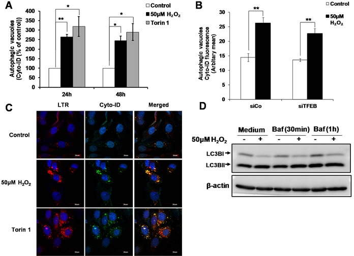
A. Autophagic vacuoles in L6 cells exposed to 50μM H2O2 and 100nM Torin1 for 24h and 48h were detected using Cyto-ID staining, statistical analysis was done by comparing treatment (H2O2 or Torin1) to untreated control at respective time point (24h or 48h). Values represent mean +/- SEM, *P < 0.05, **P < 0.005; n = 4 (t-test). B. Detection of autophagic vacuoles in cells transfected with siTFEB or negative control siRNA (siCo), and exposed to 50μM H2O2 for 24h using Cyto-ID staining. Values represent mean +/- SEM, **P < 0.005; n = 5 (t-test). The effect of siTFEB was not statistically significant, P>0.05 (mixed model). C. Detection and co-localization of lysosomes and autophagic vacuoles using LTR and Cyto-ID respectively in L6 cells treated with 50μM H2O2 and 100nM Torin1 for 24h. Results are shown as confocal microscopy image. Scale = 30μm. D. L6 cells were treated with 50μM H2O2 for 24h. Cells were then incubated with 200nM Bafilomycin for 30min/1h before cells were harvested for LC3II Western Blot analysis.