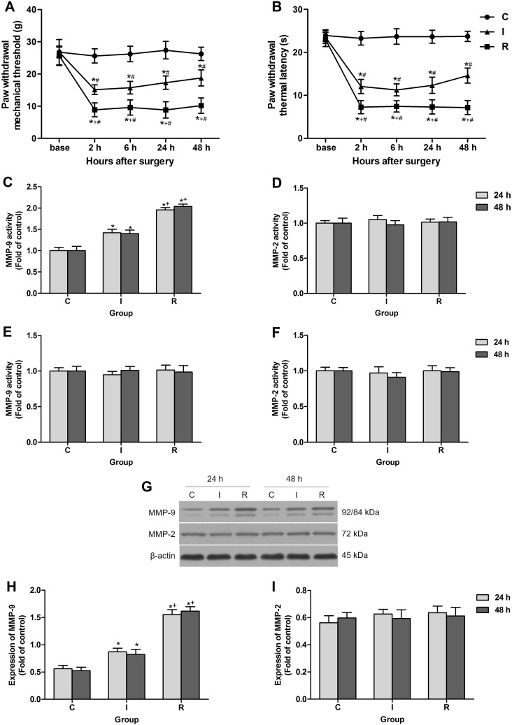
Figure 1. Intraoperative subcutaneous remifentanil infusion increased MMP-9 activity and expression in ipsilateral DRGs.
(A and B) Remifentanil-induced postoperative mechanical allodynia presented as PWMT and PWTL of right hind paw (n = 8). (C and D) Colorimetric quantitative detection showed that MMP-9 was significantly activated in ipsilateral lumbar DRGs at 24 h and 48 h after intraoperative remifentanil infusion, and the activity of MMP-2 remained unchanged (n = 5). (E and F) Neither MMP-9 nor MMP-2 activity was changed in ipsilateral spinal cord dorsal horn at 24 h and 48 h after surgery (n = 5). (G–I) Western blotting showed that the expression of MMP-9 was up-regulated in ipsilateral lumbar DRGs at 24 h and 48 h after intraoperative remifentanil infusion, and MMP-2 remained unchanged. Representative bands for MMP-9, MMP-2 and β-actin resulted in products of 92/84, 72, 43 kDa (G) and data summary (H and I) are shown (n = 5). β-actin was used as a loading control. Values expressed as mean ± SD. Group C: sham surgery; Group I: subcutaneous infusion of saline during incisional surgery; Group R: subcutaneous infusion of remifentanil during incisional surgery. Significant difference in pain behaviors was revealed after Repeated measures ANOVA, and significant difference in the results of western blotting and Colorimetric quantitative detection was revealed after One-way ANOVA (*P < 0.05 compared with group C, +P < 0.05 compared with group I, #P < 0.05 compared with baseline, Bonferroni post hoc tests).

