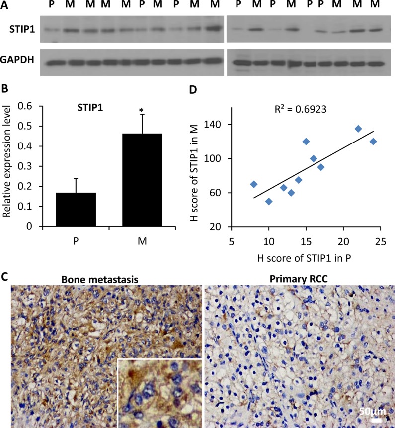
Figure 2. STIP1 protein expression in clinical samples.
(A) STIP1 protein was examined in 19 protein specimens from primary RCC tumors (P, n = 7) and bone metastatic samples (M, n = 12). Proteins were electrophoresed in two 10% SDS-PAGE gels, and were subsequently transferred to two PVDF membranes (10 and 9 specimens for gels 1and 2, respectively). (B) The intensity of STIP1 in each lane was normalized with the intensity of GAPDH. *p < 0.05. Experiments were duplicated, and western blot images shown have been cropped to show the protein of interest, and all blots were performed under the same experimental conditions. (C) Representative immunohistochemistry staining of STIP1 in primary RCC and bone metastasis tumors. Both intracellular and extracellular STIP1 immunoreactivity was examined as shown in the inset. Images were taken under 20× objective. Scale bar: 50 μm. (D) Correlation between the H scores of STIP1 in primary RCC and bone metastasis tumors of the 10 pairs of matched samples. R2 = 0.6923.

