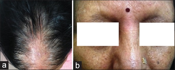
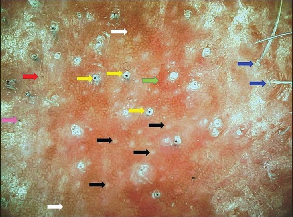
A 67-year-old postmenopausal lady presented with frontal scalp hair loss and loss of eyebrows of 1-year duration. Examination revealed frontal cicatricial alopecia with temporoparietal extension, peripheral perifollicular scaling, and eyebrow madarosis [Figure 1]. Polarized videodermoscopy (EScope; Nakoda, ×20) revealed honeycombing of the scalp skin and multiple white dots representing absence of follicular openings, follicular hyperkeratosis and plugging, perifollicular scaling, and erythema [Figure 2]. Cicatricial white patches were not seen. Some interfollicular scaling and erythema were present, suggestive of mild seborrheic dermatitis.
Figure 1.

(a) Cicatricial alopecia of the frontal scalp with temporoparietal extension and (b) eyebrow madarosis
Figure 2.

Dermoscopy of the alopecic area revealing honeycombing of the scalp skin (white arrow), multiple white dots representing absence of follicular openings (black arrows), prominent follicular hyperkeratosis and plugging encompassing black dots (yellow arrows), perifollicular scaling (blue arrows), and perifollicular erythema (green arrow). In addition, a black dot without surrounding hyperkeratosis/plugging (pink arrow) and yellow dot (red arrow) are visible. (polarizing mode, ×20)
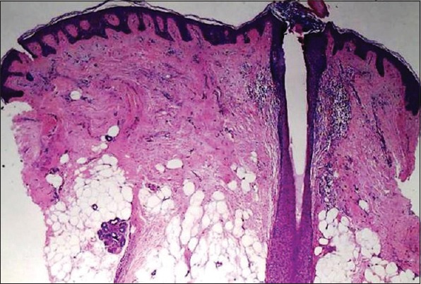
Few scattered yellow and several black dots were appreciable [Figure 2]. Miniaturized or vellus hairs were absent. Histopathology revealed perifollicular lymphocytic infiltrate, papillary dermal fibroplasia, and focal replacement of follicles with early fibrotic changes [Figure 3], confirming the diagnosis of active frontal fibrosing alopecia (FFA).
Figure 3.

Histopathology revealing partial flattening of epidermal rete ridges, perifollicular lymphocytic infiltrate, papillary dermal fibroplasias, marked reduction in the number of hair follicles, and focal replacement of follicles with early fibrotic changes (H and E, ×100)
FFA, a cicatricial alopecia typically encountered in post-menopausal women, is considered to be a distinct variant of lichen planopilaris.[1] Trichoscopy conveniently differentiates between the causes of alopecia of the scalp margin as FFA, alopecia areata (AA), traction alopecia, and cicatricial marginal alopecia (CMA).[2] While the trichoscopic features easily ruled out AA, lack of miniaturized hairs and fractured hair shafts excluded traction alopecia. The absence of follicular openings, absence of vellus hairs, presence of follicular hyperkeratosis and plugs, and perifollicular scales are dermoscopic hallmarks of FFA.[2,3,4] Absence of cicatricial white patches and presence of perifollicular erythema further suggest disease activity.[3,4,5] These typical dermoscopic features and the typical histopathology confidently ruled out CMA. Honeycombing of the scalp skin is a normal trichoscopic finding in the skin of color chronically exposed to sunlight. The most common trichoscopic features of FFA include absence of follicular openings, followed by follicular hyperkeratosis, perifollicular erythema, and follicular plugs.[3] In this case, we also observed the presence of few scattered yellow and several black dots, an inconsistent and uncommon feature of FFA.
Financial support and sponsorship
Nil.
Conflicts of interest
There are no conflicts of interest.
References
- 1.Kossard S, Lee MS, Wilkinson B. Postmenopausal frontal fibrosing alopecia: A frontal variant of lichen planopilaris. J Am Acad Dermatol. 1997;36:59–66. doi: 10.1016/s0190-9622(97)70326-8. [DOI] [PubMed] [Google Scholar]
- 2.Rubegni P, Mandato F, Fimiani M. Frontal Fibrosing Alopecia: Role of Dermoscopy in Differential Diagnosis. Case Rep Dermatol. 2010;2:40–5. doi: 10.1159/000298283. [DOI] [PMC free article] [PubMed] [Google Scholar]
- 3.Fernández-Crehuet P, Rodrigues-Barata AR, Vañó-Galván S, Serrano-Falcón C, Molina-Ruiz AM, Arias-Santiago S, et al. Trichoscopic features of frontal fibrosing alopecia: Results in 249 patients. J Am Acad Dermatol. 2015;72:357–9. doi: 10.1016/j.jaad.2014.10.039. [DOI] [PubMed] [Google Scholar]
- 4.Duque-Estrada B, Tamler C, Sodré CT, Barcaui CB, Pereira FB. Dermoscopy patterns of cicatricial alopecia resulting from discoid lupus erythematosus and lichen planopilaris. An Bras Dermatol. 2010;85:179–83. doi: 10.1590/s0365-05962010000200008. [DOI] [PubMed] [Google Scholar]
- 5.Toledo-Pastrana T, Hernández MJ, Camacho Martínez FM. Perifollicular erythema as a trichoscopy sign of progression in frontal fibrosing alopecia. Int J Trichol. 2013;5:151–3. doi: 10.4103/0974-7753.125616. [DOI] [PMC free article] [PubMed] [Google Scholar]


