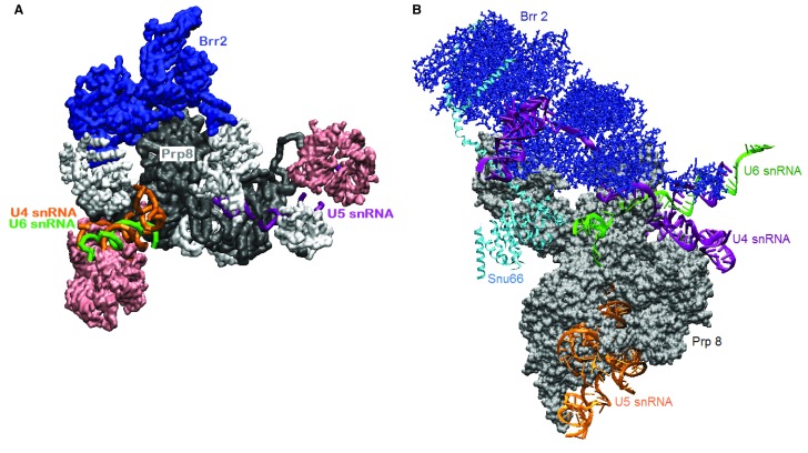
Figure 3. Two tri-small nuclear ribonucleoprotein particle (snRNP) structures trap different states of Brr2.
A. Human tri-snRNP cryo-electron microscopy (cryo-EM) at 7 Å resolution 72 shows Brr2 sitting on Prp8 (PDB ID 3jcr). A U4/U6 snRNA duplex is visible. Sm and Lsm rings are pink; other proteins are white. B. In a yeast tri-snRNP complex 70, (PDB ID 5GAN), U4 snRNA is threaded through Brr2 in the RNA-binding tunnel. These structures might correspond to the tri-snRNP in the nucleus ( A) and the tri-snRNP poised for activation by Brr2 as it joins the pre-spliceosome ( B). Visualized with visual molecular dynamics (VMD).

