Abstract
Peroxisome proliferator–activated receptors (PPARs) regulate energy metabolism and hence are therapeutic targets in metabolic diseases such as type 2 diabetes and non-alcoholic fatty liver disease. While they share anti-inflammatory activities, the PPAR isotypes distinguish themselves by differential actions on lipid and glucose homeostasis. In this Review we discuss the complementary and distinct metabolic effects of the PPAR isotypes together with the underlying cellular and molecular mechanisms, as well as the synthetic PPAR ligands that are used in the clinic or under development. We highlight the potential of new PPAR ligands with improved efficacy and safety profiles in the treatment of complex metabolic disorders.
Introduction
Metabolic syndrome (MetS) is a pathophysiologic condition characterized by increased visceral adiposity, dyslipidemia, prediabetes, and hypertension. This cluster of risk factors predisposes to type 2 diabetes (T2D) and nonalcoholic fatty liver disease (NAFLD) and increases the risk of microvascular complications and cardiovascular (CV) events. With the global increase in obesity, the prevalence of MetS has reached epidemic proportions. The pathophysiology of MetS and its comorbidities is complex and includes alterations in lipid and glucose metabolism accompanied by multi-organ inflammation; because of this complexity, current treatments address the individual components (1).
Over the last decades, the PPARs, which are members of the nuclear receptor superfamily of transcription factors (TFs), have been targeted to fight MetS and its complications. Three PPAR isotypes with different tissue distribution, ligand specificity, and metabolic regulatory activities exist in mammals: PPARα (NR1C1), PPARβ/δ (NR1C2), and PPARγ (NR1C3). PPARs regulate many metabolic pathways upon activation by endogenous ligands, such as fatty acids (FAs) and derivatives, or synthetic agonists, which bind to the ligand-binding domain of the receptor, triggering a conformational change. Subsequent recruitment of coactivators to the PPAR/retinoid X receptor heterodimer assembled at specific DNA response elements called PPAR response elements (PPREs) results in transactivation of target genes. In addition, PPAR activation attenuates the expression of pro-inflammatory genes, mostly through transrepressive mechanisms (2). This Review focuses on the metabolic effects of PPAR isotypes as well as synthetic PPAR ligands that are currently used in the clinic or are under development.
Endogenous PPAR ligands
PPARs are activated by FAs and their derivatives, and the level of physiologic receptor activation depends on the balance between ligand production and inactivation. Endogenous PPAR ligands originate from three main sources: diet, de novo lipogenesis (DNL), and lipolysis, all of which are processes that integrate changes in nutritional status and circadian rhythms (3). PPARs control these metabolic processes to maintain metabolic flexibility, a prerequisite for the preservation of health.
Dietary lipids regulate PPAR activity, as evidenced by the increased target gene expression of PPARα in liver (4) and PPARβ/δ in skeletal muscle (SKM) (5) upon high-fat diet (HFD) feeding in mice. Tissue-specific deficiency of FA synthase — a key enzyme in DNL — impairs PPARα activity and identifies DNL as another source of PPAR ligands (6, 7). PPARα ligands originating from DNL are not only simple FAs but include more complex molecules such as phosphatidylcholines (8). Lipolysis is a third source of endogenous PPAR activators. Angiopoietin-like (ANGPTL) proteins are secreted glycoproteins that inhibit lipoprotein lipase (LPL), thereby controlling the plasma lipid pool according to lipid availability and cellular fuel demand. ANGPTL4 expression is induced in several tissues including adipose tissue, liver, and SKM by circulating FAs via PPARs, leading to inhibition of LPL and decreased plasma triglyceride-derived FA uptake, thus forming a negative feedback loop (9). Intracellular lipolysis also provides PPAR ligands. Deficiency of adipose triglyceride lipase, which lipolyzes lipid droplet triglycerides, decreases PPAR target gene expression in various tissues (10–13). Ligand availability is also modulated by FA degradation in peroxisomes, which are regulated by PPARs (14). Thus, PPAR activity relies on a careful balance between ligand production and degradation to meet fluctuating energy demands.
Contrasting metabolic effects of ligand-activated PPARα and PPARγ
Although they share similarities in function and mechanism of action, PPAR isotypes display important physiologic and pharmacologic differences. This section discusses the clinical and genetic evidence of contrasting PPARα and PPARγ effects, and sheds light on the cellular and molecular mechanisms underlying these differences.
Clinical effects of PPARα and PPARγ activation.
Fibrates are synthetic PPARα ligands used to treat dyslipidemia. Except for the weak pan-agonist bezafibrate, all clinically used fibrates are specific activators of PPARα. Fibrate outcome trials such as the Helsinki Heart Study (HHS) (15), Veterans Affairs High-Density Lipoprotein Cholesterol Intervention Trial (VA-HIT) (16), Bezafibrate Infarction Prevention (BIP) (17), Fenofibrate Intervention and Event Lowering in Diabetes (FIELD) (18), and Action to Control Cardiovascular Risk in Diabetes (ACCORD) (19) consistently show beneficial effects on plasma lipids, particularly in normalizing the typical MetS dyslipidemia characterized by an “atherogenic lipid triad” (high LDL cholesterol [LDL-C] and triglycerides, low HDL cholesterol [HDL-C]). Fibrate therapy significantly decreases triglycerides and increases HDL-C, whereas LDL-C decreases except in patients with severe hypertriglyceridemia and low baseline LDL-C. Fibrate therapy, however, does not change circulating FA concentrations (20). Although both the FIELD and ACCORD trials showed a trend towards decreased CV risk (primary endpoint) in T2D, post-hoc and meta-analysis revealed that dyslipidemic patients (high triglyceride and low HDL-C levels) show the highest CV reduction (21, 22). Fibrates do not improve glucose homeostasis in people with T2D (18, 19, 23). However, PPARα activation improves glucose homeostasis in prediabetic patients (24) and may prevent conversion of prediabetes to overt T2D. Fibrates exert few adverse effects. Most compounds induce mild hypercreatininemia and hyperhomocysteinemia, but these effects are pharmacodynamic markers of PPARα activation rather than indicators of renal dysfunction (25). Hepatic carcinogenesis has been observed in rodents treated with fibrates but not in humans or non-human primates, likely due to lower peroxisome and peroxisomal β-oxidation levels in human liver (26).
Thiazolidinediones (TZDs, also referred to as glitazones), synthetic PPARγ ligands, are anti-diabetic drugs with potent insulin-sensitizing effects that confer long-term glycemic control (27). However, their clinical use has been challenged due to side effects such as body weight gain, edema, and bone fractures (2). The increase in body weight upon TZD administration is due to PPARγ-dependent white adipose tissue (WAT) expansion (28) and fluid retention caused by PPARγ activation in the kidney collecting ducts (29). The increased fracture risk in TZD-treated patients results from a PPARγ-driven rebalancing of bone remodeling in favor of net bone loss. Indeed, PPARγ activation in bone marrow stimulates mesenchymal progenitor differentiation into the adipocyte lineage, suppressing osteoblast and hence bone formation through pathways involving protein phosphatase PP5 (30, 31). Moreover, pharmacologic, but not physiologic, PPARγ activation promotes osteoclast formation thereby increasing bone resorption (32, 33). Rosiglitazone and pioglitazone increase plasma levels of the insulin-sensitizing adipokine adiponectin (2). They also increase HDL-C and reduce circulating FA levels (34), but have differential effects on triglyceride and LDL-C levels and CV risk. Pioglitazone, a full PPARγ agonist with modest PPARα-activating properties (35), lowers triglycerides, increases HDL-C, and reduces CV events in people with T2D (36) or who are insulin resistant (37). In contrast, the pure PPARγ agonist rosiglitazone does not decrease CV risk in people with T2D but does increase both HDL-C and LDL-C (38). Hence, the beneficial effects of pioglitazone on triglycerides and CV events are likely due to combined PPARα and PPARγ activation. In summary, activation of PPARα improves the lipid profile, whereas activation of PPARγ improves glycemic control and insulin sensitivity.
Genetic evidence of contrasting PPARα and PPARγ functions.
The different phenotypes of patients carrying SNPs and mutations in PPARα or PPARγ coding sequences highlight their contrasting functions. PPARA variants are associated with perturbations of lipid metabolism (39) and CV risk (40). PPARA SNPs also associate with conversion from impaired glucose tolerance to T2D (41). PPARA gene variation also influences the age of onset and progression of T2D (42). In contrast, dominant-negative mutations in the ligand-binding domain of PPARγ result in severe insulin resistance (43). Accordingly, rare variants in PPARG with decreased adipogenic properties are associated with increased T2D risk (44). GWAS have also revealed an association between PPARG SNPs and T2D, although not all studies concur (45, 46). A recently developed functional assay identified PPARG variants with altered PPARγ function (47). SNPs within DNA recognition motifs for PPARγ or cooperating factors that alter PPARγ recruitment to chromatin modulate the response to anti-diabetic drugs (48). Additionally, SNPs in PPARγ DNA-binding sites are highly enriched among SNPs associated with triglyceride and HDL-C levels in GWAS (48). Taken together, these genetic data confirm the functional dichotomy between PPARα and PPARγ in humans, underscoring the effects of PPARα on lipid metabolism and conversion from impaired glucose tolerance to T2D and the role of PPARγ in T2D and the regulation of glucose homeostasis.
Cellular and molecular mechanisms underlying PPARα and PPARγ functions.
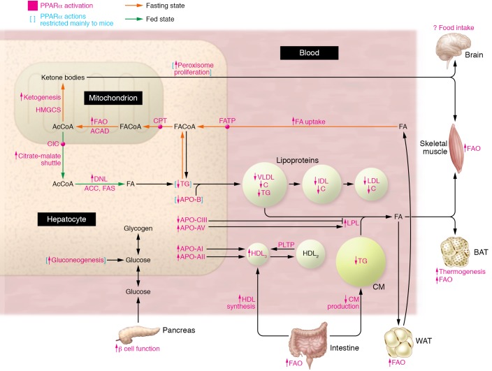
The function of PPARα (Figure 1) is best characterized in the liver, where it regulates genes involved in lipid and plasma lipoprotein metabolism during the nutritional transition phases (49, 50). During fasting, PPARα increases hepatic uptake and mitochondrial transport of FA originating from adipose tissue lipolysis through transcriptional upregulation of FA transport proteins and carnitine palmitoyltransferases. PPARα induces expression of mitochondrial acyl-CoA dehydrogenases, hence stimulating hepatic FA oxidation (FAO) and increasing acetyl-CoA production. Upon prolonged fasting, acetyl-CoA is preferentially converted into ketone bodies to provide energy for extrahepatic tissues. PPARα also upregulates mitochondrial hydroxymethylglutaryl-CoA synthase (HMGS), a rate-limiting ketogenesis enzyme (51, 52). Glucagon receptor signaling (53) and the IRE1α/XBP1 pathway (54) cooperate with PPARα to control metabolic pathways during fasting. In the fed state, PPARα coordinates DNL to supply FAs, which are stored as hepatic triglycerides and used in periods of starvation. A crucial step in DNL is the citrate-malate shuttle, which controls the efflux of acetyl-CoA from the mitochondria to the cytosol, where it serves as a precursor for FA synthesis. Citrate carrier, an essential component of this shuttle system, is a direct PPARα target gene in hepatocytes (55). Additionally, PPARα increases protein levels of the lipogenic factor SREBP1c by promoting proteolytic cleavage of its precursor (56), hence stimulating transcription of its target genes (57). In these postprandial conditions, mTORC1, activated through the insulin-dependent PI3K pathway, inhibits PPARα-mediated hepatic ketogenesis (58). Thus, PPARα contributes to the maintenance of metabolic flexibility by adapting fuel utilization to fuel availability, and its expression decreases in conditions of metabolic inflexibility such as NAFLD (59). PPARα activity is also dysregulated by microRNA-10b (60), microRNA-21 (61), and JNK (62), all of which are upregulated in NAFLD.
Figure 1. PPARα activation stimulates FA and triglyceride metabolism.
During fasting (yellow), FAs released from WAT are taken up by the liver and transported to mitochondria, where FAO takes place, to produce acetyl-CoA (AcCoA), which can be further converted to ketone bodies and serve as fuel for peripheral tissues. In the fed state (green), acetyl-CoA is shuttled to the cytosol, where DNL takes place. The effects of PPARα activation and PPARα target genes are indicated in pink. FAO is also stimulated by PPARα in WAT and SKM. By regulating hepatic apolipoprotein synthesis, PPARα activation decreases plasma levels of triglycerides (TG) and LDL-C and increases HDL-C. PPARα also acts on BAT, gut, and pancreas, but its central effects are unclear. Blue brackets indicate PPARα actions that are mainly restricted to mice and do not occur (e.g., peroxisome proliferation, reduced liver fat content) or occur to a lesser extent (e.g., reduced APO-B production) in humans. ACAD, acyl-CoA dehydrogenase; ACC, acetyl-CoA carboxylase; CM, chylomicron; CPT, carnitine palmitoyltransferase; FACoA, fatty acyl-CoA; FAS, fatty acid synthase; FATP, fatty acid transport protein.
PPARα activation reduces plasma triglyceride-rich lipoproteins by enhancing FA uptake and FAO and increasing the activity of LPL, which hydrolyzes lipoprotein triglycerides. PPARα stimulation of LPL enzyme activity is both direct, through PPRE-dependent activation of LPL (63), as well as indirect, through decreasing the expression of the LPL inhibitor and pro-atherogenic APO-CIII (64, 65) and increasing the expression of the LPL activator APO-AV (66). Reduced VLDL production contributes to the triglyceride-lowering effects of PPARα activation mainly in rodents and, likely to a lesser extent, in humans. Interestingly, a SNP in the TM6SF7 gene reduces VLDL production and lowers circulating triglyceride levels while promoting hepatic steatosis (67), an effect not observed in PPARα agonist–treated patients (68). In line with this, administration of fenofibrate to people with MetS increases the fractional catabolic rate of VLDL-APOB, intermediate-density lipoprotein–APOB (IDL-APOB), and LDL-APOB without affecting VLDL-APOB production (69). The rise in plasma HDL-C upon PPARα activation is linked to increased synthesis of major HDL-C constituents, apolipoproteins APO-AI and APO-AII (70), and induction of phospholipid transfer protein (PLTP) (71). Of note, differences between rodents and humans with respect to apolipoprotein regulation exist, as APO-AI and APO-AV are direct positive PPARα target genes in human but not murine liver (49). Through FAO, PPARα activation leads to energy dissipation not only in the liver but also in SKM (72) and WAT (73). In brown adipose tissue (BAT) PPARα stimulates lipid oxidation as well as thermogenesis in synergy with PPARγ coactivator-1α (PGC1A) (74). While PPARα activation reduces weight gain in rodents (73), there is no evidence of PPARα effects on body mass in humans (18, 19).
The inability of fibrates to improve glucose homeostasis in people with T2D (18, 19) may result from several mechanisms. Glucose handling in liver and peripheral tissues is reduced as a consequence of increased FAO (75). PPARα activation also reduces pyruvate kinase (PK) and induces PDK4 expression in the liver, decreases glycolysis, and enhances gluconeogenesis in mice (76). As discussed above, clinical and genetic data have revealed a role for PPARα in preventing conversion from impaired glucose tolerance to overt T2D. This effect of PPARα might stem from pancreatic β cell protection from lipotoxicity (77) and decreases in insulin clearance mediated by the biliary glycoprotein CEACAM1 (78).
PPARγ is highly expressed in WAT, where it controls FA uptake and lipogenesis. Target genes contributing to this activity include FA binding protein-4 and the FA translocase CD36 (79). Additionally, PPARγ is a master regulator of white adipocyte differentiation. Multiple TFs including the glucocorticoid receptor (GR) and STAT5A cooperatively induce PPARγ during adipogenesis (28), while other TFs such as C/EBPα cooperate with PPARγ to stimulate genomic binding and transcription of target genes (80), thereby regulating both housekeeping and adipocyte-specific functions (81). These PPARγ-mediated changes in gene expression are preceded by chromatin remodeling involving both adipocyte-specific TFs such as C/EBPβ (82) as well as ubiquitous TFs such as CCCTC-binding factor (CTCF) (83). Interestingly, promotion of adipogenesis by the mTORC1 complex occurs through stimulation of PPARγ translation (84) and transcriptional activity (85), which contrasts with the inhibitory effect of mTORC1 on PPARα (discussed above) (58).
In contrast to WAT, PPARγ target genes in BAT encode thermogenic proteins and inducers of mitochondrial biogenesis such as PGC1A and uncoupling protein-1 (UCP1, also known as thermogenin). PPARγ promotes brown adipocyte differentiation, but additional TFs including PPARα are required to switch on their thermogenic program (74).
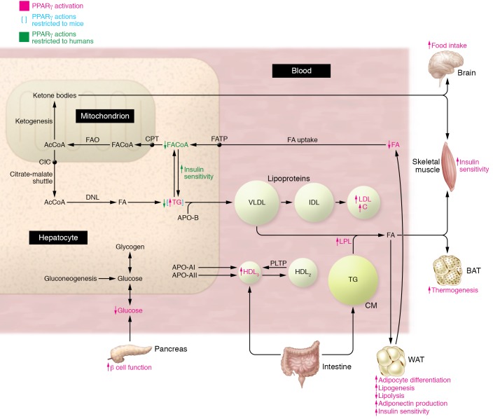
PPARγ enhances whole body insulin sensitivity through multiple mechanisms (Figure 2). By augmenting WAT expandability, PPARγ shifts lipids from liver and SKM to WAT, thereby indirectly increasing glucose utilization in liver and peripheral tissues. As a result of this “lipid stealing,” lipotoxicity, which impairs insulin signaling, is alleviated. PPARγ also regulates the expression of adipocyte hormones that modulate liver and SKM insulin sensitivity such as adiponectin and leptin (86, 87). Results of a Mendelian randomization study refuted a causal role for adiponectin in CV disease (88), which may explain why pure PPARγ agonists, such as rosiglitazone, are not cardioprotective. Finally, PPARγ activation improves pancreatic β cell function and survival by preventing FA-induced impairment of insulin secretion (77) and enhancing the unfolded protein response (89). Thus, whereas PPARα activation leads to energy dissipation, activation of PPARγ stimulates energy storage in WAT, thereby sensitizing liver and peripheral tissues to insulin.
Figure 2. PPARγ activation increases whole-body insulin sensitivity.
In WAT, PPARγ activation (effects are indicated in pink) enhances FA uptake and storage, lipogenesis, and adipogenesis (lipid steal action). PPARγ activation lowers circulating FA levels, alleviating lipotoxicity and increasing insulin sensitivity. PPARγ agonism induces adiponectin production by WAT, further enhancing insulin sensitivity and lowering blood glucose. PPARγ also exerts metabolic effects on BAT, brain, and pancreas. Increased hepatic steatosis upon PPARγ activation occurs in mice but not in humans (blue brackets), who display increased hepatic insulin sensitivity due to reduced FA flux from WAT.
The contrasting mechanisms of action of PPARα and PPARγ are also illustrated by their opposite function on hepatic lipid metabolism. Reduced hepatic steatosis due to increased FAO in hepatocytes occurs upon PPARα activation in rodent models of NAFLD (90, 91), while PPARγ activation in rodents (but not humans) increases liver fat accumulation by enhancing hepatic expression of PPARγ-dependent genes involved in lipogenesis (79, 92). Interestingly, hepatic PPARγ expression levels determine liver steatosis: mice with low hepatic PPARγ expression are resistant to diet-induced development of fatty liver when treated with rosiglitazone, whereas liver steatosis is exacerbated in obese mice expressing high hepatic levels of PPARγ (93). In mice, PPARγ expression in liver is regulated by the dimeric AP-1 protein complex, thereby controlling hepatic steatosis (94). However, in humans with NAFLD, PPARγ expression is unaltered (59) and TZD treatment decreases hepatic steatosis, likely due to decreased FA flux from WAT to liver (95, 96).
Energy homeostasis is also regulated by inter-organ communications involving the brain and the gut. Neuronal PPARγ deletion in mice diminishes food intake and energy expenditure, thus reducing weight gain upon HFD feeding, suggesting that brain PPARγ exerts hyperphagic effects and promotes obesity (97). Similarly, central PPARα activation may also increase food intake (6), although not all studies concur (98). In the intestine, PPARα activation suppresses postprandial hyperlipidemia by enhancing intestinal epithelial cell FAO (99). Furthermore, intestinal PPARα activation reduces cholesterol esterification, suppresses chylomicron production, and increases HDL synthesis by enterocytes (100).
Molecular basis for differential activities of PPARα and PPARγ.
The exact mechanisms through which the different PPAR isotypes — which share similar DNA-binding motifs — bind and regulate different genes remain to be established. Several explanations and hypotheses have been put forward. First, PPARα is predominantly expressed in the liver, whereas PPARγ expression is highest in WAT (2). The different PPARs emerged during evolution from gene duplications, but subsequent sequence variations of their promoters and 3′-UTRs have contributed to acquisition of differential expression patterns and functions (101). Tissue-specific chromatin and TF environments also play a role by restricting PPAR recruitment to selective enhancers and therefore specifying PPAR target genes (28). This is illustrated by the tissue-specific PPARγ cistromes in white adipocytes and macrophages, both of which express high PPARγ levels. The macrophage-specific PPARγ cistrome is defined by the pioneer TF PU.1 (102), which induces nucleosome remodeling and histone modifications, promoting the recruitment of additional TFs (103). In white adipocytes, however, these macrophage-specific binding regions are marked with repressive histone modifications, thus disabling PPARγ binding (104). Furthermore, PPARγ cistromes differ between white adipocyte depots (epididymal vs. inguinal) in association with depot-specific gene expression patterns (105).
Nutritional status also contributes to differential PPAR regulation. PPARα is a metabolic sensor, switching its activity from coordination of lipogenesis in the fed state to promotion of FA uptake and FAO during a fasting state (49). PPARα activation during fasting involves PGC1α coactivator induction by the fasting-induced TF EB (106). In addition to PPARα itself (107), circadian transcription of genes encoding acyl-CoA thioesterases coordinates cyclic intracellular production of FA ligands (108). The TF CREBH, a circadian regulator of hepatic lipid metabolism, rhythmically interacts with PPARα and regulates its activity (109). Adjustment of PPARα transcriptional activity to nutritional status is also controlled by kinases phosphorylating PPARα or its coregulators. In the fed state, PPARα activity is enhanced through insulin-activated MAPK and glucose-activated PKC, while glucagon-activated PKA and AMPK increase PPARα signaling in fasting (49). Moreover, the fasting response is co-controlled by PPARα and GRα, which show extensive chromatin colocalization and interact to induce lipid metabolism genes upon prolonged fasting through genomic AMPK recruitment (110). Conversely, GRβ antagonizes glucocorticoid signaling during fasting via inhibition of GRα and PPARα, thus increasing inflammation and hepatic lipid accumulation (111).
PPARγ activity is higher in the fed state, in line with its role in lipid synthesis and storage. PPARγ activity in WAT is repressed during fasting via mechanisms involving SIRT1 (112) or AMPK (113). In mice, the amplitude of hepatic circadian clock gene expression is reduced by HFD feeding (114), whereas circadian rhythmicity of PPARγ and genes containing the PPARγ binding site is induced (115). Thus, the HFD-induced transcriptional reprogramming relies at least in part on changes in expression, oscillation pattern, and chromatin recruitment of PPARγ. Gut microbiota, which also exhibit circadian activity (116), are drivers of HFD-induced hepatic transcriptional reprogramming by PPARγ in mice (117). Nutritional status also links PPARs to FGF21 signaling, as fasting increases PPARα-dependent FGF21 expression in liver, further enhancing FAO and ketogenesis (118). In WAT, PPARγ induces FGF21 expression (119), where it acts as an autocrine factor in the fed state, regulating PPARγ activity through a feedforward mechanism (120). In the pancreas, PPARγ agonism reverses high glucose–induced islet dysfunction by enhancing FGF21 signaling (121). FGF1 is also induced by PPARγ in WAT, and the PPARγ/FGF1 axis is critical for maintaining metabolic homeostasis and insulin sensitization (122).
Combating inflammation: a shared function of PPARα and PPARγ
MetS is accompanied by a low-grade inflammatory state in different metabolic tissues — termed meta-inflammation — characterized by increased secretion of pro-inflammatory chemokines and cytokines, many of which (including TNF-α, IL-1, and IL-6) influence lipid metabolism and insulin resistance (123). Besides differentially regulating lipid and glucose metabolism, PPARα and PPARγ also counter inflammation. However, the anti-inflammatory effects of PPARα and PPARγ activation are likely distinct due to differences in tissue and cell type expression.
In WAT, fenofibrate and rosiglitazone reduce the expression of several pro-inflammatory mediators, including IL-6 and the chemokines CXCL10 and MCP1 (124). PPARγ also inhibits pro-inflammatory cytokine production by WAT-resident macrophages and modulates macrophage polarization (125). Although innate immune cells such as macrophages were initially thought to be the main drivers of WAT inflammation and metabolic dysregulation, important roles of the adaptive immune system, including WAT Tregs, have recently emerged (126). PPARγ acts as a molecular orchestrator of WAT Treg accumulation, phenotype, and function (127, 128). Indeed, the WAT Treg transcriptome alterations in obese mice depend on PPARγ phosphorylation by cyclin-dependent kinase 5 (CDK5) (127). In addition, PPARγ expression in WAT Tregs is necessary for complete restoration of insulin sensitivity in obese mice upon pioglitazone treatment (128). On the other hand, activation of CD4+ T cells is accompanied by mTORC1-dependent PPARγ induction and enhanced expression of FA uptake genes, enabling rapid T cell proliferation and optimal immune responses (129). PPARα and PPARγ also modulate the inflammatory response in liver and vascular wall (130, 131).
Inhibition of pro-inflammatory gene expression is the main process underlying the anti-inflammatory properties of PPARα and PPARγ. Several mechanisms have been proposed for transcriptional repression by PPARs that are not mutually exclusive. These include direct physical interaction of PPARα or PPARγ with several pro-inflammatory TFs including AP-1 and NF-κB (132, 133). Repression of inflammation independently of direct PPARα DNA binding results in anti-inflammatory and anti-fibrotic effects in a mouse model of non-alcoholic steatohepatitis (NASH) (134). In addition to this PPRE-independent transrepression mechanism, interaction between NF-κB and PPRE-bound PPARα also occurs, leading to repression of TNF-α–mediated upregulation of complement C3 gene expression and protein secretion during acute inflammation (135). Moreover, simultaneous activation of PPARα and GRα increases the repression of NF-κB–driven genes, thereby decreasing cytokine production (136). Transcriptional repression of pro-inflammatory genes by PPARγ may include ligand-activated PPARγ sumoylation, which targets the receptor to corepressor complexes assembled at inflammatory gene promoters. This prevents promoter recruitment of the proteasome machinery that normally mediates the inflammatory signal–dependent removal of corepressor complexes required for gene activation. As a result, these complexes are not cleared from the promoters and inflammatory genes are maintained in a repressed state (137). In addition to downregulating the expression of pro-inflammatory genes, PPARα (138) and PPARγ (139) also suppress inflammation by upregulating genes with anti-inflammatory properties, such as IL-1Ra, suggesting a possible cooperation between PPAR-dependent transactivation and transrepression to counter inflammation.
The anti-inflammatory properties of PPARα likely contribute to the improved lobular inflammation and hepatocellular ballooning observed in NAFLD patients treated with pioglitazone (140) or elafibranor (141), a dual PPARα/β(δ) agonist. Pioglitazone reduces hepatic steatosis in NAFLD patients (140), likely due to PPARγ activation. The pure PPARγ agonist rosiglitazone also lowers liver fat in humans (96), whereas the pure PPARα agonist fenofibrate does not (68). Administration of fenofibrate to people with dyslipidemia lowers plasma levels of atypical deoxysphingolipids (142), which increase upon the transition from simple steatosis to NASH (143). Thus, activation of both PPARα and PPARγ appears to be beneficial in human NAFLD, although the underlying mechanisms clearly differ. Whereas the effects of PPARα agonism on inflammation and ballooning are due to direct PPARα activation in the liver, the effects of PPARγ on hepatic steatosis are likely mediated by indirect mechanisms such as suppression of FA flux to the liver; this is in line with the low expression and absence of PPARγ induction in human fatty liver (59).
PPARβ/δ, the clinically enigmatic third PPAR
Selective synthetic PPARβ/δ agonists are not yet clinically available; however, beneficial effects of PPARβ/δ activation on various MetS components have been reported and include both differences and similarities to PPARα and PPARγ, such as reduced inflammation (144–146).
PPARD variants are associated with cholesterol metabolism (147), insulin sensitivity (148), T2D risk (149), and CV risk (40). In obese men, administration of the synthetic PPARβ/δ agonist GW501516 lowers liver fat content and plasma levels of insulin, FAs, triglycerides, and LDL-C (150). These beneficial effects on plasma lipids are also observed in overweight patients treated with seladelpar (MBX-8025), a novel PPARβ/δ agonist (151). Thus, PPARβ/δ agonism combines the metabolic effects of PPARα and PPARγ activation on lipid metabolism and glucose homeostasis, respectively. Preclinical studies support this conclusion, as the administration of GW501516 to overweight monkeys (152) or obese rats (153) lowered serum LDL-C and raised HDL-C while improving insulin sensitivity.
PPARβ/δ activation protects from diet-induced or genetically induced obesity in mice by increasing energy expenditure (154). In BAT, activation of PPARβ/δ induces the expression of thermogenic genes, including UCP1, and FAO genes (154). PPARβ/δ agonism also promotes FAO in SKM (155), WAT (156), and liver (157). PPARβ/δ in brain controls energy expenditure, as neuron-specific PPARβ/δ deletion increases susceptibility to diet-induced obesity (158). Thus, similar to PPARα, PPARβ/δ activation induces energy dissipation. Interestingly, both isotypes crosstalk in liver, where PPARβ/δ stimulates the production of the PPARα ligand 16:0/18:0-phosphatidylcholine as well as PPARα expression and DNA-binding activity, thereby increasing hepatic FAO (159). Enhanced FAO upon PPARβ/δ activation contributes to its plasma lipid-lowering effects, together with decreased cholesterol absorption (160) and increased trans-intestinal cholesterol efflux (161). PPARβ/δ also raises HDL-C by increasing hepatic APO-AII (162) and PLTP (163) expression.
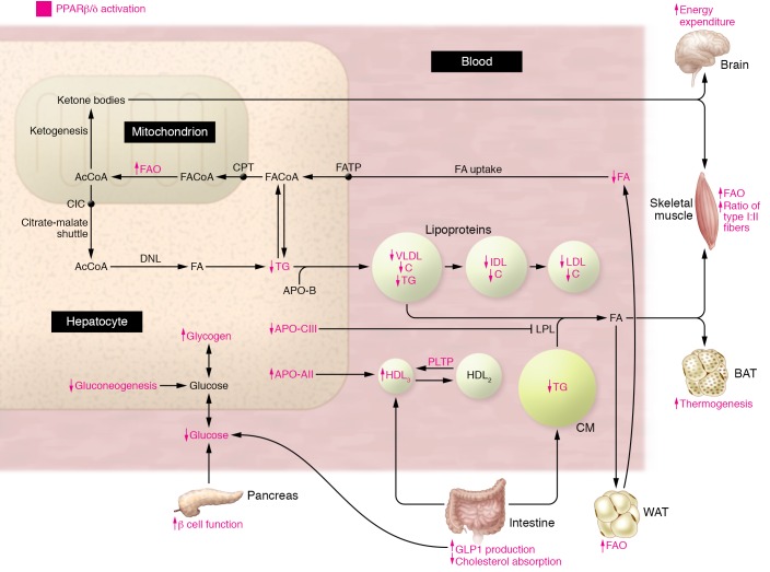
PPARβ/δ agonism improves insulin sensitivity through several mechanisms (Figure 3). In SKM, PPARβ/δ activation favors fiber type switching, from type II fast-twitch glycolytic to type I slow-twitch oxidative fibers (164), via mechanisms involving PGC1α (165) and an estrogen-related receptor γ/microRNA regulatory circuit (166), thereby improving glucose handling (167). The type I fiber fraction is reduced in people with T2D (168), which may contribute to altered glucose homeostasis. Mice with myocyte-selective PPARβ/δ deficiency exhibit decreased type I fiber count, which precedes the development of a diabetic phenotype (165). PPARβ/δ also improves glucose handling and insulin sensitivity in the liver. GW501516 treatment suppresses hepatic glucose output and enhances glucose disposal by increasing glucose flux through the pentose phosphate pathway (169). Liver-restricted PPARβ/δ overexpression reduces fasting glucose levels and stimulates hepatic glycogen production via upregulation of glucose utilization pathways (170). Additionally, stress-induced JNK signaling is reduced, contributing to improved hepatic insulin sensitivity (170). PPARβ/δ agonism promotes pancreatic β cell mitochondrial function and ATP production, thereby improving glucose-stimulated insulin secretion (171). Furthermore, PPARβ/δ increases intestinal production of the incretin glucagon-like peptide 1 (GLP1) (172).
Figure 3. PPARβ/δ activation enhances glucose and lipid homeostasis.
In SKM, PPARβ/δ activation (effects are indicated in pink) favors fiber type switching toward type I oxidative fibers, which have a higher glucose-handling capacity compared with type II fibers. PPARβ/δ also augments FAO in SKM, liver, and WAT and enhances hepatic glucose metabolism and pancreatic β cell function. PPARβ/δ activation decreases FAs, triglycerides, and LDL-C and increases HDL-C levels in blood. Metabolic effects of PPARβ/δ agonism also take place in brain and gut.
In summary, the mechanisms underlying the metabolic effects of PPARβ/δ resemble those of PPARα, which promotes energy dissipation, as opposed to PPARγ, which promotes energy storage. PPARβ/δ normalizes plasma lipids through enhanced FAO in several tissues, coupled to actions on hepatic apolipoprotein metabolism and intestinal cholesterol homeostasis. In contrast to PPARα and similar to PPARγ, activation of PPARβ/δ enhances insulin sensitivity. The mechanisms underlying PPARβ/δ-mediated improvement in glucose handling are not similar to PPARγ, but instead involve PPARβ/δ-specific actions on SKM fiber type distribution, hepatic glucose metabolism, and pancreatic islet function.
Current state of PPAR-targeted therapies
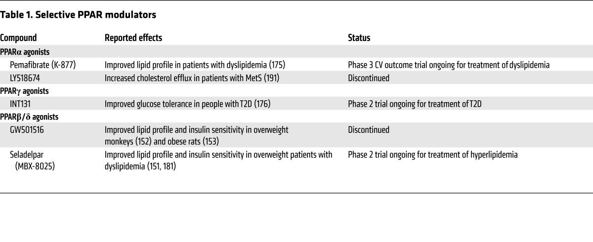
Currently used PPAR agonists display weak potencies (PPARα) or are associated with important side effects (PPARγ). Optimization of therapeutic efficacy may be achieved through the development of selective PPAR modulators that retain the beneficial effects of PPAR activation while diminishing unwanted side effects (ref. 173 and Table 1). The selective PPARα agonist pemafibrate (K-877) (174) exhibited greater lipid modifying efficacy than fenofibrate in a phase 2 trial, with little or no effect on serum creatinine and homocysteine levels (175). This compound is undergoing a phase 3 CV prevention trial, PROMINENT (Pemafibrate to Reduce Cardiovascular Outcomes by Reducing Triglycerides in Diabetic Patients), in patients with high triglyceride and low HDL-C levels. The non-TZD PPARγ modulator INT131, which improves glucose tolerance in people with T2D without adverse effects on body weight or hemodilution (176), is in phase 2 development. Several compounds that are not direct PPARγ agonists but that inhibit CDK5-mediated PPARγ phosphorylation also exert anti-diabetic activities in obese mice (177–180); whether this will eventually translate to clinical efficacy is unclear. The PPARβ/δ agonist seladelpar (MBX-8025) decreases plasma triglycerides, increases HDL-C, and improves insulin sensitivity and liver function in overweight people with dyslipidemia (151, 181).
Table 1. Selective PPAR modulators.
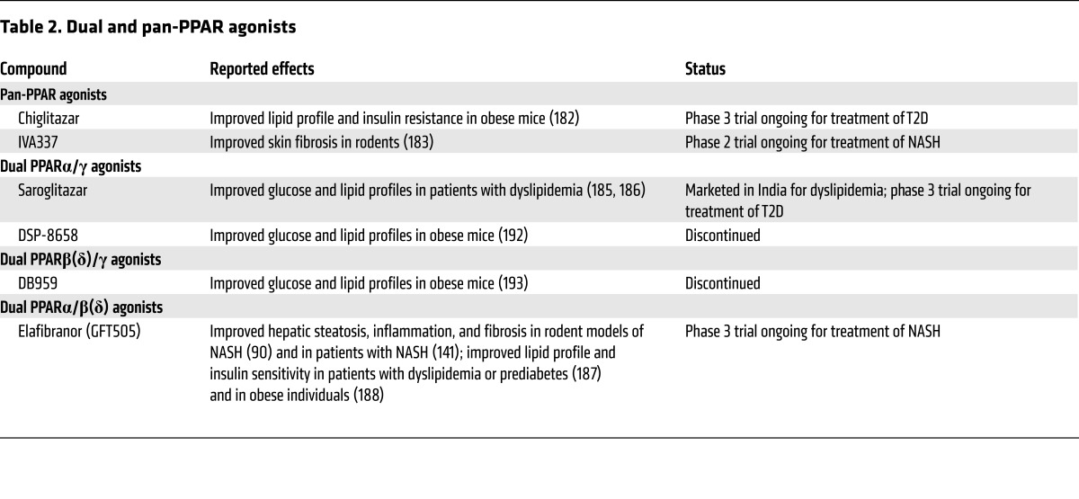
Dual PPAR agonists (which activate two PPAR isotypes) and pan-PPAR agonists (which activate all three PPARs) have been developed with the goal of combining the beneficial effects of each receptor isotype (Table 2). The pan-agonist chiglitazar (CS038) improves lipid profiles and insulin sensitivity without increasing body weight in animal models of obesity (182). IVA337, a pan-agonist that prevents and reverses skin fibrosis (183), is currently entering phase 2 trials for the treatment of NASH. Many dual PPARα/γ agonists, termed glitazars, showed improved efficacy on glucose and lipid metabolism in clinical trials, although safety concerns often halted further development (184). Two phase 3 trials with saroglitazar showed improved glucose and lipid profiles in patients with diabetic dyslipidemia compared with pioglitazone (185) or placebo (186). In contrast to the other PPARγ-dominant glitazars, saroglitazar predominantly activates PPARα with only moderate PPARγ agonism, which may explain the lack of typical PPARγ side effects. Elafibranor (GFT505), a dual PPARα/β(δ) agonist, demonstrated protective effects against hepatic steatosis, inflammation, and fibrosis in animal models of NAFLD/NASH (90). In phase 2a trials, elafibranor improved lipid and glucose profiles in dyslipidemic and prediabetic patients (187) and obese individuals (188). The GOLDEN-505 phase 2b study in people with NASH showed that elafibranor treatment induces NASH resolution without worsening fibrosis in a higher proportion of patients compared with placebo (141). The drug was well tolerated and improved glucose homeostasis and CV risk profile, and has since entered phase 3 development for NASH (the RESOLVE-IT trial; NCT02704403).
Table 2. Dual and pan-PPAR agonists.
PPARs are still valuable targets for metabolic diseases
Over the last decades, market withdrawals and failed drug development programs have cast doubts on the clinical value of PPAR-activating compounds. However, this issue is not black and white. The pure PPARγ agonist rosiglitazone as well as dual PPAR agonists with predominant PPARγ-activating properties all displayed important adverse effects that led to restricted use or halted development. However, most of these side effects were either drug specific and hence off-target (189) or related to excessive PPARγ activation. Several fibrate trials, including FIELD and ACCORD, failed to meet the primary endpoint of reduced CV risk; however, such negative outcomes are likely linked to inappropriate patient selection, since subgroup analyzes revealed significant CV risk reduction in those patients with marked dyslipidemia upon trial enrolment (21). Furthermore, in several of these fibrate trials, including BIP and FIELD, the proportion of patients who received statin therapy was unbalanced between placebo and treatment groups. Correction for this nonrandomized statin drop-in in the FIELD study estimated that fenofibrate reduces relative CV risk by 19% (190).
It has become increasingly clear that PPARα and PPARγ agonism display contrasting metabolic effects with different mechanisms of action. Whereas PPARβ/δ agonism is more related to PPARα, subtle differences exist (e.g., in regulation of glucose homeostasis). These findings are in line with the enhanced metabolic actions and improved safety profiles of novel compounds such as dual PPARα/β(δ) ligands, which target both lipid (via PPARα and PPARβ/δ) and glucose (via PPARβ/δ) abnormalities in people with MetS without displaying PPARγ-related adverse effects. Altogether, we are convinced that targeting PPARs in metabolic disorders remains a valuable and promising approach with a future ahead.
Acknowledgments
BS is a member of the Institut Universitaire de France. This work was supported by grants from the European Genomic Institute for Diabetes (grant ANR-10-LABX-46), the European Commission (RESOLVE contract FP7-305707), and the Fondation de France and Fondation pour la Recherche Médicale (contract DEQ20150331724).
Footnotes
Conflict of interest: B. Staels is an advisor for Genfit SA.
Reference information:J Clin Invest. 2017;127(4):1202–1214.https://doi.org/10.1172/JCI88894.
Contributor Information
Vanessa Dubois, Email: vanessa.dubois@inserm.fr.
Jérôme Eeckhoute, Email: jerome.eeckhoute@inserm.fr.
Philippe Lefebvre, Email: philippe-claude.lefebvre@inserm.fr.
Bart Staels, Email: Bart.Staels@pasteur-lille.fr.
References
- 1.O’Neill S, O’Driscoll L. Metabolic syndrome: a closer look at the growing epidemic and its associated pathologies. Obes Rev. 2015;16(1):1–12. doi: 10.1111/obr.12229. [DOI] [PubMed] [Google Scholar]
- 2.Gross B, Pawlak M, Lefebvre P, Staels B. PPARs in obesity-induced T2DM, dyslipidaemia and NAFLD. Nat Rev Endocrinol. 2017;13(1):36–49. doi: 10.1038/nrendo.2016.135. [DOI] [PubMed] [Google Scholar]
- 3.Woller A, Duez H, Staels B, Lefranc M. A mathematical model of the liver circadian clock linking feeding and fasting cycles to clock function. Cell Rep. 2016;17(4):1087–1097. doi: 10.1016/j.celrep.2016.09.060. [DOI] [PubMed] [Google Scholar]
- 4.Patsouris D, Reddy JK, Müller M, Kersten S. Peroxisome proliferator-activated receptor alpha mediates the effects of high-fat diet on hepatic gene expression. Endocrinology. 2006;147(3):1508–1516. doi: 10.1210/en.2005-1132. [DOI] [PubMed] [Google Scholar]
- 5.Garcia-Roves P, et al. Raising plasma fatty acid concentration induces increased biogenesis of mitochondria in skeletal muscle. Proc Natl Acad Sci U S A. 2007;104(25):10709–10713. doi: 10.1073/pnas.0704024104. [DOI] [PMC free article] [PubMed] [Google Scholar]
- 6.Chakravarthy MV, et al. Brain fatty acid synthase activates PPARalpha to maintain energy homeostasis. J Clin Invest. 2007;117(9):2539–2552. doi: 10.1172/JCI31183. [DOI] [PMC free article] [PubMed] [Google Scholar]
- 7.Chakravarthy MV, et al. “New” hepatic fat activates PPARalpha to maintain glucose, lipid, and cholesterol homeostasis. Cell Metab. 2005;1(5):309–322. doi: 10.1016/j.cmet.2005.04.002. [DOI] [PubMed] [Google Scholar]
- 8.Chakravarthy MV, et al. Identification of a physiologically relevant endogenous ligand for PPARα in liver. Cell. 2009;138(3):476–488. doi: 10.1016/j.cell.2009.05.036. [DOI] [PMC free article] [PubMed] [Google Scholar]
- 9.Dijk W, Kersten S. Regulation of lipid metabolism by angiopoietin-like proteins. Curr Opin Lipidol. 2016;27(3):249–256. doi: 10.1097/MOL.0000000000000290. [DOI] [PubMed] [Google Scholar]
- 10.Haemmerle G, et al. ATGL-mediated fat catabolism regulates cardiac mitochondrial function via PPAR-α and PGC-1. Nat Med. 2011;17(9):1076–1085. doi: 10.1038/nm.2439. [DOI] [PMC free article] [PubMed] [Google Scholar]
- 11.Jha P, et al. Role of adipose triglyceride lipase (PNPLA2) in protection from hepatic inflammation in mouse models of steatohepatitis and endotoxemia. Hepatology. 2014;59(3):858–869. doi: 10.1002/hep.26732. [DOI] [PubMed] [Google Scholar]
- 12.Biswas D, Ghosh M, Kumar S, Chakrabarti P. PPARα-ATGL pathway improves muscle mitochondrial metabolism: implication in aging. FASEB J. 2016;30(11):3822–3834. doi: 10.1096/fj.201600571RR. [DOI] [PubMed] [Google Scholar]
- 13.Schreiber R, et al. Hypophagia and metabolic adaptations in mice with defective ATGL-mediated lipolysis cause resistance to HFD-induced obesity. Proc Natl Acad Sci U S A. 2015;112(45):13850–13855. doi: 10.1073/pnas.1516004112. [DOI] [PMC free article] [PubMed] [Google Scholar]
- 14.Fan CY, Pan J, Usuda N, Yeldandi AV, Rao MS, Reddy JK. Steatohepatitis, spontaneous peroxisome proliferation and liver tumors in mice lacking peroxisomal fatty acyl-CoA oxidase. Implications for peroxisome proliferator-activated receptor alpha natural ligand metabolism. J Biol Chem. 1998;273(25):15639–15645. doi: 10.1074/jbc.273.25.15639. [DOI] [PubMed] [Google Scholar]
- 15.Manninen V, et al. Joint effects of serum triglyceride and LDL cholesterol and HDL cholesterol concentrations on coronary heart disease risk in the Helsinki Heart Study. Implications for treatment. Circulation. 1992;85(1):37–45. doi: 10.1161/01.CIR.85.1.37. [DOI] [PubMed] [Google Scholar]
- 16.Rubins HB, et al. Gemfibrozil for the secondary prevention of coronary heart disease in men with low levels of high-density lipoprotein cholesterol. Veterans Affairs High-Density Lipoprotein Cholesterol Intervention Trial Study Group. N Engl J Med. 1999;341(6):410–418. doi: 10.1056/NEJM199908053410604. [DOI] [PubMed] [Google Scholar]
- 17.Bezafibrate Infarction Prevention (BIP) study. Secondary prevention by raising HDL cholesterol and reducing triglycerides in patients with coronary artery disease. Circulation. 2000;102(1):21–27. doi: 10.1161/01.CIR.102.1.21. [DOI] [PubMed] [Google Scholar]
- 18.Keech A, et al. Effects of long-term fenofibrate therapy on cardiovascular events in 9795 people with type 2 diabetes mellitus (the FIELD study): randomised controlled trial. Lancet. 2005;366(9500):1849–1861. doi: 10.1016/S0140-6736(05)67667-2. [DOI] [PubMed] [Google Scholar]
- 19.ACCORD Study Group. et al. Effects of combination lipid therapy in type 2 diabetes mellitus. N Engl J Med. 2010;362(17):1563–1574. doi: 10.1056/NEJMoa1001282. [DOI] [PMC free article] [PubMed] [Google Scholar]
- 20.Vega GL, Cater NB, Hadizadeh DR, Meguro S, Grundy SM. Free fatty acid metabolism during fenofibrate treatment of the metabolic syndrome. Clin Pharmacol Ther. 2003;74(3):236–244. doi: 10.1016/S0009-9236(03)00170-X. [DOI] [PubMed] [Google Scholar]
- 21.Scott R, et al. Effects of fenofibrate treatment on cardiovascular disease risk in 9,795 individuals with type 2 diabetes and various components of the metabolic syndrome: the Fenofibrate Intervention and Event Lowering in Diabetes (FIELD) study. Diabetes Care. 2009;32(3):493–498. doi: 10.2337/dc08-1543. [DOI] [PMC free article] [PubMed] [Google Scholar]
- 22.Jun M, et al. Effects of fibrates on cardiovascular outcomes: a systematic review and meta-analysis. Lancet. 2010;375(9729):1875–1884. doi: 10.1016/S0140-6736(10)60656-3. [DOI] [PubMed] [Google Scholar]
- 23.Black RN, Ennis CN, Young IS, Hunter SJ, Atkinson AB, Bell PM. The peroxisome proliferator-activated receptor alpha agonist fenofibrate has no effect on insulin sensitivity compared to atorvastatin in type 2 diabetes mellitus; a randomised, double-blind controlled trial. J Diabetes Complicat. 2014;28(3):323–327. doi: 10.1016/j.jdiacomp.2014.01.001. [DOI] [PubMed] [Google Scholar]
- 24.Krysiak R, Gdula-Dymek A, Bachowski R, Okopien B. Pleiotropic effects of atorvastatin and fenofibrate in metabolic syndrome and different types of pre-diabetes. Diabetes Care. 2010;33(10):2266–2270. doi: 10.2337/dc10-0272. [DOI] [PMC free article] [PubMed] [Google Scholar]
- 25.Bonds DE, et al. Fenofibrate-associated changes in renal function and relationship to clinical outcomes among individuals with type 2 diabetes: the Action to Control Cardiovascular Risk in Diabetes (ACCORD) experience. Diabetologia. 2012;55(6):1641–1650. doi: 10.1007/s00125-012-2524-2. [DOI] [PMC free article] [PubMed] [Google Scholar]
- 26.Bentley P, Calder I, Elcombe C, Grasso P, Stringer D, Wiegand HJ. Hepatic peroxisome proliferation in rodents and its significance for humans. Food Chem Toxicol. 1993;31(11):857–907. doi: 10.1016/0278-6915(93)90225-N. [DOI] [PubMed] [Google Scholar]
- 27.Kahn SE, et al. Glycemic durability of rosiglitazone, metformin, or glyburide monotherapy. N Engl J Med. 2006;355(23):2427–2443. doi: 10.1056/NEJMoa066224. [DOI] [PubMed] [Google Scholar]
- 28.Lefterova MI, Haakonsson AK, Lazar MA, Mandrup S. PPARγ and the global map of adipogenesis and beyond. Trends Endocrinol Metab. 2014;25(6):293–302. doi: 10.1016/j.tem.2014.04.001. [DOI] [PMC free article] [PubMed] [Google Scholar]
- 29.Zhang H, Zhang A, Kohan DE, Nelson RD, Gonzalez FJ, Yang T. Collecting duct-specific deletion of peroxisome proliferator-activated receptor gamma blocks thiazolidinedione-induced fluid retention. Proc Natl Acad Sci U S A. 2005;102(26):9406–9411. doi: 10.1073/pnas.0501744102. [DOI] [PMC free article] [PubMed] [Google Scholar]
- 30.Akune T, et al. PPARgamma insufficiency enhances osteogenesis through osteoblast formation from bone marrow progenitors. J Clin Invest. 2004;113(6):846–855. doi: 10.1172/JCI19900. [DOI] [PMC free article] [PubMed] [Google Scholar]
- 31.Stechschulte LA, Ge C, Hinds TD, Sanchez ER, Franceschi RT, Lecka-Czernik B. Protein phosphatase PP5 controls bone mass and the negative effects of rosiglitazone on bone through reciprocal regulation of PPARγ (Peroxisome Proliferator-activated Receptor γ) and RUNX2 (Runt-related Transcription Factor 2) J Biol Chem. 2016;291(47):24475–24486. doi: 10.1074/jbc.M116.752493. [DOI] [PMC free article] [PubMed] [Google Scholar] [Retracted]
- 32.Wan Y, Chong LW, Evans RM. PPAR-γ regulates osteoclastogenesis in mice. Nat Med. 2007;13(12):1496–1503. doi: 10.1038/nm1672. [DOI] [PubMed] [Google Scholar]
- 33.Zou W, et al. PPAR-γ regulates pharmacological but not physiological or pathological osteoclast formation. Nat Med. 2016;22(11):1203–1205. doi: 10.1038/nm.4208. [DOI] [PMC free article] [PubMed] [Google Scholar]
- 34.Deeg MA, et al. Pioglitazone and rosiglitazone have different effects on serum lipoprotein particle concentrations and sizes in patients with type 2 diabetes and dyslipidemia. Diabetes Care. 2007;30(10):2458–2464. doi: 10.2337/dc06-1903. [DOI] [PubMed] [Google Scholar]
- 35.Sakamoto J, et al. Activation of human peroxisome proliferator-activated receptor (PPAR) subtypes by pioglitazone. Biochem Biophys Res Commun. 2000;278(3):704–711. doi: 10.1006/bbrc.2000.3868. [DOI] [PubMed] [Google Scholar]
- 36.Dormandy JA, et al. Secondary prevention of macrovascular events in patients with type 2 diabetes in the PROactive Study (PROspective pioglitAzone Clinical Trial In macroVascular Events): a randomised controlled trial. Lancet. 2005;366(9493):1279–1289. doi: 10.1016/S0140-6736(05)67528-9. [DOI] [PubMed] [Google Scholar]
- 37.Kernan WN, et al. Pioglitazone after ischemic stroke or transient ischemic attack. N Engl J Med. 2016;374(14):1321–1331. doi: 10.1056/NEJMoa1506930. [DOI] [PMC free article] [PubMed] [Google Scholar]
- 38.Home PD, et al. Rosiglitazone evaluated for cardiovascular outcomes in oral agent combination therapy for type 2 diabetes (RECORD): a multicentre, randomised, open-label trial. Lancet. 2009;373(9681):2125–2135. doi: 10.1016/S0140-6736(09)60953-3. [DOI] [PubMed] [Google Scholar]
- 39.Fan W, Shen C, Wu M, Zhou ZY, Guo ZR. Association and interaction of PPARα, δ, and γ gene polymorphisms with low-density lipoprotein-cholesterol in a Chinese Han population. Genet Test Mol Biomarkers. 2015;19(7):379–386. doi: 10.1089/gtmb.2015.0002. [DOI] [PMC free article] [PubMed] [Google Scholar]
- 40.Qian Y, et al. Association between peroxisome proliferator-activated receptor-alpha, delta, and gamma polymorphisms and risk of coronary heart disease: a case-control study and meta-analysis. Medicine (Baltimore) 2016;95(32):e4299. doi: 10.1097/MD.0000000000004299. [DOI] [PMC free article] [PubMed] [Google Scholar]
- 41.Andrulionyte L, Kuulasmaa T, Chiasson JL, Laakso M, STOP-NIDDM Study Group Single nucleotide polymorphisms of the peroxisome proliferator-activated receptor-α gene (PPARA) influence the conversion from impaired glucose tolerance to type 2 diabetes: the STOP-NIDDM trial. Diabetes. 2007;56(4):1181–1186. doi: 10.2337/db06-1110. [DOI] [PubMed] [Google Scholar]
- 42.Flavell DM, et al. Peroxisome proliferator-activated receptor alpha gene variation influences age of onset and progression of type 2 diabetes. Diabetes. 2005;54(2):582–586. doi: 10.2337/diabetes.54.2.582. [DOI] [PubMed] [Google Scholar]
- 43.Barroso I, et al. Dominant negative mutations in human PPARγ associated with severe insulin resistance, diabetes mellitus and hypertension. Nature. 1999;402(6764):880–883. doi: 10.1038/47254. [DOI] [PubMed] [Google Scholar]
- 44.Majithia AR, et al. Rare variants in PPARG with decreased activity in adipocyte differentiation are associated with increased risk of type 2 diabetes. Proc Natl Acad Sci U S A. 2014;111(36):13127–13132. doi: 10.1073/pnas.1410428111. [DOI] [PMC free article] [PubMed] [Google Scholar]
- 45.Scott LJ, et al. A genome-wide association study of type 2 diabetes in Finns detects multiple susceptibility variants. Science. 2007;316(5829):1341–1345. doi: 10.1126/science.1142382. [DOI] [PMC free article] [PubMed] [Google Scholar]
- 46.Villegas R, et al. Genetic variation and insulin resistance in middle-aged Chinese men [published online ahead of print August 7, 2015] Ann Hum Genet. doi: 10.1111/ahg.12124. https://doi.org/10.1111/ahg.12124. [DOI] [PMC free article] [PubMed] [Google Scholar]
- 47.Majithia AR, et al. Prospective functional classification of all possible missense variants in PPARG. Nat Genet. 2016;48(12):1570–1575. doi: 10.1038/ng.3700. [DOI] [PMC free article] [PubMed] [Google Scholar]
- 48.Soccio RE, et al. Genetic variation determines PPARγ function and anti-diabetic drug response in vivo. Cell. 2015;162(1):33–44. doi: 10.1016/j.cell.2015.06.025. [DOI] [PMC free article] [PubMed] [Google Scholar]
- 49.Pawlak M, Lefebvre P, Staels B. Molecular mechanism of PPARα action and its impact on lipid metabolism, inflammation and fibrosis in non-alcoholic fatty liver disease. J Hepatol. 2015;62(3):720–733. doi: 10.1016/j.jhep.2014.10.039. [DOI] [PubMed] [Google Scholar]
- 50.Montagner A, et al. Liver PPARα is crucial for whole-body fatty acid homeostasis and is protective against NAFLD. Gut. 2016;65(7):1202–1214. doi: 10.1136/gutjnl-2015-310798. [DOI] [PMC free article] [PubMed] [Google Scholar]
- 51.Kersten S. Integrated physiology and systems biology of PPARα. Mol Metab. 2014;3(4):354–371. doi: 10.1016/j.molmet.2014.02.002. [DOI] [PMC free article] [PubMed] [Google Scholar]
- 52.Janssen AW, et al. The impact of PPARα activation on whole genome gene expression in human precision cut liver slices. BMC Genomics. 2015;16:760. doi: 10.1186/s12864-015-1969-3. [DOI] [PMC free article] [PubMed] [Google Scholar]
- 53.Longuet C, et al. The glucagon receptor is required for the adaptive metabolic response to fasting. Cell Metab. 2008;8(5):359–371. doi: 10.1016/j.cmet.2008.09.008. [DOI] [PMC free article] [PubMed] [Google Scholar]
- 54.Shao M, et al. Hepatic IRE1α regulates fasting-induced metabolic adaptive programs through the XBP1s-PPARα axis signalling. Nat Commun. 2014;5:3528. doi: 10.1038/ncomms4528. [DOI] [PubMed] [Google Scholar]
- 55.Damiano F, Gnoni GV, Siculella L. Citrate carrier promoter is target of peroxisome proliferator-activated receptor α and γ in hepatocytes and adipocytes. Int J Biochem Cell Biol. 2012;44(4):659–668. doi: 10.1016/j.biocel.2012.01.003. [DOI] [PubMed] [Google Scholar]
- 56.Knight BL, et al. A role for PPARα in the control of SREBP activity and lipid synthesis in the liver. Biochem J. 2005;389(pt 2):413–421. doi: 10.1042/BJ20041896. [DOI] [PMC free article] [PubMed] [Google Scholar]
- 57.Patel DD, Knight BL, Wiggins D, Humphreys SM, Gibbons GF. Disturbances in the normal regulation of SREBP-sensitive genes in PPARα-deficient mice. J Lipid Res. 2001;42(3):328–337. [PubMed] [Google Scholar]
- 58.Sengupta S, Peterson TR, Laplante M, Oh S, Sabatini DM. mTORC1 controls fasting-induced ketogenesis and its modulation by ageing. Nature. 2010;468(7327):1100–1104. doi: 10.1038/nature09584. [DOI] [PubMed] [Google Scholar]
- 59.Francque S, et al. PPARα gene expression correlates with severity and histological treatment response in patients with non-alcoholic steatohepatitis. J Hepatol. 2015;63(1):164–173. doi: 10.1016/j.jhep.2015.02.019. [DOI] [PubMed] [Google Scholar]
- 60.Zheng L, Lv GC, Sheng J, Yang YD. Effect of miRNA-10b in regulating cellular steatosis level by targeting PPAR-α expression, a novel mechanism for the pathogenesis of NAFLD. J Gastroenterol Hepatol. 2010;25(1):156–163. doi: 10.1111/j.1440-1746.2009.05949.x. [DOI] [PubMed] [Google Scholar]
- 61.Loyer X, et al. Liver microRNA-21 is overexpressed in non-alcoholic steatohepatitis contributes to the disease in experimental models by inhibiting PPARα expression [published online ahead print September 3, 2015] Gut. doi: 10.1136/gutjnl-2014-308883. https://doi.org/10.1136/gutjnl-2014-308883. [DOI] [PMC free article] [PubMed] [Google Scholar]
- 62.Vernia S, et al. The PPARα-FGF21 hormone axis contributes to metabolic regulation by the hepatic JNK signaling pathway. Cell Metab. 2014;20(3):512–525. doi: 10.1016/j.cmet.2014.06.010. [DOI] [PMC free article] [PubMed] [Google Scholar]
- 63.Schoonjans K, et al. PPARα and PPARγ activators direct a distinct tissue-specific transcriptional response via a PPRE in the lipoprotein lipase gene. EMBO J. 1996;15(19):5336–5348. [PMC free article] [PubMed] [Google Scholar]
- 64.Staels B, et al. Fibrates downregulate apolipoprotein C-III expression independent of induction of peroxisomal acyl coenzyme A oxidase. A potential mechanism for the hypolipidemic action of fibrates. J Clin Invest. 1995;95(2):705–712. doi: 10.1172/JCI117717. [DOI] [PMC free article] [PubMed] [Google Scholar]
- 65.TG HDL Working Group of the Exome Sequencing Project, National Heart, Lung, Blood Institute. et al. Loss-of-function mutations in APOC3, triglycerides, and coronary disease. N Engl J Med. 2014;371(1):22–31. doi: 10.1056/NEJMoa1307095. [DOI] [PMC free article] [PubMed] [Google Scholar]
- 66.Vu-Dac N, et al. Apolipoprotein A5, a crucial determinant of plasma triglyceride levels, is highly responsive to peroxisome proliferator-activated receptor α activators. J Biol Chem. 2003;278(20):17982–17985. doi: 10.1074/jbc.M212191200. [DOI] [PubMed] [Google Scholar]
- 67.Dongiovanni P, et al. Transmembrane 6 superfamily member 2 gene variant disentangles nonalcoholic steatohepatitis from cardiovascular disease. Hepatology. 2015;61(2):506–514. doi: 10.1002/hep.27490. [DOI] [PubMed] [Google Scholar]
- 68.Fernández-Miranda C, Pérez-Carreras M, Colina F, López-Alonso G, Vargas C, Solís-Herruzo JA. A pilot trial of fenofibrate for the treatment of non-alcoholic fatty liver disease. Dig Liver Dis. 2008;40(3):200–205. doi: 10.1016/j.dld.2007.10.002. [DOI] [PubMed] [Google Scholar]
- 69.Watts GF, et al. Differential regulation of lipoprotein kinetics by atorvastatin and fenofibrate in subjects with the metabolic syndrome. Diabetes. 2003;52(3):803–811. doi: 10.2337/diabetes.52.3.803. [DOI] [PubMed] [Google Scholar]
- 70.Vu-Dac N, et al. Fibrates increase human apolipoprotein A-II expression through activation of the peroxisome proliferator-activated receptor. J Clin Invest. 1995;96(2):741–750. doi: 10.1172/JCI118118. [DOI] [PMC free article] [PubMed] [Google Scholar]
- 71.Bouly M, et al. Induction of the phospholipid transfer protein gene accounts for the high density lipoprotein enlargement in mice treated with fenofibrate. J Biol Chem. 2001;276(28):25841–25847. doi: 10.1074/jbc.M101160200. [DOI] [PubMed] [Google Scholar]
- 72.Muoio DM, et al. Peroxisome proliferator-activated receptor-alpha regulates fatty acid utilization in primary human skeletal muscle cells. Diabetes. 2002;51(4):901–909. doi: 10.2337/diabetes.51.4.901. [DOI] [PubMed] [Google Scholar]
- 73.Goto T, et al. Activation of peroxisome proliferator-activated receptor-alpha stimulates both differentiation and fatty acid oxidation in adipocytes. J Lipid Res. 2011;52(5):873–884. doi: 10.1194/jlr.M011320. [DOI] [PMC free article] [PubMed] [Google Scholar]
- 74.Seale P. Transcriptional regulatory circuits controlling brown fat development and activation. Diabetes. 2015;64(7):2369–2375. doi: 10.2337/db15-0203. [DOI] [PMC free article] [PubMed] [Google Scholar]
- 75.Hue L, Taegtmeyer H. The Randle cycle revisited: a new head for an old hat. Am J Physiol Endocrinol Metab. 2009;297(3):E578–E591. doi: 10.1152/ajpendo.00093.2009. [DOI] [PMC free article] [PubMed] [Google Scholar]
- 76.Peeters A, Baes M. Role of PPARα in hepatic carbohydrate metabolism. PPAR Res. 2010;2010:572405. doi: 10.1155/2010/572405. [DOI] [PMC free article] [PubMed] [Google Scholar]
- 77.Lalloyer F, et al. Peroxisome proliferator-activated receptor alpha improves pancreatic adaptation to insulin resistance in obese mice and reduces lipotoxicity in human islets. Diabetes. 2006;55(6):1605–1613. doi: 10.2337/db06-0016. [DOI] [PubMed] [Google Scholar]
- 78.Ramakrishnan SK, et al. Fenofibrate decreases insulin clearance and insulin secretion to maintain insulin sensitivity. J Biol Chem. 2016;291(46):23915–23924. doi: 10.1074/jbc.M116.745778. [DOI] [PMC free article] [PubMed] [Google Scholar]
- 79.Morán-Salvador E, et al. Role for PPARγ in obesity-induced hepatic steatosis as determined by hepatocyte- and macrophage-specific conditional knockouts. FASEB J. 2011;25(8):2538–2550. doi: 10.1096/fj.10-173716. [DOI] [PubMed] [Google Scholar]
- 80.Madsen MS, Siersbæk R, Boergesen M, Nielsen R, Mandrup S. Peroxisome proliferator-activated receptor γ and C/EBPα synergistically activate key metabolic adipocyte genes by assisted loading. Mol Cell Biol. 2014;34(6):939–954. doi: 10.1128/MCB.01344-13. [DOI] [PMC free article] [PubMed] [Google Scholar]
- 81.Oger F, et al. Peroxisome proliferator-activated receptor γ regulates genes involved in insulin/insulin-like growth factor signaling and lipid metabolism during adipogenesis through functionally distinct enhancer classes. J Biol Chem. 2014;289(2):708–722. doi: 10.1074/jbc.M113.526996. [DOI] [PMC free article] [PubMed] [Google Scholar]
- 82.Siersbæk R, et al. Molecular architecture of transcription factor hotspots in early adipogenesis. Cell Rep. 2014;7(5):1434–1442. doi: 10.1016/j.celrep.2014.04.043. [DOI] [PMC free article] [PubMed] [Google Scholar]
- 83.Dubois-Chevalier J, et al. A dynamic CTCF chromatin binding landscape promotes DNA hydroxymethylation and transcriptional induction of adipocyte differentiation. Nucleic Acids Res. 2014;42(17):10943–10959. doi: 10.1093/nar/gku780. [DOI] [PMC free article] [PubMed] [Google Scholar]
- 84.Le Bacquer O, et al. Elevated sensitivity to diet-induced obesity and insulin resistance in mice lacking 4E-BP1 and 4E-BP2. J Clin Invest. 2007;117(2):387–396. doi: 10.1172/JCI29528. [DOI] [PMC free article] [PubMed] [Google Scholar]
- 85.Kim JE, Chen J. regulation of peroxisome proliferator-activated receptor-gamma activity by mammalian target of rapamycin and amino acids in adipogenesis. Diabetes. 2004;53(11):2748–2756. doi: 10.2337/diabetes.53.11.2748. [DOI] [PubMed] [Google Scholar]
- 86.Yu JG, et al. The effect of thiazolidinediones on plasma adiponectin levels in normal, obese, and type 2 diabetic subjects. Diabetes. 2002;51(10):2968–2974. doi: 10.2337/diabetes.51.10.2968. [DOI] [PubMed] [Google Scholar]
- 87.Kallen CB, Lazar MA. Antidiabetic thiazolidinediones inhibit leptin (ob) gene expression in 3T3-L1 adipocytes. Proc Natl Acad Sci U S A. 1996;93(12):5793–5796. doi: 10.1073/pnas.93.12.5793. [DOI] [PMC free article] [PubMed] [Google Scholar]
- 88.Borges MC, et al. Role of adiponectin in coronary heart disease risk: a Mendelian Randomization Study. Circ Res. 2016;119(3):491–499. doi: 10.1161/CIRCRESAHA.116.308716. [DOI] [PMC free article] [PubMed] [Google Scholar]
- 89.Maganti AV, et al. Peroxisome proliferator-activated receptor-γ activation augments the β-cell unfolded protein response and rescues early glycemic deterioration and β cell death in non-obese diabetic mice. J Biol Chem. 2016;291(43):22524–22533. doi: 10.1074/jbc.M116.741694. [DOI] [PMC free article] [PubMed] [Google Scholar]
- 90.Staels B, et al. Hepatoprotective effects of the dual peroxisome proliferator-activated receptor α/δ agonist, GFT505, in rodent models of nonalcoholic fatty liver disease/nonalcoholic steatohepatitis. Hepatology. 2013;58(6):1941–1952. doi: 10.1002/hep.26461. [DOI] [PubMed] [Google Scholar]
- 91.Ip E, Farrell G, Hall P, Robertson G, Leclercq I. Administration of the potent PPARα agonist, Wy-14,643, reverses nutritional fibrosis and steatohepatitis in mice. Hepatology. 2004;39(5):1286–1296. doi: 10.1002/hep.20170. [DOI] [PubMed] [Google Scholar]
- 92.Wolf Greenstein A, Majumdar N, Yang P, Subbaiah PV, Kineman RD, Cordoba-Chacon J. Hepatocyte-specific, PPARγ-regulated mechanisms to promote steatosis in adult mice. J Endocrinol. 2017;232(1):107–121. doi: 10.1530/JOE-16-0447. [DOI] [PMC free article] [PubMed] [Google Scholar]
- 93.Gao M, Ma Y, Alsaggar M, Liu D. Dual outcomes of rosiglitazone treatment on fatty liver. AAPS J. 2016;18(4):1023–1031. doi: 10.1208/s12248-016-9919-9. [DOI] [PubMed] [Google Scholar]
- 94.Hasenfuss SC, Bakiri L, Thomsen MK, Williams EG, Auwerx J, Wagner EF. Regulation of steatohepatitis and PPARγ signaling by distinct AP-1 dimers. Cell Metab. 2014;19(1):84–95. doi: 10.1016/j.cmet.2013.11.018. [DOI] [PMC free article] [PubMed] [Google Scholar]
- 95.Sanyal AJ, et al. Pioglitazone, vitamin E, or placebo for nonalcoholic steatohepatitis. N Engl J Med. 2010;362(18):1675–1685. doi: 10.1056/NEJMoa0907929. [DOI] [PMC free article] [PubMed] [Google Scholar]
- 96.Ratziu V, et al. Rosiglitazone for nonalcoholic steatohepatitis: one-year results of the randomized placebo-controlled Fatty Liver Improvement with Rosiglitazone Therapy (FLIRT) Trial. Gastroenterology. 2008;135(1):100–110. doi: 10.1053/j.gastro.2008.03.078. [DOI] [PubMed] [Google Scholar]
- 97.Lu M, et al. Brain PPAR-γ promotes obesity and is required for the insulin-sensitizing effect of thiazolidinediones. Nat Med. 2011;17(5):618–622. doi: 10.1038/nm.2332. [DOI] [PMC free article] [PubMed] [Google Scholar]
- 98.Fu J, et al. Oleylethanolamide regulates feeding and body weight through activation of the nuclear receptor PPAR-α. Nature. 2003;425(6953):90–93. doi: 10.1038/nature01921. [DOI] [PubMed] [Google Scholar]
- 99.Kimura R, Takahashi N, Goto T, Murota K, Kawada T. Activation of peroxisome proliferator-activated receptor-α (PPARα) in proximal intestine improves postprandial lipidemia in obese diabetic KK-Ay mice. Obes Res Clin Pract. 2013;7(5):e353–e360. doi: 10.1016/j.orcp.2013.05.005. [DOI] [PubMed] [Google Scholar]
- 100.Colin S, et al. Activation of intestinal peroxisome proliferator-activated receptor-α increases high-density lipoprotein production. Eur Heart J. 2013;34(32):2566–2574. doi: 10.1093/eurheartj/ehs227. [DOI] [PMC free article] [PubMed] [Google Scholar]
- 101.Zhou T, et al. Evolutionary pattern and regulation analysis to support why diversity functions existed within PPAR gene family members. Biomed Res Int. 2015;2015:613910. doi: 10.1155/2015/613910. [DOI] [PMC free article] [PubMed] [Google Scholar]
- 102.Lefterova MI, et al. Cell-specific determinants of peroxisome proliferator-activated receptor γ function in adipocytes and macrophages. Mol Cell Biol. 2010;30(9):2078–2089. doi: 10.1128/MCB.01651-09. [DOI] [PMC free article] [PubMed] [Google Scholar]
- 103.Heinz S, et al. Simple combinations of lineage-determining transcription factors prime cis-regulatory elements required for macrophage and B cell identities. Mol Cell. 2010;38(4):576–589. doi: 10.1016/j.molcel.2010.05.004. [DOI] [PMC free article] [PubMed] [Google Scholar]
- 104.Dispirito JR, Fang B, Wang F, Lazar MA. Pruning of the adipocyte peroxisome proliferator-activated receptor γ cistrome by hematopoietic master regulator PU.1. Mol Cell Biol. 2013;33(16):3354–3364. doi: 10.1128/MCB.00599-13. [DOI] [PMC free article] [PubMed] [Google Scholar]
- 105.Siersbæk MS, et al. Genome-wide profiling of peroxisome proliferator-activated receptor γ in primary epididymal, inguinal, and brown adipocytes reveals depot-selective binding correlated with gene expression. Mol Cell Biol. 2012;32(17):3452–3463. doi: 10.1128/MCB.00526-12. [DOI] [PMC free article] [PubMed] [Google Scholar]
- 106.Settembre C, et al. TFEB controls cellular lipid metabolism through a starvation-induced autoregulatory loop. Nat Cell Biol. 2013;15(6):647–658. doi: 10.1038/ncb2718. [DOI] [PMC free article] [PubMed] [Google Scholar]
- 107.Yang X, et al. Nuclear receptor expression links the circadian clock to metabolism. Cell. 2006;126(4):801–810. doi: 10.1016/j.cell.2006.06.050. [DOI] [PubMed] [Google Scholar]
- 108.Gachon F, et al. Proline- and acidic amino acid-rich basic leucine zipper proteins modulate peroxisome proliferator-activated receptor alpha (PPARα) activity. Proc Natl Acad Sci U S A. 2011;108(12):4794–4799. doi: 10.1073/pnas.1002862108. [DOI] [PMC free article] [PubMed] [Google Scholar]
- 109.Zheng Z, et al. CREBH couples circadian clock with hepatic lipid metabolism. Diabetes. 2016;65(11):3369–3383. doi: 10.2337/db16-0298. [DOI] [PMC free article] [PubMed] [Google Scholar]
- 110.Ratman D, et al. Chromatin recruitment of activated AMPK drives fasting response genes co-controlled by GR and PPARα. Nucleic Acids Res. 2016;44(22):10539–10553. doi: 10.1093/nar/gkw742. [DOI] [PMC free article] [PubMed] [Google Scholar]
- 111.Marino JS, Stechschulte LA, Stec DE, Nestor-Kalinoski A, Coleman S, Hinds TD. Glucocorticoid receptor β induces hepatic steatosis by augmenting inflammation and inhibition of the peroxisome proliferator-activated receptor (PPAR) α. J Biol Chem. 2016;291(50):25776–25788. doi: 10.1074/jbc.M116.752311. [DOI] [PMC free article] [PubMed] [Google Scholar]
- 112.Picard F, et al. Sirt1 promotes fat mobilization in white adipocytes by repressing PPAR-γ. Nature. 2004;429(6993):771–776. doi: 10.1038/nature02583. [DOI] [PMC free article] [PubMed] [Google Scholar]
- 113.Kajita K, et al. Effect of fasting on PPARγ and AMPK activity in adipocytes. Diabetes Res Clin Pract. 2008;81(2):144–149. doi: 10.1016/j.diabres.2008.05.003. [DOI] [PubMed] [Google Scholar]
- 114.Hatori M, et al. Time-restricted feeding without reducing caloric intake prevents metabolic diseases in mice fed a high-fat diet. Cell Metab. 2012;15(6):848–860. doi: 10.1016/j.cmet.2012.04.019. [DOI] [PMC free article] [PubMed] [Google Scholar]
- 115.Eckel-Mahan KL, et al. Reprogramming of the circadian clock by nutritional challenge. Cell. 2013;155(7):1464–1478. doi: 10.1016/j.cell.2013.11.034. [DOI] [PMC free article] [PubMed] [Google Scholar]
- 116.Liang X, Bushman FD, FitzGerald GA. Rhythmicity of the intestinal microbiota is regulated by gender and the host circadian clock. Proc Natl Acad Sci U S A. 2015;112(33):10479–10484. doi: 10.1073/pnas.1501305112. [DOI] [PMC free article] [PubMed] [Google Scholar]
- 117.Murakami M, Tognini P, Liu Y, Eckel-Mahan KL, Baldi P, Sassone-Corsi P. Gut microbiota directs PPARγ-driven reprogramming of the liver circadian clock by nutritional challenge. EMBO Rep. 2016;17(9):1292–1303. doi: 10.15252/embr.201642463. [DOI] [PMC free article] [PubMed] [Google Scholar]
- 118.Inagaki T, et al. Endocrine regulation of the fasting response by PPARalpha-mediated induction of fibroblast growth factor 21. Cell Metab. 2007;5(6):415–425. doi: 10.1016/j.cmet.2007.05.003. [DOI] [PubMed] [Google Scholar]
- 119.Zhang X, et al. Serum FGF21 levels are increased in obesity and are independently associated with the metabolic syndrome in humans. Diabetes. 2008;57(5):1246–1253. doi: 10.2337/db07-1476. [DOI] [PubMed] [Google Scholar]
- 120.Dutchak PA, et al. Fibroblast growth factor-21 regulates PPARγ activity and the antidiabetic actions of thiazolidinediones. Cell. 2012;148(3):556–567. doi: 10.1016/j.cell.2011.11.062. [DOI] [PMC free article] [PubMed] [Google Scholar]
- 121.So WY, et al. High glucose represses β-klotho expression and impairs fibroblast growth factor 21 action in mouse pancreatic islets: involvement of peroxisome proliferator-activated receptor γ signaling. Diabetes. 2013;62(11):3751–3759. doi: 10.2337/db13-0645. [DOI] [PMC free article] [PubMed] [Google Scholar]
- 122.Jonker JW, et al. A PPARγ-FGF1 axis is required for adaptive adipose remodelling and metabolic homeostasis. Nature. 2012;485(7398):391–394. doi: 10.1038/nature10998. [DOI] [PMC free article] [PubMed] [Google Scholar]
- 123.Donath MY, Shoelson SE. Type 2 diabetes as an inflammatory disease. Nat Rev Immunol. 2011;11(2):98–107. doi: 10.1038/nri2925. [DOI] [PubMed] [Google Scholar]
- 124.Massaro M, et al. Therapeutic potential of the dual peroxisome proliferator activated receptor (PPAR)α/γ agonist aleglitazar in attenuating TNF-α-mediated inflammation and insulin resistance in human adipocytes. Pharmacol Res. 2016;107:125–136. doi: 10.1016/j.phrs.2016.02.027. [DOI] [PubMed] [Google Scholar]
- 125.Bouhlel MA, et al. PPARγ activation primes human monocytes into alternative M2 macrophages with anti-inflammatory properties. Cell Metab. 2007;6(2):137–143. doi: 10.1016/j.cmet.2007.06.010. [DOI] [PubMed] [Google Scholar]
- 126.Bapat SP, et al. Depletion of fat-resident Treg cells prevents age-associated insulin resistance. Nature. 2015;528(7580):137–141. doi: 10.1038/nature16151. [DOI] [PMC free article] [PubMed] [Google Scholar]
- 127.Cipolletta D, Cohen P, Spiegelman BM, Benoist C, Mathis D. Appearance and disappearance of the mRNA signature characteristic of Treg cells in visceral adipose tissue: age, diet, and PPARγ effects. Proc Natl Acad Sci U S A. 2015;112(2):482–487. doi: 10.1073/pnas.1423486112. [DOI] [PMC free article] [PubMed] [Google Scholar]
- 128.Cipolletta D, et al. PPAR-γ is a major driver of the accumulation and phenotype of adipose tissue Treg cells. Nature. 2012;486(7404):549–553. doi: 10.1038/nature11132. [DOI] [PMC free article] [PubMed] [Google Scholar]
- 129.Angela M, et al. Fatty acid metabolic reprogramming via mTOR-mediated inductions of PPARγ directs early activation of T cells. Nat Commun. 2016;7:13683. doi: 10.1038/ncomms13683. [DOI] [PMC free article] [PubMed] [Google Scholar]
- 130.Mansouri RM, Baugé E, Staels B, Gervois P. Systemic and distal repercussions of liver-specific peroxisome proliferator-activated receptor-α control of the acute-phase response. Endocrinology. 2008;149(6):3215–3223. doi: 10.1210/en.2007-1339. [DOI] [PubMed] [Google Scholar]
- 131.Chinetti-Gbaguidi G, Staels B. Lipid ligand-activated transcription factors regulating lipid storage and release in human macrophages. Biochim Biophys Acta. 2009;1791(6):486–493. doi: 10.1016/j.bbalip.2009.01.009. [DOI] [PubMed] [Google Scholar]
- 132.Delerive P, et al. Peroxisome proliferator-activated receptor alpha negatively regulates the vascular inflammatory gene response by negative cross-talk with transcription factors NF-κB and AP-1. J Biol Chem. 1999;274(45):32048–32054. doi: 10.1074/jbc.274.45.32048. [DOI] [PubMed] [Google Scholar]
- 133.Chung SW, et al. Oxidized low density lipoprotein inhibits interleukin-12 production in lipopolysaccharide-activated mouse macrophages via direct interactions between peroxisome proliferator-activated receptor-γ and nuclear factor-κB. J Biol Chem. 2000;275(42):32681–32687. doi: 10.1074/jbc.M002577200. [DOI] [PubMed] [Google Scholar]
- 134.Pawlak M, et al. The transrepressive activity of peroxisome proliferator-activated receptor α is necessary and sufficient to prevent liver fibrosis in mice. Hepatology. 2014;60(5):1593–1606. doi: 10.1002/hep.27297. [DOI] [PubMed] [Google Scholar]
- 135.Mogilenko DA, et al. Peroxisome proliferator-activated receptor α positively regulates complement C3 expression but inhibits tumor necrosis factor α-mediated activation of C3 gene in mammalian hepatic-derived cells. J Biol Chem. 2013;288(3):1726–1738. doi: 10.1074/jbc.M112.437525. [DOI] [PMC free article] [PubMed] [Google Scholar]
- 136.Bougarne N, et al. PPARα blocks glucocorticoid receptor α-mediated transactivation but cooperates with the activated glucocorticoid receptor α for transrepression on NF-κB. Proc Natl Acad Sci U S A. 2009;106(18):7397–7402. doi: 10.1073/pnas.0806742106. [DOI] [PMC free article] [PubMed] [Google Scholar]
- 137.Pascual G, et al. A SUMOylation-dependent pathway mediates transrepression of inflammatory response genes by PPAR-γ. Nature. 2005;437(7059):759–763. doi: 10.1038/nature03988. [DOI] [PMC free article] [PubMed] [Google Scholar]
- 138.Stienstra R, et al. The Interleukin-1 receptor antagonist is a direct target gene of PPARα in liver. J Hepatol. 2007;46(5):869–877. doi: 10.1016/j.jhep.2006.11.019. [DOI] [PubMed] [Google Scholar]
- 139.Meier CA, Chicheportiche R, Juge-Aubry CE, Dreyer MG, Dayer JM. Regulation of the interleukin-1 receptor antagonist in THP-1 cells by ligands of the peroxisome proliferator-activated receptor gamma. Cytokine. 2002;18(6):320–328. doi: 10.1006/cyto.2002.1945. [DOI] [PubMed] [Google Scholar]
- 140.Belfort R, et al. A placebo-controlled trial of pioglitazone in subjects with nonalcoholic steatohepatitis. N Engl J Med. 2006;355(22):2297–2307. doi: 10.1056/NEJMoa060326. [DOI] [PubMed] [Google Scholar]
- 141.Ratziu V, et al. Elafibranor, an agonist of the peroxisome proliferator-activated receptor-α and -δ, induces resolution of nonalcoholic steatohepatitis without fibrosis worsening. Gastroenterology. 2016;150(5):1147–1159.e5. doi: 10.1053/j.gastro.2016.01.038. [DOI] [PubMed] [Google Scholar]
- 142.Othman A, et al. Fenofibrate lowers atypical sphingolipids in plasma of dyslipidemic patients: a novel approach for treating diabetic neuropathy? J Clin Lipidol. 2015;9(4):568–575. doi: 10.1016/j.jacl.2015.03.011. [DOI] [PubMed] [Google Scholar]
- 143.Gorden DL, et al. Biomarkers of NAFLD progression: a lipidomics approach to an epidemic. J Lipid Res. 2015;56(3):722–736. doi: 10.1194/jlr.P056002. [DOI] [PMC free article] [PubMed] [Google Scholar]
- 144.Barroso E, et al. PPARβ/δ ameliorates fructose-induced insulin resistance in adipocytes by preventing Nrf2 activation. Biochim Biophys Acta. 2015;1852(5):1049–1058. doi: 10.1016/j.bbadis.2015.02.010. [DOI] [PubMed] [Google Scholar]
- 145.Kang K, et al. Adipocyte-derived Th2 cytokines and myeloid PPARdelta regulate macrophage polarization and insulin sensitivity. Cell Metab. 2008;7(6):485–495. doi: 10.1016/j.cmet.2008.04.002. [DOI] [PMC free article] [PubMed] [Google Scholar]
- 146.Odegaard JI, et al. Alternative M2 activation of Kupffer cells by PPARdelta ameliorates obesity-induced insulin resistance. Cell Metab. 2008;7(6):496–507. doi: 10.1016/j.cmet.2008.04.003. [DOI] [PMC free article] [PubMed] [Google Scholar]
- 147.Skogsberg J, Kannisto K, Cassel TN, Hamsten A, Eriksson P, Ehrenborg E. Evidence that peroxisome proliferator-activated receptor delta influences cholesterol metabolism in men. Arterioscler Thromb Vasc Biol. 2003;23(4):637–643. doi: 10.1161/01.ATV.0000064383.88696.24. [DOI] [PubMed] [Google Scholar]
- 148.Vänttinen M, et al. Single nucleotide polymorphisms in the peroxisome proliferator-activated receptor δ gene are associated with skeletal muscle glucose uptake. Diabetes. 2005;54(12):3587–3591. doi: 10.2337/diabetes.54.12.3587. [DOI] [PubMed] [Google Scholar]
- 149.Andrulionyte L, Peltola P, Chiasson JL, Laakso M, STOP-NIDDM Study Group Single nucleotide polymorphisms of PPARD in combination with the Gly482Ser substitution of PGC-1A and the Pro12Ala substitution of PPARG2 predict the conversion from impaired glucose tolerance to type 2 diabetes: the STOP-NIDDM trial. Diabetes. 2006;55(7):2148–2152. doi: 10.2337/db05-1629. [DOI] [PubMed] [Google Scholar]
- 150.Risérus U, et al. Activation of peroxisome proliferator-activated receptor (PPAR)delta promotes reversal of multiple metabolic abnormalities, reduces oxidative stress, and increases fatty acid oxidation in moderately obese men. Diabetes. 2008;57(2):332–339. doi: 10.2337/db07-1318. [DOI] [PubMed] [Google Scholar]
- 151.Bays HE, et al. MBX-8025, a novel peroxisome proliferator receptor-delta agonist: lipid and other metabolic effects in dyslipidemic overweight patients treated with and without atorvastatin. J Clin Endocrinol Metab. 2011;96(9):2889–2897. doi: 10.1210/jc.2011-1061. [DOI] [PubMed] [Google Scholar]
- 152.Oliver WR, et al. A selective peroxisome proliferator-activated receptor delta agonist promotes reverse cholesterol transport. Proc Natl Acad Sci U S A. 2001;98(9):5306–5311. doi: 10.1073/pnas.091021198. [DOI] [PMC free article] [PubMed] [Google Scholar]
- 153.Li X, et al. Treatment with PPARδ agonist alleviates non-alcoholic fatty liver disease by modulating glucose and fatty acid metabolic enzymes in a rat model. Int J Mol Med. 2015;36(3):767–775. doi: 10.3892/ijmm.2015.2270. [DOI] [PubMed] [Google Scholar]
- 154.Wang YX, et al. Peroxisome-proliferator-activated receptor delta activates fat metabolism to prevent obesity. Cell. 2003;113(2):159–170. doi: 10.1016/S0092-8674(03)00269-1. [DOI] [PubMed] [Google Scholar]
- 155.Tanaka T, et al. Activation of peroxisome proliferator-activated receptor delta induces fatty acid β-oxidation in skeletal muscle and attenuates metabolic syndrome. Proc Natl Acad Sci U S A. 2003;100(26):15924–15929. doi: 10.1073/pnas.0306981100. [DOI] [PMC free article] [PubMed] [Google Scholar]
- 156.Roberts LD, Murray AJ, Menassa D, Ashmore T, Nicholls AW, Griffin JL. The contrasting roles of PPARδ and PPARγ in regulating the metabolic switch between oxidation and storage of fats in white adipose tissue. Genome Biol. 2011;12(8):R75. doi: 10.1186/gb-2011-12-8-r75. [DOI] [PMC free article] [PubMed] [Google Scholar]
- 157.Bojic LA, et al. PPARδ activation attenuates hepatic steatosis in Ldlr–/– mice by enhanced fat oxidation, reduced lipogenesis, and improved insulin sensitivity. J Lipid Res. 2014;55(7):1254–1266. doi: 10.1194/jlr.M046037. [DOI] [PMC free article] [PubMed] [Google Scholar]
- 158.Kocalis HE, et al. Neuron-specific deletion of peroxisome proliferator-activated receptor delta (PPARδ) in mice leads to increased susceptibility to diet-induced obesity. PLoS One. 2012;7(8):e42981. doi: 10.1371/journal.pone.0042981. [DOI] [PMC free article] [PubMed] [Google Scholar]
- 159.Barroso E, et al. The PPARβ/δ activator GW501516 prevents the down-regulation of AMPK caused by a high-fat diet in liver and amplifies the PGC-1α-Lipin 1-PPARα pathway leading to increased fatty acid oxidation. Endocrinology. 2011;152(5):1848–1859. doi: 10.1210/en.2010-1468. [DOI] [PubMed] [Google Scholar]
- 160.van der Veen JN, et al. Reduced cholesterol absorption upon PPARdelta activation coincides with decreased intestinal expression of NPC1L1. J Lipid Res. 2005;46(3):526–534. doi: 10.1194/jlr.M400400-JLR200. [DOI] [PubMed] [Google Scholar]
- 161.Vrins CL, et al. Peroxisome proliferator-activated receptor delta activation leads to increased transintestinal cholesterol efflux. J Lipid Res. 2009;50(10):2046–2054. doi: 10.1194/jlr.M800579-JLR200. [DOI] [PMC free article] [PubMed] [Google Scholar]
- 162.Thulin P, Glinghammar B, Skogsberg J, Lundell K, Ehrenborg E. PPARδ increases expression of the human apolipoprotein A-II gene in human liver cells. Int J Mol Med. 2008;21(6):819–824. [PubMed] [Google Scholar]
- 163.Chehaibi K, et al. PPAR-β/δ activation promotes phospholipid transfer protein expression. Biochem Pharmacol. 2015;94(2):101–108. doi: 10.1016/j.bcp.2015.01.016. [DOI] [PubMed] [Google Scholar]
- 164.Luquet S, et al. Peroxisome proliferator-activated receptor delta controls muscle development and oxidative capability. FASEB J. 2003;17(15):2299–2301. doi: 10.1096/fj.03-0269fje. [DOI] [PubMed] [Google Scholar]
- 165.Schuler M, et al. PGC1alpha expression is controlled in skeletal muscles by PPARβ, whose ablation results in fiber-type switching, obesity, and type 2 diabetes. Cell Metab. 2006;4(5):407–414. doi: 10.1016/j.cmet.2006.10.003. [DOI] [PubMed] [Google Scholar]
- 166.Gan Z, et al. Nuclear receptor/microRNA circuitry links muscle fiber type to energy metabolism. J Clin Invest. 2013;123(6):2564–2575. doi: 10.1172/JCI67652. [DOI] [PMC free article] [PubMed] [Google Scholar]
- 167.Albers PH, et al. Human muscle fiber type-specific insulin signaling: impact of obesity and type 2 diabetes. Diabetes. 2015;64(2):485–497. doi: 10.2337/db14-0590. [DOI] [PubMed] [Google Scholar]
- 168.Oberbach A, et al. Altered fiber distribution and fiber-specific glycolytic and oxidative enzyme activity in skeletal muscle of patients with type 2 diabetes. Diabetes Care. 2006;29(4):895–900. doi: 10.2337/diacare.29.04.06.dc05-1854. [DOI] [PubMed] [Google Scholar]
- 169.Lee CH, et al. PPARdelta regulates glucose metabolism and insulin sensitivity. Proc Natl Acad Sci U S A. 2006;103(9):3444–3449. doi: 10.1073/pnas.0511253103. [DOI] [PMC free article] [PubMed] [Google Scholar]
- 170.Liu S, et al. Role of peroxisome proliferator-activated receptor δ/β in hepatic metabolic regulation. J Biol Chem. 2011;286(2):1237–1247. doi: 10.1074/jbc.M110.138115. [DOI] [PMC free article] [PubMed] [Google Scholar]
- 171.Tang T, Abbott MJ, Ahmadian M, Lopes AB, Wang Y, Sul HS. Desnutrin/ATGL activates PPARδ to promote mitochondrial function for insulin secretion in islet β cells. Cell Metab. 2013;18(6):883–895. doi: 10.1016/j.cmet.2013.10.012. [DOI] [PMC free article] [PubMed] [Google Scholar]
- 172.Daoudi M, et al. PPARβ/δ activation induces enteroendocrine L cell GLP-1 production. Gastroenterology. 2011;140(5):1564–1574. doi: 10.1053/j.gastro.2011.01.045. [DOI] [PubMed] [Google Scholar]
- 173.Sahebkar A, Chew GT, Watts GF. New peroxisome proliferator-activated receptor agonists: potential treatments for atherogenic dyslipidemia and non-alcoholic fatty liver disease. Expert Opin Pharmacother. 2014;15(4):493–503. doi: 10.1517/14656566.2014.876992. [DOI] [PubMed] [Google Scholar]
- 174.Hennuyer N, et al. The novel selective PPARα modulator (SPPARMα) pemafibrate improves dyslipidemia, enhances reverse cholesterol transport and decreases inflammation and atherosclerosis. Atherosclerosis. 2016;249:200–208. doi: 10.1016/j.atherosclerosis.2016.03.003. [DOI] [PubMed] [Google Scholar]
- 175.Ishibashi S, et al. Effects of K-877, a novel selective PPARα modulator (SPPARMα), in dyslipidaemic patients: A randomized, double blind, active- and placebo-controlled, phase 2 trial. Atherosclerosis. 2016;249:36–43. doi: 10.1016/j.atherosclerosis.2016.02.029. [DOI] [PubMed] [Google Scholar]
- 176.Dunn FL, Higgins LS, Fredrickson J, DePaoli AM, INT131-004 Study Group Selective modulation of PPARγ activity can lower plasma glucose without typical thiazolidinedione side-effects in patients with Type 2 diabetes. J Diabetes Complicat. 2011;25(3):151–158. doi: 10.1016/j.jdiacomp.2010.06.006. [DOI] [PubMed] [Google Scholar]
- 177.Banks AS, et al. An ERK/Cdk5 axis controls the diabetogenic actions of PPARγ. Nature. 2015;517(7534):391–395. doi: 10.1038/nature13887. [DOI] [PMC free article] [PubMed] [Google Scholar]
- 178.Choi JH, et al. Thrap3 docks on phosphoserine 273 of PPARγ and controls diabetic gene programming. Genes Dev. 2014;28(21):2361–2369. doi: 10.1101/gad.249367.114. [DOI] [PMC free article] [PubMed] [Google Scholar]
- 179.Choi SS, et al. PPARγ antagonist gleevec improves insulin sensitivity and promotes the browning of white adipose tissue. Diabetes. 2016;65(4):829–839. doi: 10.2337/db15-1382. [DOI] [PMC free article] [PubMed] [Google Scholar]
- 180.Choi SS, et al. A novel non-agonist peroxisome proliferator-activated receptor γ (PPARγ) ligand UHC1 blocks PPARγ phosphorylation by cyclin-dependent kinase 5 (CDK5) and improves insulin sensitivity. J Biol Chem. 2014;289(38):26618–26629. doi: 10.1074/jbc.M114.566794. [DOI] [PMC free article] [PubMed] [Google Scholar]
- 181.Choi YJ, et al. Effects of the PPAR-δ agonist MBX-8025 on atherogenic dyslipidemia. Atherosclerosis. 2012;220(2):470–476. doi: 10.1016/j.atherosclerosis.2011.10.029. [DOI] [PubMed] [Google Scholar]
- 182.He BK, et al. In vitro and in vivo characterizations of Chiglitazar, a newly identified PPAR pan-agonist. PPAR Res. 2012;2012:546548. doi: 10.1155/2012/546548. [DOI] [PMC free article] [PubMed] [Google Scholar]
- 183.Ruzehaji N, et al. Pan PPAR agonist IVA337 is effective in prevention and treatment of experimental skin fibrosis. Ann Rheum Dis. 2016;75(12):2175–2183. doi: 10.1136/annrheumdis-2015-208029. [DOI] [PMC free article] [PubMed] [Google Scholar]
- 184.Rosenson RS, Wright RS, Farkouh M, Plutzky J. Modulating peroxisome proliferator-activated receptors for therapeutic benefit? Biology, clinical experience, and future prospects. Am Heart J. 2012;164(5):672–680. doi: 10.1016/j.ahj.2012.06.023. [DOI] [PMC free article] [PubMed] [Google Scholar]
- 185.Pai V, et al. A multicenter, prospective, randomized, double-blind study to evaluate the safety and efficacy of saroglitazar 2 and 4 mg compared to pioglitazone 45 mg in diabetic dyslipidemia (PRESS V) J Diabetes Sci Technol. 2014;8(1):132–141. doi: 10.1177/1932296813518680. [DOI] [PMC free article] [PubMed] [Google Scholar]
- 186.Jani RH, et al. A multicenter, prospective, randomized, double-blind study to evaluate the safety and efficacy of Saroglitazar 2 and 4 mg compared with placebo in type 2 diabetes mellitus patients having hypertriglyceridemia not controlled with atorvastatin therapy (PRESS VI) Diabetes Technol Ther. 2014;16(2):63–71. doi: 10.1089/dia.2013.0253. [DOI] [PMC free article] [PubMed] [Google Scholar]
- 187.Cariou B, Zaïr Y, Staels B, Bruckert E. Effects of the new dual PPAR α/δ agonist GFT505 on lipid and glucose homeostasis in abdominally obese patients with combined dyslipidemia or impaired glucose metabolism. Diabetes Care. 2011;34(9):2008–2014. doi: 10.2337/dc11-0093. [DOI] [PMC free article] [PubMed] [Google Scholar]
- 188.Cariou B, et al. Dual peroxisome proliferator-activated receptor α/δ agonist GFT505 improves hepatic and peripheral insulin sensitivity in abdominally obese subjects. Diabetes Care. 2013;36(10):2923–2930. doi: 10.2337/dc12-2012. [DOI] [PMC free article] [PubMed] [Google Scholar]
- 189.Wright MB, Bortolini M, Tadayyon M, Bopst M. Minireview: Challenges and opportunities in development of PPAR agonists. Mol Endocrinol. 2014;28(11):1756–1768. doi: 10.1210/me.2013-1427. [DOI] [PMC free article] [PubMed] [Google Scholar]
- 190.Staels B, Maes M, Zambon A. Fibrates and future PPARα agonists in the treatment of cardiovascular disease. Nat Clin Pract Cardiovasc Med. 2008;5(9):542–553. doi: 10.1038/ncpcardio1278. [DOI] [PubMed] [Google Scholar]
- 191.Khera AV, Millar JS, Ruotolo G, Wang MD, Rader DJ. Potent peroxisome proliferator-activated receptor-α agonist treatment increases cholesterol efflux capacity in humans with the metabolic syndrome. Eur Heart J. 2015;36(43):3020–3022. doi: 10.1093/eurheartj/ehv291. [DOI] [PMC free article] [PubMed] [Google Scholar]
- 192.Goto T, et al. Effects of DSP-8658, a novel selective peroxisome proliferator-activated receptors a/γ modulator, on adipogenesis and glucose metabolism in diabetic obese mice. Exp Clin Endocrinol Diabetes. 2015;123(8):492–499. doi: 10.1055/s-0035-1549965. [DOI] [PubMed] [Google Scholar]
- 193. Delmedico MK, et al. DB959 is a novel, dual PPAR δ/γ agonist which controls glucose regulates triglycerides HDLc in animal models of T2D dyslipidemia. 69th Annual Scientific Sessions of the American Diabetes Association. June 5, 2009. Abstr. 365-OR. [Google Scholar]


