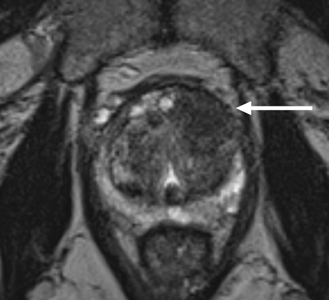
Figure 1a:
Multiparametric 3-T MR imaging was performed in a 73-year-old man with a history of prior biopsy-proven PCa. (a) Axial T2-weighted MR image and (c) apparent diffusion coefficient (ADC) map show a lesion (arrow) in the anterior transition zone with a longest diameter of about 1.7 cm, which is irregular, and uniform hypointensity on the T2-weighted image (a). (b) DWI image and ADC map (c) show focal high and low signal intensity, respectively, at 700 × 10−6 mm2/sec. (d) Enhancement curve shows early and intense enhancement with immediate washout. PI-RADS assessment category is 5. (e) Photograph illustrates that for in-bore biopsy, the patient was placed in the prone position. The DynaTRIM system (InVivo) includes the foam base pad, the clamp stand, and the three adjustable blue screws (arrowheads) for biopsy needle positioning during biopsy planning. The biopsy gun was inserted (arrow).

