

Abstract
Objectives:
The aim of this study was to assess the effect of tube current, kilovoltage peak (kVp), metal type, and the position of metal objects on metal artifacts in cone-beam computed tomography (CBCT) images.
Materials and Methods:
Titanium and cobalt-chromium rods were fabricated and placed in a dry human mandible. Samples were scanned using a Promax 3D CBCT unit with different milli-amperages and kVp. The artifacts induced by metal objects were evaluated using the Image J software in four regions of interest (ROIs) on each image.
Results:
A higher kVp decreased artifacts of the buccal surface of the rods in 97% of the cases (P=0.046) but did not affect the severity of artifacts between the two metal objects (P>0.05). Increasing the tube current had no effect on metal artifacts in 93% of the cases (P>0.05). Artifacts induced by a cobalt-chromium alloy were more severe than those with titanium (P<0.001). Artifacts were more intense in the buccal surface of anterior rods compared to the posterior rods (P<0.003).
Conclusions:
Tube voltage, metal type and the position of metal objects affected the severity of metal artifacts on CBCT images. The metal type had the greatest effect on metal artifact intensity in this study.
Keywords: Artifacts, Cone-Beam Computed Tomography, Metals
INTRODUCTION
With the introduction of cone-beam computed tomography (CBCT) to dentistry as a valuable imaging modality, this modality is increasingly used since it can provide three dimensional (3D), high resolution accurate information of hard tissues with a relatively low radiation dose [1]. It enhances diagnosis, treatment planning and follow up of patients in various fields of dentistry including implantology, surgery, endodontics and orthodontics [2–8].
Many parameters such as field of view, X-ray beam quality and quantity, pixel size and rotation arc affect the CBCT image diagnostic quality and image characteristics that may include noise, contrast resolution and artifacts [1,9]. Metal artifact is among the factors degrading image quality. Artifacts are problematic, especially in the dentoalveolar area due to metal objects like metallic restorative materials, posts, cores, and dental implants. These artifacts are produced because of the high density of the metal, which is beyond the normal range that a computer can measure. Since metals severely attenuate X-ray beams, beam attenuation in structures adjacent to the metallic structures is not recorded properly. Due to image reconstruction techniques in 3D modalities like computed tomography (CT) and CBCT, presence of metal in scanned areas may lead to production of dark and light bands that significantly reduce image quality [10].
Fan-shaped beams in multi-detector computed tomography result in streak artifacts in the gantry path in horizontal direction. However, the cone-shaped beams in CBCT lead to artifacts in all dimensions around the metallic objects [10,11]. As one goal in use of CBCT is accurate measurement and observation of anatomical structural details, evaluation of methods that can reduce metal artifacts is of great importance. There have been studies in this context, however, most of these studies have focused on using artifact-reducing algorithms [12–15]. Although these types of software programs would eliminate streaks far from the metallic object, the details around the metal-tissue interface, which might be the main region of interest (ROI), still may not be visible to the clinicians [16]. Among the considered effective factors in image quality, exposure parameters are adjustable in some CBCT units. Despite this, just a few studies have concerned the effect of exposure parameters on metallic artifacts [11,17, 18]. The effects of metallic objects positioned in the jaw [11,19] and the type of metal [17,20] have been evaluated less often. Therefore, this study was designed to evaluate the effects of current intensity and kilovoltage peak (kVp) of the CBCT unit, position of metallic objects in the jaw, and the metal type on metal artifacts.
MATERIALS AND METHODS
In this experimental study, impacts of current intensity and kVp of a CBCT unit, position of metallic objects in the jaw, and metal type on metal artifacts were evaluated. This study was conducted on a dry human mandible. In order to fabricate samples, three cobalt-chromium rods and three titanium rods with 3mm diameter and 8mm length were constructed. The titanium rods were used to evaluate artifacts induced by dental implants, and cobalt-chromium alloy was selected to assess the artifacts from base-metal, cast restorations. In order to reach maximum accuracy and similarity in dimensions, a computer aided design/computer aided manufacturing unit was used for preparation of metal rods.
Three parallel holes with 3mm diameter and 8mm height were placed in the mandible. The anterior hole was drilled at the canine site, the middle hole was created at the second premolar site, and the posterior hole was created at the distal root of the first molar site. The distance from the anterior and posterior holes to the middle hole was the same. In order to simulate the beam attenuation effect of soft tissue, the bone surface was covered with layers of soft base plate wax with a total thickness of 15 mm (Fig. 1). Titanium rods were first placed into the holes and the mandible was scanned using the Promax 3D unit (Planmeca, Helsinki, Finland) with different currents (8, 12, and 16 mA) and different voltages (70, 78, and 84 kVp), with 8×8cm field of view and 0.16 mm voxel size. Next, the cobalt-chromium rods were placed into the holes and the same process was repeated (Fig. 2).
Fig. 1:
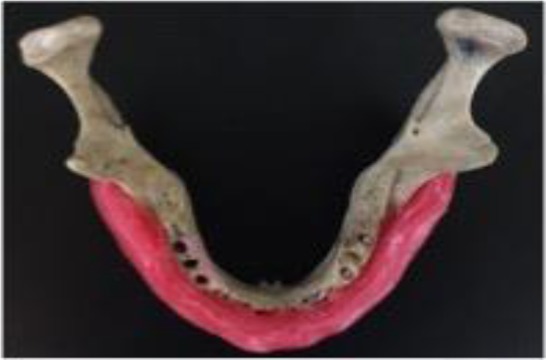
The mandibular bone covered by soft base plate wax.
Fig. 2:

The mandibular bone positioned in ProMax 3D CBCT unit.
The axial plane, which was perpendicular to the middle rod and located 4mm from its superior surface was selected as the reference axial plane (Fig. 3) and was saved as the two-dimensional form (Fig. 4).
Fig. 3:
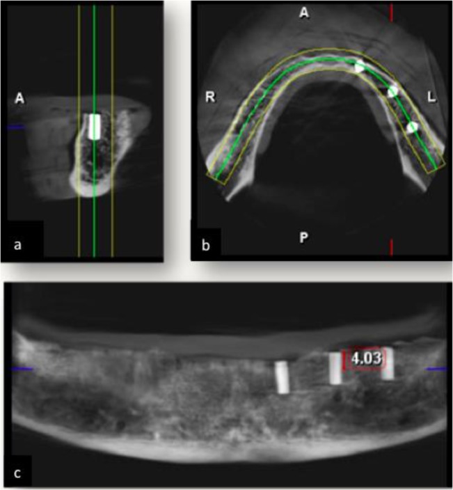
(a) Sagittal, (b) axial and (c) panoramic images.
Fig. 4:

Axial images of all samples
Two-dimensional images were numbered without any specific order and were given to two observers. Four square-shaped 25-pixel areas were considered as the ROI. These regions included a region adjacent to the buccal surface of the anterior implant, the region adjacent to the buccal surface of the posterior implant, a region in the middle of an imaginary line connecting the anterior and middle implants, and a region in the middle of an imaginary line connecting the middle and posterior implants (Fig. 5). The images were evaluated using the Image J software and the mean gray value in four regions on each image was measured. Observers evaluated each image twice. To evaluate the inter-observer and intra-observer agreements, an intra-class correlation coefficient (type: absolute agreement, model: two-way random) was calculated and the inter-observer and intra-observer agreements were found to be 99.9%.
Fig. 5:

Four ROIs as used for evaluation of gray values
Statistical analysis:
To evaluate the effects of four factors (voltage, current intensity, metal type and metal object position) on metal artifacts, interactions were calculated; double, triple, and quadruple interactions were significant in most cases (the significance level was considered less than 0.20). Therefore, to compare the material factor effect (dichotomous variable), an independent t-test was used. To compare other factors that had more than two categories, one-way ANOVA was used (followed by Tukey’s test or Games-Howell test for pairwise comparisons). The significance level was considered to be less than 0.05.
RESULTS
In this study, gray value was measured as an index for metal artifact evaluation in different conditions in terms of four factors. As stated earlier, these factors consisted of tube voltage (70, 78 and 84 kVp), tube current (8, 12 and 16 mA), material (titanium and cobalt-chromium alloy) and position of metal object (adjacent to the posterior and anterior rods, an area between the posterior and middle rods, an area between middle and anterior rods), which provided 288 conditions of comparison. Because the effect of each variable was not consistent in all situations, number of conditions in which metal artifacts increased or decreased was reported for better interpretation of the results.
Effect of voltage change on artifacts:
The change in tube voltage did not have a significant effect in 35 out of 36 (97%) conditions (P>0.05) in the positions “between the posterior and middle rods” and “between the middle and anterior rods”. Increasing the kVp resulted in a significant reduction in metal artifacts in 35 out of 36 (97%) conditions (P=0.046) in positions adjacent to the anterior and posterior rods (Fig. 6).
Fig. 6:

Mean and standard deviations of gray values in CBCT images due to metal artifacts in different tube current intensities and voltages irrespective of metal type and metal object position.
Effect of current intensity change on artifacts:
The tube current intensity change did not affect artifacts in 67 out of 72 (93%) conditions (P>0.05, Fig. 6).
Effect of metal type on artifacts:
Cobalt-chromium alloy produced significantly more intense artifacts than titanium in all 36 conditions (P<0.001, Fig. 7).
Fig. 7:

Mean and standard deviations of gray values in CBCT images due to metal artifacts induced by different metal types in different metal object positions irrespective of kVp and mA. (Position 1: between the posterior and middle rods, 2: between middle and anterior rods, 3: buccal surface of posterior rod, 4: buccal surface of anterior rod).
Effect of metal object position on artifacts:
The gray value adjacent to the anterior rod was higher than the posterior rod and two intermediate positions in all 54 conditions. In addition, the gray value adjacent to the posterior rod was significantly higher than the two intermediate positions in all 36 conditions (P<0.003).
When assessing the artifacts induced by cobalt-chromium rods, there were no significant differences comparing the position “between the posterior and middle rods” with the position “between the middle and anterior rods” in all nine conditions (P>0.05).
In the samples with titanium rods in five out of nine conditions (55.5%), artifacts in the area “between the anterior and middle rods” were significantly more intense than those “between the posterior and middle rods” (P=0.038). In four out of nine conditions (44.5%), the difference was not significant (P>0.05, Fig. 7).
DISCUSSION
Presence of metal artifacts complicates CBCT interpretations. In general, when a polychromatic X-ray beam passes through an object, low energy photons are absorbed more than high energy photons. This leads to an increase in the mean X-ray beam energy and beam hardening. Less X-ray beam energy, more density, and an irradiated substance with a higher atomic number lead to more beam hardening [9,21]. Therefore, artifacts are more severe when metal is present. In this study, the current intensity and voltage of the CBCT unit, the position of metallic objects in the jaw, and the type of metal were assessed to determine their effects on metal artifacts. According to the results of this study, a higher voltage generally resulted in significant reductions in metal artifacts adjacent to the buccal surfaces of the rods. However, it did not affect artifact severity in positions between the rods. According to previous studies [11,16,20,22], increasing the voltage results in higher energy and greater penetration of the X-ray beam. Therefore, in higher voltages, there is less beam hardening and less metal artifacts. In studies by Schulze et al, [11] and Chindasombatjareon et al, [20] increasing the voltage led to a reduction in metal artifacts. In studies by Barret and Keat [16] and Kataoka et al, [22] concerning CT artifacts, use of high kVp techniques in order to decrease metal artifacts was recommended. Other factors that could affect beam hardening include the amount of machine rotation, the X-ray beam configuration, and the algorithms used for processing information [23–25].
In this study, increasing the current intensity did not affect metal artifacts in most cases similar to the study by Pauwels et al [17]. In a study by Chindasombatjareon et al, [20] the current intensity had no consistent effect on metal artifacts. Pauwels et al, [9] in their review article in 2015 stated that changes in mA affected noise but not beam hardening. In the study by Kataoka et al, [22] using CT, higher tube currents decreased metal artifacts. However, we must consider that the range of current intensity used in the study by Kataoka et al, [22] was different from that used in the present study. In the current study, the difference between the maximum and minimum mA was 8; whereas, in the study by Kataoka et al, [22] the current intensity reached 500 from 100 mA. The narrow range of changes in mA value in CBCT can explain the insignificant effect of mA change on metal artifacts.
Comparisons between titanium and cobalt-chromium revealed that in all four positions and all exposure parameters, cobalt-chromium artifacts were more intense than those observed with titanium. The missing value artifacts induced by cobalt-chromium were more severe than with titanium in the region between the posterior and middle rods and also between the middle and anterior rods. Thus, the gray value between the two cobalt-chromium inserts with different exposure parameters equaled zero or nearly zero. Photoelectric absorption relates to the cube of the atomic number of the irradiated substance. Since the atomic number of chromium (24) and cobalt (27) is higher than that of titanium (22), and there are metals with higher atomic numbers used in cobalt-chromium alloys (e.g., molybdenum and tungsten), X-ray beam absorption and beam hardening were more than with titanium.
In studies by Pauwels et al, [17] and Kataoka et al, [22] lead and stainless steel produced more artifacts than titanium. Kuusisto et al. [26] reported that zirconia and titanium produced severe artifacts on CBCT images, and the artifacts were more severe in composite models with higher amounts of BaAlSiO2. When the composite opacifier reached 68%, artifacts were similar to titanium. In the study by Chindasombatjareon et al, [20] the artifacts produced by four metals were assessed and it was noted that type IV gold alloy caused the largest artifact areas followed by the cobalt-chromium alloy, titanium, and aluminum, respectively.
Beam hardening results in two kinds of artifacts; one is distortion of the metal structure due to X-ray differential absorption, which is known as a cupping artifact. The other artifact is dark bands between two metal objects known as an extinction artifact or missing value artifact [27]. According to our results, the gray value adjacent to the buccal surface of the posterior and anterior inserts was significantly higher than the positions between the two rods, which was a result of different afore-mentioned artifacts. Distortion of a rod’s image leads to higher gray values in the adjacent area; whereas, extinction artifacts and less gray values are seen between the two metal rods. This is due to the fact that when X-ray beams pass through two metal rods simultaneously, beam hardening would be more severe [26,27].
In this study, metal artifacts seen adjacent to the anterior rod were more intense than those with the posterior rod in all samples, which might be due to different projection paths, information processing, and reconstruction techniques. It is possible to have different results in different CBCT units as seen in studies by Schulze et al, [11] and Benic et al, [19] where the implant site did not affect the artifact severity. In the study by Schulze et al, [11] gray values were measured in a region adjacent to the rods, which was located on the imaginary line connecting the rods. Whereas, in the current study, regions adjacent to the buccal surfaces of the rods were evaluated. The results of this study revealed that, in cobalt-chromium samples, the difference in artifact severity of intermediate areas was not significant. However, in 55% of titanium samples, artifacts between the middle and anterior rods were more intense than the region between the posterior and middle rods. As mentioned, extinction artifacts from the cobalt-chromium inserts were very severe, and it seems that this is the reason why the different positions did not affect artifact severity in these areas. However, in titanium rods, which showed less beam hardening, the results of comparison of intermediate positions were similar to the results of the comparison of positions adjacent to the rods.
It is recommended to provide CBCT units that enable operators to choose the proper exposure settings in order to reduce artifacts and also consider a balance between the image quality and the radiation dose.
CONCLUSION
A higher kVp decreased metal artifacts in the buccal surface of the rods but did not affect the severity of artifacts between the two metal objects. Artifacts induced by cobalt-chromium alloy were more severe than those induced by titanium, and artifacts were more intense in the buccal surface of anterior rods compared to the posterior rods. Increasing the tube current had no effect on metal artifacts. The metal type had the greatest effect on the metal artifact intensity in this study.
REFERENCES
- 1-. Brullmann D, Schulze RK. Spatial resolution in CBCT machines for dental/maxillofacial applications-what do we know today? Dentomaxillofac Radiol. 2015; 44 (1): 20140204. [DOI] [PMC free article] [PubMed] [Google Scholar]
- 2-. Bornstein MM, Scarfe WC, Vaughn VM, Jacobs R. Cone beam computed tomography in implant dentistry: a systematic review focusing on guidelines, indications, and radiation dose risks. Int J Oral Maxillofac Implants. 2014. January; 29 Suppl: 55– 77 [DOI] [PubMed] [Google Scholar]
- 3-. Benavides E, Rios HF, Ganz SD, An CH, Resnik R, Reardon GT, et al. Use of cone beam computed tomography in implant dentistry: the International Congress of Oral Implantologists consensus report. Implant Dent. 2012. April; 21 (2): 78– 86. [DOI] [PubMed] [Google Scholar]
- 4-. Harris D, Horner K, Grondahl K, Jacobs R, Helmrot E, Benic GI, et al. E.A.O. guidelines for the use of diagnostic imaging in implant dentistry 2011. A consensus workshop organized by the European Association for Osseointegration at the Medical University of Warsaw. Clin Oral Implants Res. 2012. November; 23 (11): 1243– 53. [DOI] [PubMed] [Google Scholar]
- 5-. American Association of Endodontists. American Academy of Oral and Maxillofacial Radiology Use of cone-beam computed tomography in endodontics Joint Position Statement of the American Association of Endodontists and the American Academy of Oral and Maxillofacial Radiology. Oral Surg Oral Med Oral Pathol Oral Radiol Endod. 2011. February; 111 (2): 234– 7. [DOI] [PubMed] [Google Scholar]
- 6-. Scarfe WC, Levin MD, Gane D, Farman AG. Use of cone beam computed tomography in endodontics. Int J Dent. 2009; 2009: 634567. [DOI] [PMC free article] [PubMed] [Google Scholar]
- 7-. Garib DG, Calil LR, Leal CR, Janson G. Is there a consensus for CBCT use in Orthodontics? Dental Press J Orthod. 2014. Sep-Oct; 19 (5): 136– 149. [DOI] [PMC free article] [PubMed] [Google Scholar]
- 8-. Ahmad M, Jenny J, Downie M. Application of cone beam computed tomography in oral and maxillofacial surgery. Aust Dent J. 2012. March; 57 Suppl 1: 82– 94. [DOI] [PubMed] [Google Scholar]
- 9-. Pauwels R, Araki K, Siewerdsen JH, Thongvigitmanee SS. Technical aspects of dental CBCT: state of the art. Dentomaxillofac Radiol. 2015; 44 (1): 20140224. [DOI] [PMC free article] [PubMed] [Google Scholar]
- 10-. Jaju PP, Jain M, Singh A, Gupta A. Artefacts in cone beam CT. Open J Stomatol. 2013. July; 3 (05): 292. [Google Scholar]
- 11-. Schulze RK, Berndt D, d’Hoedt B. On cone-beam computed tomography artifacts induced by titanium implants. Clin Oral Implants Res. 2010. January; 21 (1): 100– 7. [DOI] [PubMed] [Google Scholar]
- 12-. Bechara B, McMahan CA, Geha H, Noujeim M. Evaluation of a cone beam CT artefact reduction algorithm. Dentomaxillofac Radiol. 2012. July; 41 (5): 422– 8. [DOI] [PMC free article] [PubMed] [Google Scholar]
- 13-. Gong XY, Meyer E, Yu XJ, Sun JH, Sheng LP, Huang KH, et al. Clinical evaluation of the normalized metal artefact reduction algorithm caused by dental fillings in CT. Dentomaxillofac Radiol. 2013; 42 (4): 20120105. [DOI] [PMC free article] [PubMed] [Google Scholar]
- 14-. Ibraheem I. Reduction of artifacts in dental cone beam CT images to improve the three dimensional image reconstruction. J Biomed Sci Eng. 2012. August; 5 ( 8): 409– 15. [Google Scholar]
- 15-. Bechara BB, Moore WS, McMahan CA, Noujeim M. Metal artefact reduction with cone beam CT: an in vitro study. Dentomaxillofac Radiol. 2012. March; 41 ( 3): 248– 53. [DOI] [PMC free article] [PubMed] [Google Scholar]
- 16-. Barrett JF, Keat N. Artifacts in CT: recognition and avoidance. Radiographics. 2004. Nov-Dec; 24 (6): 1679– 91. [DOI] [PubMed] [Google Scholar]
- 17-. Pauwels R, Stamatakis H, Bosmans H, Bogaerts R, Jacobs R, Horner K, et al. Quantification of metal artifacts on cone beam computed tomography images. Clin Oral Implants Res. 2013. August; 24 Suppl A100: 94– 9. [DOI] [PubMed] [Google Scholar]
- 18-. Katsumata A, Hirukawa A, Noujeim M, Okumura S, Naitoh M, Fujishita M, et al. Image artifact in dental cone-beam CT. Oral Surg Oral Med Oral Pathol Oral Radiol Endod. 2006. May; 101 (5): 652– 7. [DOI] [PubMed] [Google Scholar]
- 19-. Benic GI, Sancho-Puchades M, Jung RE, Deyhle H, Hammerle CH. In vitro assessment of artifacts induced by titanium dental implants in cone beam computed tomography. Clin Oral Implants Res. 2013. April; 24 (4): 378– 83. [DOI] [PubMed] [Google Scholar]
- 20-. Chindasombatjareon J, Kakimoto N, Murakami S, Maeda Y, Furukawa S. Quantitative analysis of metallic artifacts caused by dental metals: comparison of cone-beam and multi-detector row CT scanners. Oral Radiol. 2011. December; 27 (2): 114– 20. [Google Scholar]
- 21-. Schulze R, Heil U, Gross D, Bruellmann DD, Dranischnikow E, Schwanecke U, et al. Artefacts in CBCT: a review. Dentomaxillofac Radiol. 2011. July; 40 (5): 265– 73. [DOI] [PMC free article] [PubMed] [Google Scholar]
- 22-. Kataoka ML, Hochman MG, Rodriguez EK, Lin PJ, Kubo S, Raptopolous VD. A review of factors that affect artifact from metallic hardware on multi-row detector computed tomography. Curr Probl Diagn Radiol. 2010. Jul-Aug; 39 (4): 125– 36. [DOI] [PubMed] [Google Scholar]
- 23-. Schulze D, Heiland M, Blake F, Rother U, Schmelzle R. Evaluation of quality of reformatted images from two cone-beam computed tomographic systems. J Craniomaxillofac Surg. 2005. February; 33 (1): 19– 23. [DOI] [PubMed] [Google Scholar]
- 24-. Hunter A, McDavid D. Analyzing the beam hardening artifact in the Planmeca Promax. Oral Surg Oral Med Oral Pathol Oral Radiol Endod. 2009. April; 107 (4): e28– e29. 19272803 [Google Scholar]
- 25-. Draenert FG, Coppenrath E, Herzog P, Muller S, Mueller-Lisse UG. Beam hardening artefacts occur in dental implant scans with the NewTom cone beam CT but not with the dental 4-row multidetector CT. Dentomaxillofac Radiol. 2007. May; 36 (4): 198– 203. [DOI] [PubMed] [Google Scholar]
- 26-. Kuusisto N, Vallittu PK, Lassila LV, Huumonen S. Evaluation of intensity of artefacts in CBCT by radio-opacity of composite simulation models of implants in vitro. Dentomaxillofac Radiol. 2015; 44 (2): 20140157. [DOI] [PMC free article] [PubMed] [Google Scholar]
- 27-. White SC, Pharoah MJ. Oral radiology: principles and interpretation. 7th Ed., St. Louis, Missouri, Elsevier-Mosby, 2014: 196. [Google Scholar]