Abstract
During dental education, dental students learn how to examine patients, make diagnosis, plan treatment and perform dental procedures perfectly and efficiently. However, progresses in computer-based technologies including virtual reality (VR) simulators, augmented reality (AR) and computer aided design/computer aided manufacturing (CAD/CAM) systems have resulted in new modalities for instruction and practice of dentistry. Virtual reality dental simulators enable repeated, objective and assessable practice in various controlled situations. Superimposition of three-dimensional (3D) virtual images on actual images in AR allows surgeons to simultaneously visualize the surgical site and superimpose informative 3D images of invisible regions on the surgical site to serve as a guide. The use of CAD/CAM systems for designing and manufacturing of dental appliances and prostheses has been well established.
This article reviews computer-based technologies, their application in dentistry and their potentials and limitations in promoting dental education, training and practice. Practitioners will be able to choose from a broader spectrum of options in their field of practice by becoming familiar with new modalities of training and practice.
Keywords: Virtual Reality Exposure Therapy, Immersion, Computer-Aided Design, Dentistry, Education
INTRODUCTION
Computer-based technologies play an important role in all aspects of our daily life as well as in dentistry. Simplified interactions between human and computer have caused a profound progress in virtual reality (VR)-based dental training. On the other hand, computer-aided design/computer aided manufacturing (CAD/CAM) of dental appliances and prostheses is now widely used around the globe [1]. Although many dentists have become familiar with CAD/CAM subgroup of computer-based technologies in the recent years, VR and augmented reality (AR) techniques, which are going to find their place in learning and instruction of dental skills, are not as much known [2].
In this article, different types of computer-based technologies including VR and AR simulators, CAD/CAM systems and their applications in dentistry are reviewed.
Virtual Reality Technology
Virtual reality technology is defined as a method by which, an environment is three dimensionally simulated or replicated, giving the user a sense of being inside it, controlling it, and personally interacting with it [3,4]. The virtual environments are almost completely generated by computers [5]. Technology has a wide range of probable benefits in many aspects of life like construction of a building by providing a highly detailed virtual three-dimensional (3D) model of the building to verify each part of the plan, the cost and layout [6]. Similarly, in designing a new product, the virtual prototype can be used instead of physical prototypes to evaluate the design from different aspects [6]. In the medical field, VR is used for instruction of surgical procedures [7,8], education of patients and training students [9–12]. It also helps in treatment of psychological disorders by providing a valid controlled virtual environment to objectively assess the behavior and rehabilitate cognitive and functional abilities [3]. It is successfully used for treatment of complex regional pain syndrome as well [13]. The 3D virtual sceneries help improve dental experience via distraction intervention [14].
Features of VR
Virtual reality depends on two basic features namely immersion and interaction.
Immersion is the sense of being present in a virtual (non-real) environment. The environment is generated by synthesizing 3D images, sound and other stimuli, which surround the users and make them feel being physically present in a non-physical (non-real) world [15]. The degree to which, the user believes being present in a virtual environment (immersion) is different in various systems ranging from fully immersive to nonimmersive, depending on the capabilities of the system [6].
Interaction is the power of user to modify the virtual environment [15]. This feature is the main difference between 3D movies and virtual environments. In VR systems, the user is able to interact with the virtual world, moving around it, seeing it from different angles, reaching it, grabbing it and reshaping it. This sort of interaction is possible by means of head mounted video goggles, wired clothing and fiber-optic data gloves. Position-tracking devices and real-time update of visual, auditory displaying systems are also necessary [16].
Types of VR Systems
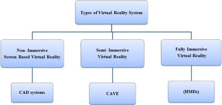
Virtual reality systems are classified into three major groups of non-immersive, semi-immersive and immersive (Fig. 1) based on the immersion and type of components utilized in the system [6].
Fig. 1:
Types of virtual reality systems
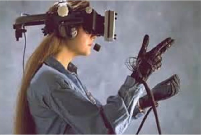
Immersive VR simulation is a technology that gives the user the psychophysical experience of being surrounded completely by a virtual computer-generated environment (Fig. 2) by using hardware, software and interaction devices. Full immersion is the highest level of immersion, which is produced by a head mounted device that displays images three dimensionally via a process known as stereoscopy. In this process, the user sees two images -one per eye- and the brain combines them into a single 3D image. The other components are data gloves, which enable the person to interact with the objects, for example, pulling, twisting or gripping them and may also give a force feedback to the user, which is known as haptic. There are also tracking devices, which track user’s head, hands, fingers, eyes and feet to enable interaction with the virtual world. Sound is displayed as well. In fully immersive virtual environments the user is completely separated from the real world [17].
Fig. 2:

Immersive virtual reality; head mounted displays (HMD) and data gloves
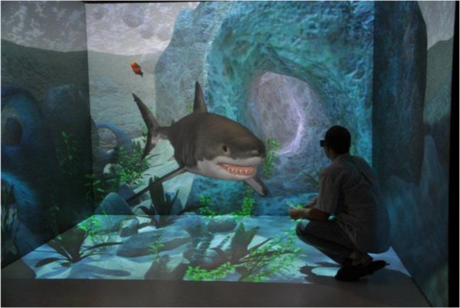
Semi-immersive VR simulation is a system in which the user stands in a room with rear projection walls, down projection floor, speakers at different angles, tracking sensors in the walls and sound/music devices (Fig. 3). By wearing eye goggles, the user sees everything three dimensionally. Since the user can still see him/herself, the system is not considered a fully immersive simulator. Cave Automatic Virtual Environment is an example of these systems [18].
Fig. 3:

Semi-immersive virtual reality; Cave Automatic Virtual Environment (CAVE)
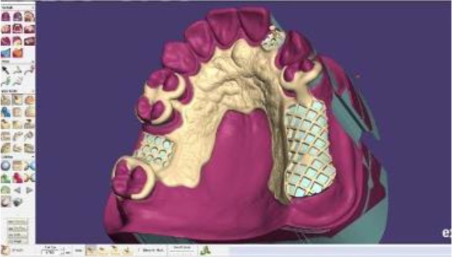
Non-immersive VR simulation is the least immersive and least expensive of all. It allows the user to be involved with a 3D environment just by incorporating a stereo display monitor and glasses. They can be run on a standard desktop computer using mouse and joystick (Fig. 4). It is mostly used in designing and CAD systems [17].
Fig. 4:

Computer aided design system
Applications of VR in Dentistry
Although the VR simulation systems are used in different aspects of medical training such as laparoscopic surgery [19], their use in dentistry is not an easy task. The reason can be sought in complexity of dental instruments in type, shape and speed, and the diversity of oral tissues, which include gingiva, multilayer teeth and bone.
To simulate tooth reduction in dental virtual reality systems, the operator uses a stylus, which with the help of worn special goggles appears as the intended instrument like high or low speed hand piece in the 3D displaying stereoscopic monitor. The process mandates accurate digitized models of the instruments and oral cavity tissues and sophisticated graphic programs for showing the reduction of tooth. Unlike the monolayer resin models generally used for preclinical training, different layers of tooth like enamel, dentin and dental pulp are modeled in VR systems. This helps dental students to avoid unintentional exposure of dental pulp during preliminary clinical practice [20].
The differentiation between various structures or speed of the instrument is only possible with the help of haptic devices. They enable the user to feel the force required for each practice (force feedback) and provide a realistic tactile sense, just like working on real structures with real instruments. In surgery, haptic devices allow surgeons to touch and feel objects such as surgical tools and human organs in a virtual environment, and to perform operations like pushing, pulling, and cutting of soft or hard tissue with realistic force feedback. Another advantage of VR simulators is that they are programmed to identify errors and assess the quality of performance. Experts determine the errors and the best performances, which would be the basis for comparison and assessment. The system is able to record and replay the performance of each user as well, which allows them to know their faults and fix them. It is also possible to practice and receive assessment as many times as required at any time, which is not possible with programmed traditional classroom instruction [21].
Augmented Reality
Augmented reality refers to superimposition of computer-generated graphics over a real-world scene [22]. In contrast to VR simulators, in AR the real environment is not completely suppressed and in fact plays a dominant role in this process (Table 1). Augmented reality aims to add synthetic additives to the real world (or to a live video of the real world) instead of engaging a person in a world, which is completely generated by a computer [23]. It is widely used in image guided surgery, where real and virtual objects need to be composed, integrated, presented or manipulated simultaneously in a single scene [24]. It is also applied in dental implantation, maxillofacial surgery, temporomandibular joint motion analysis and prosthetic surgery [8,9].
Table 1:
Comparison of virtual reality and augmented reality
| Concept | Classification | Fully synthetic virtual world | Fully real virtual world | Absolute spatial registration critical | Relative spatial registration critical | Real-Time interactivity critical |
|---|---|---|---|---|---|---|
| Augmented reality | Uses computer | No | No | Maybe | Yes | Yes |
| Virtual reality | Uses computer | Yes | No | No | Yes | Yes |
One of the main uses of AR in oral and maxillofacial surgery is in visualization of deep masked structures. Before surgery, the surgeon would be able to map the surgical plan on the 3D image of the site and consider any necessary modifications. During surgery, the surgeon sees and follows the mapped image overlaid on the surgical site by use of special glasses. This system can be developed for root canal therapy as well [24].
In some systems, both AR and VR technologies are used together in order to enhance the training capacity of the system. Developers of such systems believe that this integration improves the performance of users.
Future of VR and AR in Dentistry
The VR and robotics as new technologies will have a great impact on health care in the next decades [25]. The dynamic association of operation on a real organ with imaging data may create new modes of diagnosis and treatment for technically challenging patients. Experienced surgeons will benefit the most from such systems by extending the safe limit for more efficient operations, while less experienced surgeons may at least benefit from better visualization and orientation of critical anatomical landmarks [8]. The VR and AR systems seem promising, but there are still technological challenges that researchers and developers must face in order to fulfill these promises.
Dental CAD/CAM Systems
The evolution of dental materials and advances in computer science led to a rapid development in dental CAD/CAM technology. During the past couple of decades, many advanced chairside and laboratory CAD/CAM systems were introduced. Computers are used to collect data and design and manufacture a wide range of products in CAD/CAM systems. These systems have long been used in industries but they were not available for dental applications until the 1980s [26]. Nowadays, the term CAD/CAM in dentistry is equal to manufacturing by milling technology. However, it is not completely true, because manufacturing can either be by subtractive (milling) or additive technologies [27,28].
The CAD/CAM systems consist of three components:
A digitization tool/scanner that transforms geometry of a real world object into digital data to enable processing by a computer.
Software for data processing.
A technology, which manufactures the desired product from the digitized data set [27].
At present, many fixed prosthetic restorations are manufactured by the CAD/CAM systems, using different types of materials such as porcelain, composite resin and metallic blocks. Even materials like zirconia, which could not be manufactured by the conventional methods previously because of technical limitations, can now be fabricated by these systems [29]. The CAD/CAM systems are going to find substantial applications in implant dentistry to manufacture implant-supported prostheses, abutments and diagnostic templates [30].
The main benefit of this type of manufacturing in dentistry is that conventional impressions are not needed anymore, which is believed to save the dentist’s chair time and eliminate a time-consuming step [31]. Table 2 shows the advantages and limitations of CAD/CAM systems. The CAD/CAM techniques and rapid prototyping are extensively used in treatment of maxillofacial defects and surgeries [32]. These techniques are also used for designing and manufacturing removable partial denture metal frameworks through 3D printing [33] and by collecting 3D data from the patient’s cast, determining the path of insertion and designing the shape of the components of the frameworks digitally. The completed model data are stored as stereo lithography files, which are transferred to rapid prototyping models. Finally, metal removable partial denture frameworks are fabricated by selective laser melting technique [34].
Table 2:
Advantages and limitations of CAD/CAM systems
| Advantages of CAD/CAM systems | Limitations of CAD/CAM systems |
|---|---|
| No need for traditional impressions when intra-oral scanners are used | High cost |
| Chairside fabrication of restorations | Need mastering of technology |
| Fewer visits | Manual veneering is used most of the time |
| Needs less manual procedures in laboratory | |
| Needs less laboratory time | |
| Easier laboratory procedures | |
| Good marginal accuracy | |
| Suitable for materials like zirconia |
The CAD/CAM technology is used in orthodontic diagnosis, treatment planning [35] and fabrication of appliances (Invisalign Production Process) which include submitting of the scan or impressions and photographs to the company with the doctor’s instructions. These intraoral scans or impressions will be used to design an accurate 3D digital model for each dental arch, after that a stereolithographic model will be fabricated for each step. Finally, a clear plastic aligner will be made over each model, and the set of aligners are then sent directly to the doctor [36]. Also, it is used in determining the position of impacted maxillary canines and for the fabrication of occlusal splints. However, CAD/CAM/additive manufacturing innovations have not been used successfully on a wide scale for removable orthodontic appliances [35].
In the CAD-CAM systems, virtual articulator is a basic tool that deals primarily with the functional aspects of the occlusion and is a core tool in many diagnostic and therapeutic procedures. With the introduction of virtual articulators a bright future will be expected and a revolutionary change will occur in digital dentistry [2]. Table 3 shows the applications of computer-based technologies in dental aspects.
Table 3:
Applications of computer-based technologies in dental aspects
| Virtual reality | Augmented reality | CAD/CAM systems | |
|---|---|---|---|
| Prosthodontics | Diagnosis and treatment planning for implant placement | Fabrication of crown and bridge frameworks Fabrication of custom made abutments for implants Designing and manufacturing of implant surgical splints Designing and manufacturing RPD metal frameworks Designing complete dentures Virtual articulator which used in many diagnostic and therapeutic procedures |
|
| Maxillofacial Surgery | Training by performing virtual surgery 3D observation of surgical site |
Superimposition of radiographs over the surgical site Implant placement during surgery |
Making maxillofacial prosthesis |
| Orthodontics | Diagnosis and treatment of periodontal diseases | Diagnosis and treatment planning Determining the position of impacted maxillary canines |
|
| Periodontics | Training of scaling | Differentiating between pathological and normal conditions | |
| Restorative Dentistry | Training tooth preparation | Fabricating indirect restorations |
CONCLUSION
At present, computer-based technologies are well established in designing and manufacturing dental appliances and prostheses. However, simulation systems for the instruction of dental skills are the new modalities not much known or widely used and only a few dental schools are using them at the time being. These systems are under constant progress and development. They are still too expensive and their maintenance and repair costs are high like any other new technology. On the other hand, computer-assisted skill acquisition in conjunction with traditional training would enable students to practice repeatedly, with constant assessment and force feedback, which is not routinely possible with resin models. In the surgical fields, simulation systems enable the students to practice in real mode on virtual subjects. They also offer visual information about the surgical site, which is of great use for the surgeons.
REFERENCES
- 1-.Jevremović DP, Puškar TM, Budak I, Vukelić D, Kojić V, Eggbeer D, et al. An RE/RM approach to the design and manufacture of removable partial dentures with a biocompatibility analysis of the F75 Co-Cr SLM alloy. Materiali in Tehnologije. 2012. March;46 (2):123–9. [Google Scholar]
- 2-.Gugwad R, Kore A, Basavakumar M, Arvind M, Sudhindra M, Ramesh C. Virtual articulators in prosthodontics. Int J Dent Clin. 2011;3(4):39–41. [Google Scholar]
- 3-.Schultheis MT, Rizzo AA. The application of virtual reality technology in rehabilitation. Rehabil Psychol. 2001. August;46(3):296–311. [Google Scholar]
- 4-.Ausburn LJ, Ausburn FB. Desktop Virtual Reality: A Powerful New Technology for Teaching and Research in Industrial Teacher Education JITE. 2004. Winter;41(4): 1–16. [Google Scholar]
- 5-.Connacher HI, Jayaram S, Lyons KW. Virtual assembly using virtual reality techniques. Comput Aided Des. 1997. August;29(8):575–84. [Google Scholar]
- 6-.Mujber TS, Szecsi T, Hashmi MSJ. Virtual reality applications in manufacturing process simulation. J Mater Process Technol. 2004. November;155(156):1834–8 (Confrence Proceedings). [Google Scholar]
- 7-.Botden SM1, Jakimowicz JJ. What is going on in augmented reality simulation in laparoscopic surgery? Surg Endosc. 2009. August;23(8):1693–700. [DOI] [PMC free article] [PubMed] [Google Scholar]
- 8-.Shuhaiber JH. Augmented reality in surgery. Arch Surg. 2004. February;139(2):170–4. [DOI] [PubMed] [Google Scholar]
- 9-.Dutã M, Amariei CI, Bodan CM, Popovici DM, Ionescu N, Nuca CI. An overview of virtual and augmented reality in dental education. OHDM. 2011. March;10(1):42–9. [Google Scholar]
- 10-.Rhienmora P, Haddawy P, Khanal P, Suebnukarn S, Dailey MN. A virtual reality simulator for teaching and evaluating dental procedures. Methods Inf Med. 2010;49(4):396–405. [DOI] [PubMed] [Google Scholar]
- 11-.LeBlanc VR, Urbankova A, Hadavi F, Lichtenthal RM. A Preliminary Study in Using Virtual Reality to Train Dental Students. J Dent Educ. 2004. March;68(3):378–83. [PubMed] [Google Scholar]
- 12-.Buchanan JA. Experience with virtual reality-based technology in teaching restorative dental procedures. J Dent Educ. 2004. December;68(12):1258–65. [PubMed] [Google Scholar]
- 13-.Sato K, Fukumori S, Matsusaki T, Maruo T, Ishikawa S, Nishie H, et al. Nonimmersive virtual reality mirror visual feedback therapy and its application for the treatment of complex regional pain syndrome: an open-label pilot study. Pain Med. 2010. April;11(4):622–9. [DOI] [PubMed] [Google Scholar]
- 14-.Tanja-Dijkstra K, Pahl S, White MP, Andrade J, May J, Stone RJ, et al. Can virtual nature improve patient experiences and memories of dental treatment? A study protocol for a randomized controlled trial. Trials. 2014. March 22;15:90. [DOI] [PMC free article] [PubMed] [Google Scholar]
- 15-.Ryan ML. Immersion vs. interactivity: Virtual reality and literary theory. SubStance. 1999;28(2):110–37. [Google Scholar]
- 16-.Steuer J. Defining virtual reality: Dimensions determining telepresence. J. Commun. 1992. December 1;42(4):73–93. [Google Scholar]
- 17-.Shahrbanian S, Ma X, Aghaei N, Korner-Bitensky N, Moshiri K, Simmonds MJ. Use of virtual reality (immersive vs. non immersive) for pain management in children and adults: A systematic review of evidence from randomized controlled trials. Eur J Exp Biol. 2012;2(5):1408–22. [Google Scholar]
- 18-.Ponto K, Kohlmann J, Tredinnick R. DSCVR: designing a commodity hybrid virtual reality system. Virtual Reality. 2015. March;19(1):57–70. [Google Scholar]
- 19-.Aggarwal R, Moorthy K, Darzi A. Laparoscopic skills training and assessment. Br J Surg. 2004. December;91(12):1549–58. [DOI] [PubMed] [Google Scholar]
- 20-.Mallikarjun SA, Tiwari S, Sathyanarayana S, Devi PR. Haptics in periodontics. J Indian Soc Periodontol. 2014. January;18(1):112–3. [DOI] [PMC free article] [PubMed] [Google Scholar]
- 21-.Konukseven EI, Önder ME, Mumcuoglu E, Kisnisci RS. Development of a Visio-Haptic Integrated Dental Training Simulation System. J Dent Educ. 2010. August;74(8):880–91. [PubMed] [Google Scholar]
- 22-.Van Krevelen DW, Poelman R. A survey of augmented reality technologies, applications and limitations. Int J Virtual Real. 2010. June;9(2):1. [Google Scholar]
- 23-.Bimber O, Raskar R. Spatial augmented reality: merging real and virtual worlds. AK Peters, Wellesly, CRC Press, Tylor &Francis Group; 2005. [Google Scholar]
- 24-.Takato T. New technologies in oral science. JMAJ. 2011. May-Jun;54(3):194–6. [Google Scholar]
- 25-.McCloy R, Stone R. Virtual Reality in Surgery. BMJ. 2001. October 20;323(7318):912–5. [DOI] [PMC free article] [PubMed] [Google Scholar]
- 26-.Liu PR. A Panorama of dental CAD/CAM restorative system. Compend Contin Educ Dent. 2005. July;26(7):507–12. [PubMed] [Google Scholar]
- 27-.Beuer F, Schweiger J, Edelhoff D. Digital dentistry: an overview of recent developments for CAD/CAM generated restorations. Br Dent J. 2008. May 10;204(9):505–11. [DOI] [PubMed] [Google Scholar]
- 28-.Azari A, Nikzad S. The evolution of rapid prototyping in dentistry: a review. Rapid Prototyping J 2009. May 29;15(3):216–25. [Google Scholar]
- 29-.Raigrodski AJ. Contemporary materials and technologies for all ceramic fixed partial dentures: a review of the literature. J Prosthet Dent. 2004. December;92(6):557–62. [DOI] [PubMed] [Google Scholar]
- 30-.Fuster-Torres MÁ, Albalat-Estela S, Alcañiz-Raya M, Peñarrocha-Diago M. CAD/CAM dental systems in implant dentistry: update. Med Oral Patol Oral Cir Bucal. 2009. March 1;14(3):E141–5. [PubMed] [Google Scholar]
- 31-.Daniel JP. CAD/CAM Today: A 22-Year Retrospective. Inside Dent. 2008. Nov-Dec; 4(10). [Google Scholar]
- 32-.Hughes CW, Page K, Bibb R, Taylor J, Revington P. The custom-made titanium orbital floor prosthesis in reconstruction for orbital floor fractures. Br J Oral Maxillofac Surg. 2003. February;41(1):50–3. [DOI] [PubMed] [Google Scholar]
- 33-.Bibb RJ, Eggbeer D, Williams RJ, Woodward A. Trial fitting of a removable partial denture framework made using computer-aided design and rapid prototyping techniques. Proc Inst Mech Eng H. 2006. July;220(7):793–7. [DOI] [PubMed] [Google Scholar]
- 34-.Han J, Wang Y, Lü P. A preliminary report of designing removable partial denture frameworks using a specifically developed software package. Int J Prosthodont. 2010. Jul-Aug;23(4):370–5. [PubMed] [Google Scholar]
- 35-.Al Mortadi N, Eggbeer D, Lewis J, Williams RJ. CAD/CAM/AM applications in the manufacture of dental appliances. Am J Orthod Dentofacial Orthop. 2012. November;142(5):727–33. [DOI] [PubMed] [Google Scholar]
- 36-.Proffit WR, Fields HW, Sarver DM, Ackerman JL. Contemporary orthodontics. 5th ed St. Luis, Elsevier-Mosby, 2013:355. [Google Scholar]


