



Abstract
The practice of treating PAH patients with oral or intravenous sildenafil suffers from the limitations of short dosing intervals, peripheral vasodilation, unwanted side effects, and restricted use in pediatric patients. In this study, we sought to test the hypothesis that inhalable poly(lactic-co-glycolic acid) (PLGA) particles of sildenafil prolong the release of the drug, produce pulmonary specific vasodilation, reduce the systemic exposure of the drug, and may be used as an alternative to oral sildenafil in the treatment of PAH. Thus, we prepared porous PLGA particles of sildenafil using a water-in-oil-in-water double emulsion solvent evaporation method with polyethyleneimine (PEI) as a porosigen and characterized the formulations for surface morphology, respirability, in-vitro drug release, and evaluated for in vivo absorption, alveolar macrophage uptake, and safety. PEI increased the particle porosity, drug entrapment, and produced drug release for 36 hours. Fluorescent particles showed reduced uptake by alveolar macrophages. The polymeric particles were safe to rat pulmonary arterial smooth muscle cell and to the lungs, as evidenced by the cytotoxicity assay and analyses of the injury markers in the bronchoalveolar lavage fluid, respectively. Intratracheally administered sildenafil particles elicited more pulmonary specific and sustained vasodilation in SUGEN-5416/hypoxia-induced PAH rats than oral, intravenous, or intratracheal plain sildenafil did, when administered at the same dose. Overall, true to the hypothesis, this study shows that inhaled PLGA particles of sildenafil can be administered, as a substitute for oral form of sildenafil, at a reduced dose and longer dosing interval.
Keywords: Pulmonary arterial hypertension, sildenafil, phosphodiesterase 5, PLGA, inhalation, controlled release
Graphical abstract

1. Introduction
Sildenafil, a drug used in the treatment of erectile dysfunctions, works via nitric oxide mediated relaxation of penile smooth muscles [1–4]. Sildenafil competitively inhibits enzyme phosphodiesterase type 5 (PDE5), which inactivates cyclic guanosine monophosphate (cGMP). Just as in the penile smooth muscles, PDE5 is also highly expressed in pulmonary arterial smooth muscle cells (PASMCs) [5, 6]. Because of high expression of PDE5 in human PASMCs, sildenafil reduces pulmonary arterial pressure in patients with pulmonary arterial hypertension (PAH). Sildenafil, now approved for its use in adult PAH in adult patients [7–9], reduces pulmonary arterial pressure by increasing the levels of cGMP and nitric oxide in the pulmonary vasculature [10]. Since its approval for use in PAH in 2005, sildenafil has become a widely prescribed anti-PAH drug and an important member of the three major categories of anti-PAH medications that include prostanoids, endothelin receptor antagonists, and PDE5 inhibitors [8, 9].
Currently, sildenafil is administered orally (tablets) or intravenously for the treatment of PAH [10, 11]. However, the use of oral or intravenous sildenafil in PAH is associated with some practical limitations including a large dose, short dosing-intervals, unwanted systemic side-effects due to systemic exposure and limited use in pediatric populations [12–14]. Indeed, long-term use of oral/intravenous sildenafil causes resting hypotension and nose-bleeding, elicits painful and prolonged penile erections, and worsens pulmonary vascular occlusive disorders [15]. Moreover, chronic use of sildenafil is not recommended in PAH afflicted children [16, 17].
We believe that many of the limitations of oral sildenafil can be overcome by reducing the dose and dosing frequency of the drug. In fact, as an alternative to the oral form of the drug, poly-lactic-co-glycolic acid (PLGA) particles of sildenafil have been prepared with a goal to treat PAH and other diseases [18, 19]. Nebulized sildenafil has been reported to potentiate the vasodilatory effects of nitric oxide in a sheep model of PAH [20]. However, no studies have systemically evaluated the feasibility of aerosolized formulations of sildenafil, nor have any published studies shown the advantages of inhaled prolonged-release sildenafil over oral sildenafil. In this study, we proposed to test the hypothesis that inhaled long-acting particulate formulations of sildenafil produce pulmonary preferential vasodilation at a reduced dose and dosing frequency, and reduce systemic drug exposure.
With this goal in mind, we prepared porous PLGA polymer based inhalable microparticles of sildenafil citrate by a water-in-oil-in-water (w/o/w) double emulsion solvent evaporation method. Polyethyleneimine (PEI) was used in the internal aqueous phase (IAP) as a porosigen. Sildenafil loaded particles were characterized for surface morphology, particle size, zeta potential, drug loading efficiency, aerodynamic properties, drug release in a simulated lung fluid, interactions with alveolar macrophage, and safety after aerosolization of the particles into the lungs. In addition, we monitored the pharmacokinetics of the optimized formulation in healthy animals and studied the vasodilatory effects of the formulations in an animal model of PAH.
2. Materials and methods
2.1 Materials
PLGA polymers (inherent viscosity 0.55–0.75 dl/g) were purchased from Lactel Absorbable Polymers (Birmingham, AL) and sildenafil citrate from Biotang Inc. (Lexington, MA). Male Sprague–Dawley (SD) rats (250–350 g) were supplied by Charles River Laboratories (Wilmington, MA), and for fluorescent particles, DiD oil (1,1′-Dioctadecyl-3,3,3′,3′ tetramethylindodicarbocyanine perchlorate) was purchased from Thermo Fisher Scientific Inc. (Waltham, MA). Rat PASMC cells were from Dr. Eva Nozik-Grayck’s laboratory at the University of Colorado. All other chemicals were HPLC grade and purchased from Sigma-Aldrich (St. Louis, MO). All animal studies were performed in compliance with the NIH Guideline for the Care and Use of Laboratory Animals under an approved protocol (AM-10012).
2.2 Methods
2.2.1 Preparation of microparticles
Sildenafil loaded microparticles were prepared by a water-in-oil-in-water (W1/O/W2) double emulsion–solvent evaporation method after slight modification of our previously established method [21]. Briefly, 0.5 mL of an internal aqueous phase (IAP, W1) containing sildenafil citrate (20 mg/ml) in methanol-water (20:80) was first emulsified in dichloromethane (organic phase, OP) containing 250 mg of polymer using a Branson Sonifier 450 (Branson Ultrasonics Corporation, Danbury, CT), in the absence or presence of 0.5% or 1.25% polyethyleneimine (PEI) in water. A double emulsion was prepared by homogenizing primary (W1/O) emulsion with 0.5% w/v polyvinyl alcohol (PVA) solution (EAP, W2). The organic phase was removed and particles were hardened by stirring the resulting double emulsion for 8 h at room temperature. The particles were then washed thrice with water and lyophilized for 48 h to get free-flowing powdered formulations. Each formulation was prepared in triplicate and stored at 4°C for further studies. Similarly, fluorescent particles were prepared by adding DiD oil in the OP.
2.2.2 Physical characterization of sildenafil microparticles
Sildenafil loaded microparticles were characterized for their morphology, size, aerodynamic diameter, and zeta potential. The morphology of the microparticles was examined in a Hitachi S-4300 (Freehold, NJ) scanning electron microscope (SEM). The volume-based mean diameter (Dv) and particle size distribution of the formulations were measured in a Malvern® Mastersizer 2000 (Malvern Instruments Ltd., Worcestershire, UK) particle size analyzer. To measure Dv, freeze-dried particles (~15 mg) were dispersed in deionized water by a Hydro 2000MU sample-dispersion unit and then pumped into the particle size analyzer. The zeta potential was determined in a Nano ZS90 Zetasizer (Malvern® Instruments Ltd., Worcestershire, UK) after dispersing the particles in 1×PBS buffer. The mass median aerodynamic diameters (MMAD) were measured in an eight-stage Marc-II Andersen Cascade Impactor (Westech Instruments Inc., Marietta, GA). Pre-weighed glass fiber filter papers were placed on the plates of each stage, and solid freeze-dried particles were filled in a size 4 gelatin capsule (Capsuline Inc., Pompano Beach, FL). The particles were fired into the impactor from a HandiHaler® (Pfizer, Brooklyn, NY) at a flow rate of 28.3 L/min, and the particles deposited on each stage were weighed after deducting the weights of glass fiber filter papers without the powder. Each sample was run into the impactor thrice, percent cumulative weight of the particles was plotted, on a semi-log graph paper, against the effective cut-off diameter (μm) of various stages, and the MMAD was recorded from 50% of the % cumulative weight scale.
2.2.3 Entrapment efficiency
The entrapment efficiency of the formulations was determined directly by extracting the drug from PLGA particles. Particles (~5 mg) were dissolved in a 70:30 mixture of methanol:dimethyl sulfoxide and the absorbance of the extracted drug was measured at 311 nm in a spectrophotometer (Hewlett Packard, Palo Alto, CA). The concentration of sildenafil, extracted from the particles, was read from a calibration curve of absorbance versus concentrations of sildenafil citrate in the vehicle.
2.2.4 Assay of sildenafil in the simulated lung fluid (SLF) and rat plasma
By adding 0.1% Tween 80 at pH 7.4 to the Moss formula [22] and stirring the solution at 300 rpm at 37±1 °C (Table 2), we prepared an SLF [23]. We periodically sampled SLF, diluted with the mobile phase, centrifuged at 17,000g for 15 minutes, and measured the concentration of sildenafil in SLF by an ultra-high performance liquid chromatographic (UPLC) method. For chromatographic separation, we used a Kinetex® C18 UHPLC column (50×2.1 mm, 1.3 μm; Phenomenex, Torrance, CA), gradient elution of the mobile phase comprising 1% formic acid in water and 0.1% formic acid in methanol, and a flow rate of 0.25 mL/min. Rosiglitazone maleate, at a concentration of 200 μg/mL, was used as the internal standard.
Table 2.
Compositions of simulated lung fluid
| Ion | mEq |
|---|---|
| Calcium, Ca++ | 5.0 |
| Magnesium, Mg++ | 2.0 |
| Potassium, K+ | 4.0 |
| Sodium, Na+ | 145.0 |
|
| |
| Total cations | 156.0 |
|
| |
| Bicarbonate, HCO3− | 31.0 |
| Chloride, Cl− | 114.0 |
| Citrate, H5C6O73− | 1.0 |
| Acetate,H3C2O2− | 7.0 |
| Phosphate, HPO42− | 2.0 |
| Sulfate, SO42− | 1.0 |
|
| |
| Total anions | 156.0 |
We have determined the concentration of sildenafil in rat plasma in an AB SCIEX QTRAP® 5500 tandem mass spectrometer (Framingham, MA) attached to a UPLC system and an electrospray ionization (ESI) interface. Using a protein precipitation technique with methanol, we extracted sildenafil from the plasma. For internal standard, we used deuterated sildenafil. Keeping the chromatographic conditions just as described above, we maintained the ESI source in a positive ionization mode, and quantified the drug by means of multiple-reaction-monitoring (MRM) method with the transitions of the parent ions to the product ions of m/z 475.3 → 283.2 for sildenafil and m/z 483.4 → 283.3 for deuterated sildenafil, respectively.
2.2.5 In-vitro release profiles of sildenafil
To study the release of sildenafil from the microparticles, ~10 mg freeze-dried particles were suspended in 1 mL of SLF. An aliquot of samples (100 μL) was withdrawn over a period of 36 hours at various time intervals, centrifuged and the drug concentration in the samples was quantified by the UPLC method described above.
2.2.6. Alveolar macrophage uptake
We used both qualitative and quantitative methods to assess the uptake of the formulations by NR8383 rat alveolar macrophage cells (American type cell culture, Manassas, VA), grown on DMEM:F12 (50:50) medium in the presence of 15% FBS and penicillin/streptomycin. For qualitative studies, we incubated fluorescent particles with NR8383 cells for 2 hours, washed thrice with PBS, fixed with 4% paraformaldehyde, and then sequentially incubated with 0.1% Triton™-X for 40 min, blocking solution (goat serum and Tween®-20) for 2 hours, and a monoclonal anti-β-actin primary antibody (Sigma-Aldrich, St. Louis, MO). Next day, we incubated the cells with Alexa Fluor® 647 goat anti-mouse IgG (Jackson ImmunoResearch Laboratories, Inc., West Grove, PA) for 2 hours at room temperature, stained the nuclei with 4′,6-diamidino-2-phenylindole (DAPI), and washed thrice with PBS. Finally, we fixed the cover-slips containing the cells fixed on a glass slide and examined under a fluorescence microscope (IX-81; Olympus America Inc., Center Valley, PA) [24].
Using a BD FACSVerse™ flow-cytometer (Qume Drive, San Jose, CA), we performed the flow-cytometric study to quantify particle uptake by NR8383 cells. Briefly, an aliquot of the cells was first attached on a cell culture dish, incubated in a fresh medium containing fluorescent particles for 2 hours, then washed with PBS, trypsinized for 3–5 min, centrifuged, re-suspended and washed again with PBS to obtain at least one million cells in each mL of PBS.
2.2.7 Safety studies
2.2.7.1 Cytotoxicity study
The cytotoxicity of the formulations was evaluated by an SRB (sulpho-rhodamine B) assay as described previously [25] after incubating the particles with rat PASMCs. The cells were seeded onto 96-well plates at a density of 50,000 cells in each well and incubated overnight. Next day, test samples containing suspended particles at concentrations of 0.5 and 5 mg/mL in culture medium were added to the wells and incubated at 37 °C for 24 h. Normal saline (0.9% NaCl) and 0.1% sodium dodecyl sulfate (SDS) were used as negative and positive controls, respectively. After 24 h, formulations were removed and cells were washed with PBS and fixed with 10% trichloroacetic acid at 4°C for an hour. The fixed cells were then washed and dried at 37°C for 10 minutes and incubated with 0.057% SRB for 30 minutes at room temperature. Then the supernatant was removed, the cells were washed with 1% acetic acid solution, dried for 10 minutes at 37°C, and finally incubated in 10 mM Tris base while keeping on a shaker for 2 minutes. The absorbance was measured in a microplate reader at 570 nm. Each formulation was tested in 12 replicates and the cell viability was expressed as the percentage of absorbance of test samples relative to that of saline.
2.2.7.2 Bronchoalveolar lavage (BAL) studies
BAL studies were performed to evaluate the safety of the particles after inhalation as described previously [26]. Male SD rats were anesthetized with ketamine/xylazine cocktail and randomly assigned into four groups to receive the following treatments intratracheally: (i) saline (negative control), (ii) 0.1% sodium dodecyl sulfate (positive control), (iii) formulation S2, and (iv) formulation S6. Twelve hours after administration of the formulations, animals were weighed, lungs were surgically removed, and weighed. The excised lungs were washed by instilling 5 mL of normal saline into the trachea and collecting after 30 s. The BAL fluid was centrifuged at 500 g for 10 min and the supernatant was stored at −20 °C. The enzymatic activities of lactate dehydrogenase (LDH) and alkaline phosphatase (ALP) in BAL fluid was determined by using commercial assay kits for LDH and ALP (Pointe Scientific, Canton, MI) and the enzyme levels were reported as fold increase compared with saline treated groups. To evaluate the extent of subacute inflammation after pulmonary administration of the optimized particles (S5), two inflammatory cytokines (IL-6 and TNF-α), in the BAL fluid, were measured 8 hours after intra-tracheal administration into the rats. Saline and lipopolysaccharide (LPS, 0.1 μg/mL) were used as negative and positive controls, respectively. Rat IL-6 and TNF-α were quantified using commercial ELISA kits (Ray Biotech, Inc., Norcross, GA).
2.2.8 In vivo absorption studies
In vivo absorption studies were performed in male SD rats (Charles River Laboratories, Charlotte, NC) with an average weight of 250 g. The rats were divided into four groups, 4 rats in each group, and given free access to food and water before the experiment. On the day of the experiment, the animals were anesthetized by injecting a cocktail of ketamine (90 mg/kg) and xylazine (10 mg/kg) intramuscularly and the anesthesia was maintained with additional doses of anesthetic cocktail as needed. Four groups of rats received the following treatments: (i) plain intravenous sildenafil (0.5 mg/kg), (ii) plain intra-tracheal sildenafil (0.5 mg/kg), (iii) plain oral sildenafil (0.5 mg/kg) and (i) sildenafil particles given intratracheally (equivalent to 0.5 mg/kg sildenafil). For pulmonary administration of the formulations, the particles were first dispersed in 100 μL of saline, administered using an intratracheal aerosolizer for rats (Penn Century, Wyndmoor, PA). Blood samples were collected in heparinized tubes from the tail vein at different time points and placed on ice. The plasma was collected by centrifuging blood samples at 17,000×g for 15 min and stored at−20°C until further analysis. The amount of sildenafil in the plasma samples was quantified by the LC-MS/MS method described above.
2.2.9 The hemodynamic efficacy in SUGEN-5416/hypoxia-induced PAH rats
The efficacy of sildenafil microparticles in reducing pulmonary arterial pressure was studied in a SUGEN-5416 plus hypoxia induced PAH rodent model, as we have reported previously [27, 28]. In short, we injected 20 mg/kg SUGEN-5416 (Bio-Techne, Minneapolis, MO), dispersed in 0.5% CMC w/v in 0.9% saline, into the rats and housed them in a hypoxia chamber (BioSpherix®, Lacona, NY) at 10% oxygen level for 21 days with free access to food and water. On the 21st day, we divided the rats into four groups and gave them four treatments: (i) plain intravenous sildenafil (0.5 mg/kg), (ii) plain intratracheal sildenafil (0.5 mg/kg), (iii) plain oral sildenafil (0.5 mg/kg) and (iv) sildenafil particles given intratracheally (equivalent to 0.5 mg/kg sildenafil).
To measure hemodynamic parameters (mean pulmonary arterial pressure (mPAP) and mean systemic arterial pressure (mSAP)), we anesthetized the PAH rats with an intramuscular injection of ketamine and xylazine, shaved the ventral neck area, and swabbed the shaved skin with ethyl alcohol. We then inserted polyvinyl catheter (PV-1, Tygon®; Lima, OH) into the right jugular vein, and maneuvered it to the pulmonary artery via the right ventricle to measure mPAP. But for mSAP, we inserted a Millar® PV catheter (SPR-868, Millar, Inc., Houston, TX) into the left ventricle after maneuvering through the right-carotid artery. To record mSAP and mPAP, we used a PowerLab™ 16/30 system (AD Instruments, Inc., Colorado Springs, CO) equipped with Millar® MPVS ultra system (Millar, Inc., Houston, TX) and LabChart Pro™ 7.0 software (AD Instruments, Inc., Colorado Springs, CO). We recorded mPAP using a MEMSCAP™ SP844 physiological pressure transducers (Memscap AS, Scoppum, Norway) and bridge amplifier [29], but to record mSAP, we used the MPVS ultra system. We first recorded the baseline pressure for 10 min, then treated the rats with one of the above formulations, monitored mPAP and mSAP for 3–6 hours. Setting the initial pressure at 100%, we calculated the percentage of reduction of mPAP and mSAP.
2.2.10 Data analysis
The data are presented in mean ± SD and were analyzed by one-way analysis of variance (ANOVA) followed by Tukey’s post-hoc analyses (Origin Pro 2015, OriginLab Corporation, Northampton, MA). The data presented in Fig. 7 were analyzed by two-way repeated-measure ANOVA followed by Bonferroni’s post-hoc test. p<0.05 was considered to be statistically significant. Phoenix® WinNonlin® (Certara Inc.; Princeton, NJ) was used to perform non-compartmental analysis and calculate the elimination half-life (t1/2).
Figure 7.

Changes in the mean pulmonary arterial pressure (mPAP) and the mean systemic arterial pressure (mSAP) in SUGEN-5416/hypoxia induced PAH animals after administration of 0.5 mg/kg sildenafil in the following forms: no drug, saline (A), plain intravenous (B), plain intra-tracheal (C), plain oral (D), PLGA particles of sildenafil, intra-tracheal (E) (n=3–4). The plots comparing the changes in mPAP (F) and mSAP (G), when treated with various forms of sildenafil. Data represent mean ± SD. The differences between mPAP and mSAP of sildenafil-PLGA-particle groups and that among various treatments were analyzed by two-way repeated-measure ANOVA; *p<0.05, §p<0.005, Ƶp<0.001, ¥p<0.0001. ‡ shows statistically significant differences (at p<0.05) between sildenafil-PLGA-particle treated group and other treatment groups.
3. Results and discussion
3.1 Morphology and surface characteristics
The surface textures, evaluated by SEM microscopy, of sildenafil loaded PLGA particles varied depending on the presence or absence of PEI in the IAP (Fig. 1). Particles prepared in the absence of PEI (Formulations S1 and S4) were of spherical shape with smooth surfaces, but those prepared with 1.25% PEI had highly porous surfaces (Formulations S3 and S6). The presence of PEI renders the particles porous by two possible mechanisms: (i) reducing the interfacial tension between PLGA-dichloromethane-based oil phase and PEI containing IAP, and (ii) increasing the osmotic pressure of the IAP containing PEI. In the absence of PEI, the interfacial tension between PLGA molecules in w/o/w emulsion and external aqueous phase was relatively high. Thus hydrophobic PLGA molecules oriented away from the aqueous phase and formed energetically favorable smaller and denser particles. In contrast, the presence of PEI reduced the oil-water interfacial tension and led PLGA particles to assume a relaxed state and form larger particles [30, 31]. The presence of PEI can also increase the osmotic pressure gradient between the external and internal aqueous phases. The greater osmotic pressure gradient pulled water from the external phase toward the internal phase. In both cases, relaxation of PLGA molecules and water movement from the external to the internal phase, PEI increases the volume of water in the internal phase containing the drug that give rise to porous particles upon solvent evaporation [21, 23, 32]. With the increase in PEI concentration, the porosity of the particles also increased.
Figure 1.

The morphology of the sildenafil particles prepared with different PLGA co-polymers and varying amounts of PEI in the internal aqueous phase. For compositions of the particles, please see Table 1.
3.2 Particle size, aerodynamic diameter, and zeta potential
Inhaled particles with MMADs between 1 and 5 μm are considered to be optimal for efficient deposition into the small airways and the alveolar region [33]. More recently, porous low-density particles with a diameter (Dv) greater than 5 μm are also considered to be respirable with an added advantage of long residence time in the alveolar region [34, 35]. Volume-based diameters (Dv) can affect particle aerodynamic behaviors and deposition efficiency and zeta potential (ζ) can influence particle interaction with biological membranes. We have investigated the influence of polymer type, the presence or absence of PEI, and the amount of PEI in internal aqueous phase on MMAD, Dv, and ζ. The amount of PEI in the internal aqueous phase had a statistically significant influence on the Dv of the particles (Fig. 2A). In the absence of PEI, particles were nonporous and relatively small with a Dv of 0.87±0.03 μm and 0.91±0.0 μm for S1 and S4, respectively. When PEI concentration was increased from 0.5% to 1.25%, the Dv also increased (Fig. 2A). However, particles prepared with 0.5% PEI were less porous than the particles prepared with 1.25% PEI (Fig. 1) perhaps because 1.25% PEI elevated the osmotic pressure more in the IAP than 0.5% PEI did. Indeed, PEI, when used at a concentration of 1.25%, increased the pore size, number of pores, and size of PLGA particles; this observation is consistent with our previous studies [21, 23].
Figure 2.
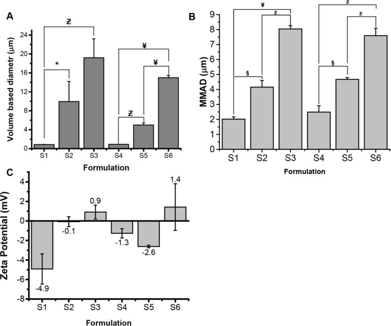
Size (A), mass median aerodynamic diameters (B) and zeta potentials (C) of PLGA particles of sildenafil (n=3). For compositions of the particles, please see Table 1. *p<0.05, §p<0.005, Ƶp<0.001, ¥p<0.0001.
MMAD is the major determinant for the extent and pattern of particle deposition in the lungs. Inhaled particles with MMADs between 1 and 5 μm are considered optimal for efficient deposition into the small airways and the alveolar region [33]. More recently, particles with an MMAD of 1–10 μm are also considered respirable [34]. Just as in the case for particle size distribution, an increase in the amounts of PEI in the IAP also increased the MMADs of the particles. Formulation S1 and S4, wherein there was no PEI in the IAP, demonstrated an MMAD of 2.02±0.16 μm and 2.49±0.42 μm, respectively. When 0.5% PEI was used in the IAP, MMAD just doubled compared with the formulations wherein no PEI was used. With a further increase in concentration in IAP, we observed an additional increase in the MMAD of formulation S3. The MMAD of all the formulations were between 1 μm and 9 μm, a range appropriate for deposition in the peripheral lungs upon inhalation (Fig. 2B).
The zeta potentials of particles also changed from negative to positive values depending on the type of polymer and concentration of PEI used (Fig. 2C). The reversal in charge sign and magnitude may have resulted from the interaction of cationic PEI with anionic PLGA polymers. Positively charged particles may increase the residence time in the respiratory mucus, which is composed of negatively charged mucins [36]. Overall, the amount of PEI in the IAP can greatly influence the surface texture, physical and aerodynamic properties, and zeta potential of the formulations. Further, the MMADs of porous particles were within the acceptable range for inhaled particles.
3.3 Entrapment efficiencies and in-vitro release profiles of sildenafil
PEI greatly influenced the drug entrapment efficiencies of the formulations: In the absence of PEI, the drug entrapment was rather low, but when PEI concentration was increased to 1.25%, the drug entrapment increased to as high as 90% (Fig. 3A). As we have discussed previously [21, 23], PEI, a polycation, may have increased the drug entrapment efficiency by increasing the stability of IAP [37] and by interacting with negatively charged sildenafil [38].
Figure 3.

Drug entrapment efficiency of sildenafil particles prepared with different PLGA co-polymers and varying concentration PEI used in the internal aqueous phase (A). Cumulative release of sildenafil from particles prepared with PLGA 50:50 (B); and PLGA 75:25 (C) (n=3) *p<0.05, §p<0.005, Ƶp<0.001, ¥p<0.0001.
Like previously reported PEI containing PLGA particles [23, 39], these particles showed a very rapid rise in the amount of drug released during the first 30 minutes, called burst release, and then a slower release for a long time (Figs. 3B and 3C). Like the impact of PEI on the physical properties of the particles, PEI had a profound influence on the release of sildenafil from PLGA particles. Particles containing PEI produced a greater cumulative release of the drug for 36 hours, but particles with no PEI released much less drug from the particles. Inclusion of PEI in the IAP produced statistically significant increases in the cumulative release of the drug for 36 hours in case of particles that are prepared with PLGA 50/50, but particles with no PEI released much less drug from the particles (Fig. 3B). A similar trend was also observed in case of the particles prepared with PLGA 75/25 although the changes were not statistically significant (Fig. 3C). As shown in Fig. 1, PEI containing particles had large surface pores, which might have eased the entry of simulated lung fluids into the particle core and thus let the particle swell and release the drug via surface erosion or diffusion of the drug from particle core. On the other hand, the absence of pores may have restricted the release of drug from the particles with no PEI [40]. The initial burst release (amount of drug released at first 30 minutes) for both non-porous and porous particles ranged between ~10% and~20%. Overall, in-vitro release data suggest that porous particles will be a better choice for efficient deposition and controlled release of sildenafil after aerosolization into the lungs for the treatment of PAH.
3.4 Uptake of the particles by rat alveolar macrophages
Inhaled particles are cleared by two major mechanisms: (i) mucociliary escalator consisting of mucus-producing goblet cells and ciliated epithelium; and (ii) the clearance by alveolar macrophages [41]. Because particles deposited in the alveolar region are quickly engulfed and eliminated by alveolar macrophages [41], we engineered particles to avoid uptakes by alveolar macrophages and thus prepared particles that are too large to be taken up by macrophages. Published studies also documented that particles with a physical diameter (Dv) of 1–5 μm are easily recognized and engulfed by macrophages [34]. Particles >10 μm can avoid uptake or are engulfed by macrophages to a lesser extent [42, 43]. Further, we assessed the influence of the particles on the uptake by alveolar macrophages, instead of their influence on the mucociliary clearance, because the latter process is less likely to play any roles in clearing particles that are deposited in deep lung or alveolar region, where mucociliary clearance mechanism has little to do [44, 45].
Further, we choose to use alveolar macrophages in uptake studies instead of respiratory epithelium or pulmonary arterial smooth muscle cells (PASMCS) because the latter two cells have no roles in clearance or absorption of the drug. The rate of drug release from PLGA matrix controls drug absorption via the air-blood barrier in the lung. PLGA particles release drugs by surface erosion of the polymeric matrix, the released drug then crosses [46] the air-blood barrier to enter the pulmonary vasculature. Free drug concentration in the epithelial lining fluid determines the extent of drug transport across the air-blood barrier.
We have used rat alveolar macrophages, NR8383, because these cells have strong phagocytic properties like other primary alveolar macrophages [47, 48]. Using both quantitative and qualitative methodologies, we have attempted to assess whether the particles can avoid uptake by macrophages (Fig. 4). Consistent with our assumption and previous studies, the extent of uptake of larger particles (Fig. 4A: S2, S3, S5, and S6) by NR8383 cells much smaller than the uptake of smaller particles (S1 and S4). Like the microscopic data, the flow cytometric analysis also indicates that large particles can avoid uptake by alveolar macrophages. Upon incubation of the particles with rat NR8383 cells, the mean fluorescent intensities for larger particles (S1, S2, S5 and S6) were significantly lesser than that for smaller particles (S1 and S4), suggesting that the uptake of smaller particles (S1 and S4) was greater than the uptake of larger particles (Fig. 4B). Both microscopic and flow cytometric data strongly point to the fact that particles ≥5 μm (S2, S3, S5 and S6) are less likely to be cleared by the alveolar macrophages and these particles can be used to control the release of sildenafil for an extended period, as we and others have shown previously [21, 23].
Figure 4.

Uptake of the different formulations by the rat alveolar macrophages: qualitative analysis with fluorescent microscopy (A), quantitative analysis using flow cytometer (B) (n=3) *p<0.05.
3.5 Safety studies
To assess the safety of the formulations, we conducted a cytotoxicity study and measured the injury markers in BAL fluid collected from rats treated with the formulations. We performed the cytotoxicity studies using sulforhodamine B and rat PASMCs. We selected this cell line for safety studies because PASMCs are the target cells for the vasodilatory effect of sildenafil. As discussed below under therapeutic efficacy study, after being absorbed via the pulmonary vascular capillary beds, anti-PAH drugs dilate pulmonary vasculature by acting on PASMCs and decreases pulmonary vascular resistance [49, 50].
The cytotoxicity data suggest that only a small percentage of cells survived, when they were treated with sodium dodecyl sulfate (SDS), the positive control (Fig. 5A), but most of the cells were alive (95%), when treated with the drug, saline or any of the formulations used at a concentration of 5 mg/mL particles (Fig. 5A). Thus, neither polymer nor PEI was cytotoxic, as reported previously by us and others [21, 23, 51, 52]. However, cytotoxicity data generated upon treating the cells for 24 hours may not be translated into the adverse effect that may develop due to long-term application of an inhaled formulation.
Figure 5.

The safety of the sildenafil particulate formulations: cytotoxicity when treated with rat pulmonary arterial smooth muscle cells (A); the enzymatic markers in the bronchoalveolar lavage fluid after intra-tracheal administration (B); release of inflammatory cytokines IL6(C) and TNF-alpha (D) 8 hours after intra-tracheal administration (n=3) *p<0.05, §p<0.005, Ƶp<0.001.
We have measured injury markers in BAL fluid collected from rats treated with formulations S2 and S6, which represents both the PLGA polymers used in formulations and contain different amount of PEIs as the porosigen in the IAP. PEI is expected to damage the lung because of high density of cations in PEI. Like the cytotoxicity data presented in Fig 5A, SDS increased the levels of LDH, ALP, and total protein concentrations in BAL fluid. The levels of the enzymes in two formulations treated rats were the same as that in saline treated rats, but were significantly different from SDS treated rats (Fig. 5B). Indeed, ALP and LDH levels in SDS treated animals were ~3-fold greater than in saline or formulation treated animals. Both cytotoxicity data and injury levels in the BAL fluid indicate that particles are safe for inhalational delivery. However, because in both cases treatments were for very short-term, one should be careful in claiming the safety of the formulation.
Particles that reside in the alveolar region and escape macrophage uptake may lead to a phenomenon called ‘frustrated phagocytosis’ which is known to cause subacute inflammation [53, 54]. Frustrated phagocytic phenomenon generates reactive oxygen species (ROS) and thus increases the levels of inflammatory cytokines [55]. To rule out the frustrated macrophage phenomenon, we determined IL6 and TNF-α in the BAL fluid, collected from rat lungs treated with saline and particles. The levels of IL6 and TNF-α in the BAL fluid of saline treated rats were no different from that in particle treated rats (Fig. 5C). However, the cytokine levels in BAL fluid in LPS treated group remarkably increased. No rise in cytokine levels suggest that the particles did not induce subacute inflammation despite their long residence time in the lung.
3.5 Pharmacokinetic studies
In both pharmacokinetic and efficacy studies, we administered the drug formulation into the rats by intratracheal aerosolization, instead of nose or head-only exposure based inhalation, the method that arguably mimic the physiological situation. However, the latter methods cannot accurately administer a small dose to rats. Although whole body, nose-only, and head-only exposure chambers allow administration of drugs directly to conscious animals, drug wastage, absorption of drugs via oral and percutaneous routes, and extensive deposition in the nasal cavity are major limitations of inhalation administration to live animals [56]. On the other hand, intratracheal instillation poorly mimics oral inhalation, induces injury upon multiple administrations, and requires animals to be anesthetized. But intratracheal instillation can accurately administer a small of amount of particles directly into the lungs. Thus, considering the limitations of both methods, we believe intratracheal aerosolization based instillation will generate valid information concerning the preclinical feasibility of the proposed formulation.
We chose formulation S5 (PLGA 75:25; 0.5% PEI in IAP) for pulmonary absorption studies, because this formulation showed an optimal MMAD (4.68±0.12 μm), high drug loading (89.28±4.44%), and sustained release properties. The absorption profile of the chosen formulation was compared with those of plain sildenafil administered orally, intratracheally (IT) and intravenously (IV). The plasma concentration of the drug rose rapidly when sildenafil was administered, at a dose of 0.5 mg/kg, intravenously and intratracheally. But the drug plasma concentration declined rapidly with an elimination half-life of ≅ 3 hours, suggesting that the absorption pattern of IT sildenafil is the same as that of IV sildenafil. Assuming that the plasma concentration represents the therapeutic efficacy of the drug, we expect that IV and IT sildenafil would be equally effective in ameliorating mPAP.
Plain sildenafil, administered orally at the same dose, did not show any detectable concentrations in the blood (Fig. 6). This is consistent with the poor availability of oral sildenafil (41% in human) due to metabolism in the gut and liver at first pass [57]. A comparison between the absorption profiles of oral and IT sildenafil gives two important pieces of information: (i) when administered at the same dose, IT sildenafil has more bioavailability than oral sildenafil has, and (ii) the dose of the drug can be substantially reduced by aerosolization of the drug into the lung. The net outcome would be reduced exposure of the drug in the systemic circulation and reduced side effects.
Figure 6.

Changes in plasma concentration of sildenafil after pulmonary administration of 0.5 mg/kg sildenafil in the following forms: (i) plain intravenous, (ii) plain intra-tracheal, (iii) plain oral and (i) PLGA particles of sildenafil intra-tracheal (n=3–4). Analysis was performed using two-way repeated measure ANOVA with point-to-point multiple comparisons. Statistical significance was presented when plain intratracheal sildenafil are compared with intratracheal PLGA particles; §p<0.005, Ƶp<0.001, ¥p<0.0001.
The absorption kinetics of PLGA particles of sildenafil, administered intratracheally at an equivalent dose of 0.5 mg/kg sildenafil, was dramatically different from those of the plain drug administered via three routes–oral, IT and IV. Like that of the in-vitro drug release patterns, the drug continues to be detectable in rat plasma for >24 hours. The elimination half-life of sildenafil from PLGA particles was 5.8-times longer than the elimination half-life of plain sildenafil, given IT (Fig. 6). The extended elimination half-life was the result of absorption-controlled elimination of the drug, the so-called flip-flop phenomena associated with some drugs and formulations [58].
However, the extent of drug absorption from PLGA particles of sildenafil was as high as that of intravenous sildenafil. Unlike plasma concentration for intravenous sildenafil, that of sildenafil loaded particle showed a high variation. In fact, the variation in absorption profile of inhaled drug formulation is a well-known limitation of the lung as a route of administration. Such variability occurs because, unlike intravenously administered drugs, inhaled drug enters the systemic circulation via an absorption phase. Other reasons for variation and high absorption include total dose administered, the absence of effective mucociliary clearance mechanism in the deep lung (whereby much of the particles deposit after intratracheal instillation), and particles’ ability to avoid macrophage uptake. Although the AUC for sildenafil was similar to that of intravenous sildenafil, there was no sharp rise in plasma concentration of the drug after intratracheal administration. The absence of sharp rise in plasma concentration but the presence of a steady plasma concentration is likely to eliminate rapid fall in systemic blood pressure associated with intravenous sildenafil. Together, the absorption kinetics of sildenafil particles is encouraging based on the assumption that sildenafil formulated in PLGA particles can possibly be administered at a reduced dose and once a day, instead of three times a day as required for oral sildenafil.
3.7 Therapeutic efficacy of the formulations
Similar to the pharmacokinetics study, we assessed the effects of oral, IT and IV plain sildenafil, and formulation S5, administered at a dose of 0.5 mg/kg drug or particles containing an equivalent amount of the drug, on mPAP and mSAP in SUGEN/hypoxia induced PAH rats. Normal saline (0.9% NaCl) was used as an additional control. As expected, saline control did not affect the pulmonary hemodynamics (Fig. 7A) in PAH rats. IV sildenafil reduced mPAP by 15%. The vasodilatory effect of IV sildenafil persisted for 120 minutes before returning to its baseline and IV sildenafil had no effect on mSAP (Fig. 7B). Like IV sildenafil, IT sildenafil also reduced mPAP but to a greater extent, a 30% reduction compared with the initial mPAP. The vasodilatory duration, in this case, was 60 minutes (Fig. 7C). But mSAP, in IT sildenafil treated animals, declined to 55% of the initial value. This systemic vasodilatory effect of IT sildenafil persisted for ≅30 minutes. IT sildenafil reduced mSAP because the drug underwent systemic absorption from the lungs and subsequently caused peripheral vasodilation. Oral sildenafil did not have any effect on mPAP or mSAP (Fig. 7D), which echoes the data presented in Fig. 6, when little or no drug was absorbed after oral administration. The lack of effect from oral sildenafil also corroborates the fact that sildenafil undergoes first-pass metabolism and have a bioavailability of 23% in rats [59].
Consistent with the extended elimination half-life, observed in pharmacokinetics studies, the effect of sildenafil loaded PLGA particles (S5) on the pulmonary hemodynamics was rather dramatic. Sildenafil PLGA particles, when administered intratracheally, reduced mPAP to 21–42% of the initial levels and maintained that level of mPAP reduction for ≅ 6 hrs (Fig. 7E). In contrast, the formulation reduced mSAP only by 15%, and mSAP returned to the initial value within an hour. Modest and short-term reduction of mSAP but pronounced and long-term reduction of mPAP suggests delivery of the drug to the systemic circulation with increased localized concentration of the drug in the lung. In terms of time and magnitude, the differences between mPAP and mSAP, determined by repeated-measure ANOVA, were statistically significant (Fig. 7E). Similarly sildenafil particles produced statistically significant reduction in mPAP when compared with other forms of sildenafil (Fig. 7F). The particles acted as a depot and released the drug slowly but continuously, which likely accounts for the prolonged pulmonary vasodilatory effect. After IV and IT administration of free sildenafil, we observed a sudden decrease in mSAP that returned to normal values at 30 minutes (Fig. 7G). However, oral sildenafil or IT particle treated groups did not show any reduction in mSAP probably because of slow absorption process that failed to increase the plasma concentration to a level to affect mSAP. This initial decrease in mSAP in IT or IV sildenafil treated groups is the result of sharp rise in plasma concentration of sildenafil, observed in absorption studies (Fig. 6). We believe that formulation S5 elicited a strong, selective, and sustained reduction in mPAP by slowly and continuously releasing the drug in the pulmonary vascular bed, the major site of action for PAH. Importantly, although mSAP profiles for various routes showed a very similar pattern, mSAP and mPAP for PLGA particles were significantly different, suggesting that the proposed formulation may eliminate off-target effects caused by oral sildenafil, which is the goal of ideal anti-PAH therapy: reduce the mPAP but not mSAP.
The mechanism by which inhaled anti-PAH drugs reduce mPAP is poorly understood. A drug may absorb systemically, reach the pulmonary circulation, and cause vasodilation. The pulmonary absorption kinetics of Ventavis® an inhaled form of anti-PAH drug, iloprost, has not yet been studied. Inhaled treprostinil (Tyvaso®) has an absolute bioavailability of 64% after inhalation. After absorption via the pulmonary vascular capillary beds, these drugs are believed to dilate pulmonary vasculature and decrease pulmonary vascular resistance [49, 50]. Regardless of the mechanism or route that takes the drug into the pulmonary vasculature, our finding is important from the patient compliance perspective. Using a smaller dose and administering at a longer dosing interval, we can eliminate multiple dosing a day required for oral sildenafil, reduce systemic exposure, and minimize peripheral vasodilation.
4. Conclusions
In this study, we reported the effects of a PLGA-based formulation of sildenafil, a frequently prescribed anti-PAH drug, on selectively reducing mPAP in PAH rats. Particles of sildenafil, prepared in this study, had favorable aerodynamic behavior properties, underwent slower macrophage uptake, released the drug for an extended period and prolonged the elimination half-life of the drug. Sildenafil-loaded inhaled PLGA particles lowered mPAP for 6 hours with minimum effects on mSAP. Thus, preparing and characterizing PLGA particles of sildenafil, monitoring pharmacokinetics in healthy rats, assessing the pharmacological efficacy in PAH rats, we have demonstrated that an inhaled long-acting formulation of sildenafil can potentially be developed as a viable alternative to oral tablets of sildenafil–the current dosage form, which prompts patients to noncompliance because of multiple dosing a day. Because both the drug and polymeric carrier (PLGA) are already FDA approved for human use, this alternative delivery system for sildenafil would be cost-effective to rapidly test and develop as a viable anti-PAH formulation.
Table 1.
Compositions of different formulations of PLGA-based particles of sildenafil.
| Formulation | Polymer | IAP | EAP |
|---|---|---|---|
| S1 | PLGA 50/50 | Water | 0.5% PVA |
| S2 | PLGA 50/50 | 0.5% PEI | 0.5% PVA |
| S3 | PLGA 50/50 | 1.25% PEI | 0.5% PVA |
| S4 | PLGA 75/25 | Water | 0.5% PVA |
| S5 | PLGA 75/25 | 0.5% PEI | 0.5% PVA |
| S6 | PLGA 75/25 | 1.25% PEI | 0.5% PVA |
Footnotes
Publisher's Disclaimer: This is a PDF file of an unedited manuscript that has been accepted for publication. As a service to our customers we are providing this early version of the manuscript. The manuscript will undergo copyediting, typesetting, and review of the resulting proof before it is published in its final citable form. Please note that during the production process errors may be discovered which could affect the content, and all legal disclaimers that apply to the journal pertain.
References
- 1.Ignarro LJ, Bush PA, Buga GM, Wood KS, Fukuto JM, Rajfer J. Nitric oxide and cyclic GMP formation upon electrical field stimulation cause relaxation of corpus cavernosum smooth muscle. Biochem Biophys Res Commun. 1990;170:843–850. doi: 10.1016/0006-291x(90)92168-y. [DOI] [PubMed] [Google Scholar]
- 2.Rajfer J, Aronson WJ, Bush PA, Dorey FJ, Ignarro LJ. Nitric oxide as a mediator of relaxation of the corpus cavernosum in response to nonadrenergic, noncholinergic neurotransmission. N Engl J Med. 1992;326:90–94. doi: 10.1056/NEJM199201093260203. [DOI] [PubMed] [Google Scholar]
- 3.Bush PA, Aronson WJ, Buga GM, Rajfer J, Ignarro LJ. Nitric oxide is a potent relaxant of human and rabbit corpus cavernosum. J Urol. 1992;147:1650–1655. doi: 10.1016/s0022-5347(17)37671-1. [DOI] [PubMed] [Google Scholar]
- 4.McCullough AR. Four-year review of sildenafil citrate. Rev Urol. 2002;4(Suppl 3):S26–38. [PMC free article] [PubMed] [Google Scholar]
- 5.Kass DA, Takimoto E, Nagayama T, Champion HC. Phosphodiesterase regulation of nitric oxide signaling. Cardiovasc Res. 2007;75:303–314. doi: 10.1016/j.cardiores.2007.02.031. [DOI] [PubMed] [Google Scholar]
- 6.Lin CS, Lin G, Xin ZC, Lue TF. Expression, distribution and regulation of phosphodiesterase 5. Curr Pharm Des. 2006;12:3439–3457. doi: 10.2174/138161206778343064. [DOI] [PubMed] [Google Scholar]
- 7.Hemnes AR, Champion HC. Sildenafil, a PDE5 inhibitor, in the treatment of pulmonary hypertension. Expert Rev Cardiovasc Ther. 2006;4:293–300. doi: 10.1586/14779072.4.3.293. [DOI] [PubMed] [Google Scholar]
- 8.Humbert M, Lau EM, Montani D, Jais X, Sitbon O, Simonneau G. Advances in therapeutic interventions for patients with pulmonary arterial hypertension. Circulation. 2014;130:2189–2208. doi: 10.1161/CIRCULATIONAHA.114.006974. [DOI] [PubMed] [Google Scholar]
- 9.Humbert M, Oleg VE, Stasch J. Pharmacotherapy of pulmonary hypertension. Springer; New York: 2013. [Google Scholar]
- 10.Galie N, Ghofrani HA, Torbicki A, Barst RJ, Rubin LJ, Badesch D, Fleming T, Parpia T, Burgess G, Branzi A, Grimminger F, Kurzyna M, Simonneau G, Sildenafil G. Use in Pulmonary Arterial Hypertension Study, Sildenafil citrate therapy for pulmonary arterial hypertension. N Engl J Med. 2005;353:2148–2157. doi: 10.1056/NEJMoa050010. [DOI] [PubMed] [Google Scholar]
- 11.Taichman DB, Ornelas J, Chung L, Klinger JR, Lewis S, Mandel J, Palevsky HI, Rich S, Sood N, Rosenzweig EB, Trow TK, Yung R, Elliott CG, Badesch DB. Pharmacologic therapy for pulmonary arterial hypertension in adults: CHEST guideline and expert panel report. Chest. 2014;146:449–475. doi: 10.1378/chest.14-0793. [DOI] [PMC free article] [PubMed] [Google Scholar]
- 12.Beghetti M, Wacker Bou Puigdefabregas J, Merali S. Sildenafil for the treatment of pulmonary hypertension in children. Expert Rev Cardiovasc Ther. 2014;12:1157–1184. doi: 10.1586/14779072.2014.958077. [DOI] [PubMed] [Google Scholar]
- 13.Montani D, Gunther S, Dorfmuller P, Perros F, Girerd B, Garcia G, Jais X, Savale L, Artaud-Macari E, Price LC, Humbert M, Simonneau G, Sitbon O. Pulmonary arterial hypertension. Orphanet J Rare Dis. 2013;8:97. doi: 10.1186/1750-1172-8-97. [DOI] [PMC free article] [PubMed] [Google Scholar]
- 14.Samiee-Zafarghandy S, Smith PB, van den Anker JN. Safety of sildenafil in infants*. Pediatr Crit Care Med. 2014;15:362–368. doi: 10.1097/PCC.0000000000000077. [DOI] [PMC free article] [PubMed] [Google Scholar]
- 15.U.F.a.D. Administration. Revatio prescribing information. Maryland: 2015. [Google Scholar]
- 16.USFDA. FDA Drug Safety Communication: FDA recommends against use of Revatio (sildenafil) in children with pulmonary hypertension. USFDA, USFDA Maryland; 2012. [Google Scholar]
- 17.Dhariwal AK, Bavdekar SB. Sildenafil in pediatric pulmonary arterial hypertension. J Postgrad Med. 2015;61:181–192. doi: 10.4103/0022-3859.159421. [DOI] [PMC free article] [PubMed] [Google Scholar]
- 18.Ghasemian E, Vatanara A, Najafabadi AR, Rouini MR, Gilani K, Darabi M. Preparation, characterization and optimization of sildenafil citrate loaded PLGA nanoparticles by statistical factorial design. Daru. 2013;21:1. doi: 10.1186/2008-2231-21-68. [DOI] [PMC free article] [PubMed] [Google Scholar]
- 19.Beck-Broichsitter M, Bohr A, Aragao-Santiago L, Klingl A, Kissel T. Formulation and process considerations for the design of sildenafil-loaded polymeric microparticles by vibrational spray-drying. Pharm Dev Technol. 2016:1–8. doi: 10.3109/10837450.2015.1098661. [DOI] [PubMed] [Google Scholar]
- 20.Ichinose F, Erana-Garcia J, Hromi J, Raveh Y, Jones R, Krim L, Clark MW, Winkler JD, Bloch KD, Zapol WM. Nebulized sildenafil is a selective pulmonary vasodilator in lambs with acute pulmonary hypertension. Crit Care Med. 2001;29:1000–1005. doi: 10.1097/00003246-200105000-00024. [DOI] [PubMed] [Google Scholar]
- 21.Rawat A, Majumder QH, Ahsan F. Inhalable large porous microspheres of low molecular weight heparin: in vitro and in vivo evaluation. J Control Release. 2008;128:224–232. doi: 10.1016/j.jconrel.2008.03.013. [DOI] [PMC free article] [PubMed] [Google Scholar]
- 22.Moss OR. Simulants of lung interstitial fluid. Health physics. 1979;36:447–448. [PubMed] [Google Scholar]
- 23.Patel B, Gupta V, Ahsan F. PEG-PLGA based large porous particles for pulmonary delivery of a highly soluble drug, low molecular weight heparin. J Control Release. 2012;162:310–320. doi: 10.1016/j.jconrel.2012.07.003. [DOI] [PubMed] [Google Scholar]
- 24.Nahar K, Absar S, Patel B, Ahsan F. Starch-coated magnetic liposomes as an inhalable carrier for accumulation of fasudil in the pulmonary vasculature. Int J Pharm. 2014;464:185–195. doi: 10.1016/j.ijpharm.2014.01.007. [DOI] [PMC free article] [PubMed] [Google Scholar]
- 25.Vichai V, Kirtikara K. Sulforhodamine B colorimetric assay for cytotoxicity screening. Nature protocols. 2006;1:1112–1116. doi: 10.1038/nprot.2006.179. [DOI] [PubMed] [Google Scholar]
- 26.Gupta V, Rawat A, Ahsan F. Feasibility study of aerosolized prostaglandin E1 microspheres as a noninvasive therapy for pulmonary arterial hypertension. J Pharm Sci. 2010;99:1774–1789. doi: 10.1002/jps.21946. [DOI] [PubMed] [Google Scholar]
- 27.Taraseviciene-Stewart L, Kasahara Y, Alger L, Hirth P, Mahon G Mc, Waltenberger J, Voelkel NF, Tuder RM. Inhibition of the VEGF receptor 2 combined with chronic hypoxia causes cell death-dependent pulmonary endothelial cell proliferation and severe pulmonary hypertension. FASEB J. 2001;15:427–438. doi: 10.1096/fj.00-0343com. [DOI] [PubMed] [Google Scholar]
- 28.Oka M, Homma N, Taraseviciene-Stewart L, Morris KG, Kraskauskas D, Burns N, Voelkel NF, McMurtry IF. Rho kinase–mediated vasoconstriction is important in severe occlusive pulmonary arterial hypertension in rats. Circ Res. 2007;100:923–929. doi: 10.1161/01.RES.0000261658.12024.18. [DOI] [PubMed] [Google Scholar]
- 29.Gupta V, Gupta N, Shaik IH, Mehvar R, McMurtry IF, Oka M, Nozik-Grayck E, Komatsu M, Ahsan F. Liposomal fasudil, a rho-kinase inhibitor, for prolonged pulmonary preferential vasodilation in pulmonary arterial hypertension. J Control Release. 2013;167:189–199. doi: 10.1016/j.jconrel.2013.01.011. [DOI] [PMC free article] [PubMed] [Google Scholar]
- 30.Dimitrov I, Trzebicka B, Müller AH, Dworak A, Tsvetanov CB. Thermosensitive water-soluble copolymers with doubly responsive reversibly interacting entities. Prog Polym Sci. 2007;32:1275–1343. [Google Scholar]
- 31.Crotts G, Park TG. Preparation of porous and nonporous biodegradable polymeric hollow microspheres. J Control Release. 1995;35:91–105. [Google Scholar]
- 32.Ravivarapu HB, Lee H, DeLuca PP. Enhancing initial release of peptide from poly(d,l-lactide-co-glycolide) (PLGA) microspheres by addition of a porosigen and increasing drug load. Pharm Dev Technol. 2000;5:287–296. doi: 10.1081/pdt-100100543. [DOI] [PubMed] [Google Scholar]
- 33.Labiris NR, Dolovich MB. Pulmonary drug delivery. Part I: physiological factors affecting therapeutic effectiveness of aerosolized medications. Br J Clin Pharmacol. 2003;56:588–599. doi: 10.1046/j.1365-2125.2003.01892.x. [DOI] [PMC free article] [PubMed] [Google Scholar]
- 34.Edwards DA, Hanes J, Caponetti G, Hrkach J, Ben-Jebria A, Eskew ML, Mintzes J, Deaver D, Lotan N, Langer R. Large porous particles for pulmonary drug delivery. Science. 1997;276:1868–1871. doi: 10.1126/science.276.5320.1868. [DOI] [PubMed] [Google Scholar]
- 35.Vanbever R, Mintzes JD, Wang J, Nice J, Chen D, Batycky R, Langer R, Edwards DA. Formulation and physical characterization of large porous particles for inhalation. Pharm Res. 1999;16:1735–1742. doi: 10.1023/a:1018910200420. [DOI] [PubMed] [Google Scholar]
- 36.Lai SK, Wang YY, Hanes J. Mucus-penetrating nanoparticles for drug and gene delivery to mucosal tissues. Adv Drug Deliv Rev. 2009;61:158–171. doi: 10.1016/j.addr.2008.11.002. [DOI] [PMC free article] [PubMed] [Google Scholar]
- 37.Nihant N, Schugens C, Grandfils C, Jerome R, Teyssie P. Polylactide microparticles prepared by double emulsion/evaporation technique. I. Effect of primary emulsion stability. Pharm Res. 1994;11:1479–1484. doi: 10.1023/a:1018912426983. [DOI] [PubMed] [Google Scholar]
- 38.Hsiang HI, Chang YL, Fang JS, Yen FS. Polyethyleneimine surfactant effect on the formation of nano-sized BaTiO 3 powder via a solid state reaction. J Alloys Compd. 2011;509:7632–7638. [Google Scholar]
- 39.Li YP, Pei YY, Zhang XY, Gu ZH, Zhou ZH, Yuan WF, Zhou JJ, Zhu JH, Gao XJ. PEGylated PLGA nanoparticles as protein carriers: synthesis, preparation and biodistribution in rats. J Control Release. 2001;71:203–211. doi: 10.1016/s0168-3659(01)00218-8. [DOI] [PubMed] [Google Scholar]
- 40.Fredenberg S, Wahlgren M, Reslow M, Axelsson A. The mechanisms of drug release in poly (lactic-co-glycolic acid)-based drug delivery systems—a review. Int J Pharm. 2011;415:34–52. doi: 10.1016/j.ijpharm.2011.05.049. [DOI] [PubMed] [Google Scholar]
- 41.Patel B, Gupta N, Ahsan F. Particle engineering to enhance or lessen particle uptake by alveolar macrophages and to influence the therapeutic outcome. Eur J Pharm Biopharm. 2015;89:163–174. doi: 10.1016/j.ejpb.2014.12.001. [DOI] [PubMed] [Google Scholar]
- 42.Edwards DA, Ben-Jebria A, Langer R. Recent advances in pulmonary drug delivery using large, porous inhaled particles. J Appl Physiol. 1998;85:379–385. doi: 10.1152/jappl.1998.85.2.379. [DOI] [PubMed] [Google Scholar]
- 43.Thomas C, Gupta V, Ahsan F. Particle size influences the immune response produced by hepatitis B vaccine formulated in inhalable particles. Pharm Res. 2010;27:905–919. doi: 10.1007/s11095-010-0094-x. [DOI] [PubMed] [Google Scholar]
- 44.Gonda I. Aerosols for delivery of therapeutic and diagnostic agents to the respiratory tract. Crit Rev Ther Drug Carrier Syst. 1990;6:273–313. [PubMed] [Google Scholar]
- 45.Newman SP, Agnew JE, Pavia D, Clarke SW. Inhaled aerosols: lung deposition and clinical applications. Clin Phys Physiol Meas. 1982;3:1–20. doi: 10.1088/0143-0815/3/1/001. [DOI] [PubMed] [Google Scholar]
- 46.Kamaly N, Yameen B, Wu J, Farokhzad OC. Degradable controlled-release polymers and polymeric nanoparticles: Mechanisms of controlling drug release. Chem Rev. 2016;116:2602–2663. doi: 10.1021/acs.chemrev.5b00346. [DOI] [PMC free article] [PubMed] [Google Scholar]
- 47.Hino M, Oda M, Yoshida A, Nakata K, Kohchi C, Nishizawa T, Inagawa H, Hori H, Makino K, Terada H, Soma G. Establishment of an in vitro model using NR8383 cells and Mycobacterium bovis Calmette-Guerin that mimics a chronic infection of Mycobacterium tuberculosis. In Vivo. 2005;19:821–830. [PubMed] [Google Scholar]
- 48.Chono S, Tanino T, Seki T, Morimoto K. Uptake characteristics of liposomes by rat alveolar macrophages: influence of particle size and surface mannose modification. J Pharm Pharmacol. 2007;59:75–80. doi: 10.1211/jpp.59.1.0010. [DOI] [PubMed] [Google Scholar]
- 49.United States Food and Drug Administration. Tyvaso prescribing information. 2009 http://www.accessdata.fda.gov/drugsatfda_docs/label/2009/022387LBL.pdf. accessed 03.11.2016.
- 50.United States Food and Drug Administration. Ventavis prescribing information. 2013 http://www.accessdata.fda.gov/drugsatfda_docs/label/2013/021779s014lbl.pdf. accessed 03.11.2016.
- 51.Bivas-Benita M, Romeijn S, Junginger HE, Borchard G. PLGA-PEI nanoparticles for gene delivery to pulmonary epithelium. Eur J Pharm Biopharm. 2004;58:1–6. doi: 10.1016/j.ejpb.2004.03.008. [DOI] [PMC free article] [PubMed] [Google Scholar]
- 52.Gupta V, Ahsan F. Influence of PEI as a core modifying agent on PLGA microspheres of PGE(1), a pulmonary selective vasodilator. Int J Pharm. 2011;413:51–62. doi: 10.1016/j.ijpharm.2011.04.017. [DOI] [PMC free article] [PubMed] [Google Scholar]
- 53.Brown DM, Kinloch IA, Bangert U, Windle A, Walter D, Walker G, Scotchford C, Donaldson K, Stone V. An in vitro study of the potential of carbon nanotubes and nanofibres to induce inflammatory mediators and frustrated phagocytosis. Carbon. 2007;45:1743–1756. [Google Scholar]
- 54.Labrousse AM, Meunier E, Record J, Labernadie A, Beduer A, Vieu C, Ben Safta T, Maridonneau-Parini I. Frustrated phagocytosis on micro-patterned immune complexes to characterize lysosome movements in live macrophages. Front Immunol. 2011;2:51. doi: 10.3389/fimmu.2011.00051. [DOI] [PMC free article] [PubMed] [Google Scholar]
- 55.Vallyathan V, Mega JF, Shi X, Dalal NS. Enhanced generation of free radicals from phagocytes induced by mineral dusts. Am J Respir Cell Mol Biol. 1992;6:404–413. doi: 10.1165/ajrcmb/6.4.404. [DOI] [PubMed] [Google Scholar]
- 56.Nahar K, Gupta N, Gauvin R, Absar S, Patel B, Gupta V, Khademhosseini A, Ahsan F. In vitro, in vivo and ex vivo models for studying particle deposition and drug absorption of inhaled pharmaceuticals. Eur J Pharm Sci. 2013;49:805–818. doi: 10.1016/j.ejps.2013.06.004. [DOI] [PubMed] [Google Scholar]
- 57.Nichols DJ, Muirhead GJ, Harness JA. Pharmacokinetics of sildenafil after single oral doses in healthy male subjects: absolute bioavailability, food effects and dose proportionality. Br J Clin Pharmacol. 2002;53:5S–12S. doi: 10.1046/j.0306-5251.2001.00027.x. [DOI] [PMC free article] [PubMed] [Google Scholar]
- 58.Duan JZ. Pharmacokinetics of Oral Absorption. In: Shargel L, Yu AC, editors. Applied Biopharmaceutics & Pharmacokinetics. 2016. [Google Scholar]
- 59.Nichols DJ, Muirhead GJ, Harness JA. Pharmacokinetics of sildenafil after single oral doses in healthy male subjects: absolute bioavailability, food effects and dose proportionality. Br J Clin Pharmacol. 2002;53(Suppl 1):5S–12S. doi: 10.1046/j.0306-5251.2001.00027.x. [DOI] [PMC free article] [PubMed] [Google Scholar]