Abstract
Objective:
To detail the presentation of calcium pyrophosphate deposition disease (CPPD) in the ankle joint. The aim of this case report is to inform health-care practitioners about the presentation of this condition in an uncommon location and discuss the diagnosis, potential treatment, and management strategies for a patient with CPPD.
Clinical Features:
A 36-year-old male patient presented to a chiropractic clinic with an acute, painful, and swollen ankle, which was later diagnosed by plain film radiograph as CPPD. A rheumatology follow-up was recommended and at-home treatment was prescribed to treat acute symptoms and monitor progress.
Outcome:
No chiropractic treatment was provided and the patient has been referred to a rheumatologist for further assessment. The diagnosis of CPPD was confirmed and he was advised to take an anti-inflammatory if symptoms recurred and booked for further follow-up in six months.
Summary:
Although the presentation is less common, CPPD can present in the ankle joint and mimic other inflammatory disorders. Conservative treatment can be applied to treat acute symptoms and referral to a rheumatologist is suggested to monitor progress of this condition.
Keywords: chiropractic, ankle, calcium pyrophosphate deposition disease
Keywords: chiropratique, cheville, chondrocalcinose articulaire
Abstract
Objectif :
Expliquer en détail la présentation de la chondrocalcinose articulaire (CCA) dans l’articulation de cheville. Cet exposé de cas a pour but d’informer les professionnels de la santé à propos de la présentation de ce trouble dans un endroit inhabituel et de discuter du diagnostic, du traitement potentiel et des stratégies de prise en charge pour un patient atteint de CCA.
Caractéristiques cliniques :
Un patient de 36 ans se présente à une clinique de chiropratique avec une cheville enflée avec douleur aiguë, trouble qu’on a plus tard diagnostiqué au moyen d’un cliché sans préparation comme étant une CCA. On a recommandé un suivi en rhumatologie et prescrit un traitement à domicile pour traiter les symptômes aigus et surveiller la progression.
Résultat :
On n’a pas fourni de traitement chiropratique et le patient a été envoyé à un rhumatologue pour une évaluation plus poussée. Le diagnostic de CCA a été confirmé; on lui a conseillé de prendre un anti-inflammatoire si les symptômes réapparaissaient et on a planifié un suivi six mois plus tard.
Résumé :
Bien que la présentation soit moins commune, la CCA peut se présenter dans l’articulation de cheville et imiter d’autres affections inflammatoires. Un traitement conservateur peut permettre de soigner les symptômes aigus et on recommande d’envoyer le patient voir un rhumatologue pour surveiller la progression du trouble.
Introduction
Calcium pyrophosphate deposition disease (CPPD) is a condition that is characterized by the deposit of pyrophosphate crystals into tendons, ligaments, cartilage and synovium.1,2 CPPD may be associated with elevated levels of calcium, pyrophosphate or local cartilage matrix changes.3 Typically, CPPD is found in older aged individuals, with an onset of 30 years of age and a peak at 60 years2, CPPD is often associated with primary and secondary osteoarthritis, which results in decreased joint congruency and degeneration due to the aging process1.
Both the cause of CPPD and the mechanism of onset of crystal deposition remain largely unknown and vastly debated throughout literature.1,2 The formation of calcium pyrophosphate crystals is extracellular, however, pyrophosphate is a by-product of several intracellular reactions thus unable to diffuse passively across the cell membrane.1 It is unclear whether crystal deposition occurs as a result of extracellular pyrophosphate synthesis by plasma membrane-bound plasma cell glycoprotein 1 or whether pyrophosphate is transported across cell membranes by ankylosis human protein.1 Causes of CPPD can be classified into the following categories: idiopathic, metabolic, hereditary and post-traumatic.1 CPPD can be linked to underlying metabolic disorders such as hemochromatosis, hyperparathyroidism, hypophosphataemia, hypomagnesaemia and hypothyroidism, all of which increase the risk for calcium pyrophosphate deposition.3 Numerous cases of CPPD have shown familial links in the ANKH gene, which functions to upregulate protein.4 When the ANKH gene becomes mutated, protein activity is enhanced and thus extracellular pyrophosphate levels increase and promote the formation of pyrophosphate crystals leading to CPPD.1,3,4 Despite attempts to determine etiology, the majority of CPPD cases remain idiopathic.3,4
This condition has not yet shown an association with gender, obesity, or lifestyle characteristics, although it is slightly more common in Caucasian individuals.1,2 CPPD has been documented most commonly in the knees, wrists, symphysis pubis and hips.1 This condition is a common rheumatologic disease in elderly individuals, and is noted most often in knees and pelvis.1 CPPD has been found to appear in one of three forms: asymptomatic, acute or chronic.1 Asymptomatic patients most often discover the deposit of pyrophosphate crystals through plain film radiographs for an alternative reason.1 Acute CPPD, a presentation that is highly suggestive of acute crystal inflammation, is associated with the rapid development of joint pain, swelling, tenderness, warmth and restricted movement, often reaching its maximum within 6–24 hours of symptom onset.1 Chronic CPPD presents similarly to osteoarthritis with progressive joint pain, chronic synovitis, crepitus and warmth at the joint line.2
Traditionally, patients presenting with suspected CPPD can be screened for chondrocalcinosis using plain-film radiographs, however, microscopic analysis of synovial fluid provides a more definitive and accurate diagnosis due to the ability to detect calcium pyrophosphate crystals.5 Chondrocalcinosis, calcification in hyaline or fibrocartilage, is considered the key identifying feature of CPPD on radiographs, however this diagnostic method is neither highly sensitive nor specific.5 Synovial fluid analysis can be used to identify weakly positively birefringent crystals using polarized light microscopy.5 More recently, ultrasonography of articular and fibrocartilage has been used to indicate the presence of CPPD crystals.6 Ultrasound is used to detect hyperechoic bands in hyaline cartilage and hyperechoic spots in fibrous cartilage.6 Further studies are required to compare the diagnostic value of ultrasound with existing diagnostic methods, however ultrasound remains a promising tool for diagnosis of CPPD.6
Strategies for treatment and management of CPPD vary depending on the symptom severity, stage and clinical manifestation of CPPD. Staging for CPPD presentation includes the following categories: asymptomatic, acute, chronic, presenting with OA or as a pseudoarthritide.1,5 Common treatments that provide symptomatic relief include conventional anti-inflammatory medications, such as non-steroidal anti-inflammatories (NSAIDs) and corticosteroids.5 Although the progression of this condition may vary between individuals, most will experience recurrent symptomatic episodes.7 During acute attacks, patients often experience severe pain.7 Optimal and safe treatment for acute pain includes ice, temporary rest, intra-articular injections and joint aspiration.7 Despite the incurable nature of this condition, the prognosis is good, as long as symptoms are controlled and patients are monitored for pre-disposing conditions that may be treatable and preventable.8
The objective of this case report is to highlight a unique incidence of CPPD in the ankle, a less common location for this condition with minimal documentation in current literature. Additionally, this case report aims to provide health-care practitioners with a detailed case presentation, radiographic findings, and potential treatment and management options for future patients with this case presentation.
Case Presentation
History and Physical Examination Findings
A 36-year-old male initially presented to the Canadian Memorial Chiropractic College (CMCC) campus clinic with a primary complaint of acute mechanical low back pain. He reported no comorbidities, medical history or medication use. At a subsequent visit, the patient reported a new complaint of an acutely swollen left ankle, with an onset of pain six days prior to his appointment. At this time, his back pain was resolved. There was no history of trauma to the ankle. Twenty-four hours after the onset of his ankle symptoms, he was woken up in the middle of the night by severe pain that resulted in the inability to fall back asleep. Over the course of the weekend, prior to his appointment, the ankle became swollen. The patient was self-medicating with ibuprofen, which helped to relieve but did not eliminate the pain. Following observation of the joint and orthopaedic testing of the ankle, all tests created pain due to palpation of the joint but no tests were positive for ligament injury or trauma. The patient was sent for diagnostic imaging in consideration of differential diagnoses of gout, pseudogout, osteoarthritis, rheumatoid arthritis and septic arthritis.
Imaging
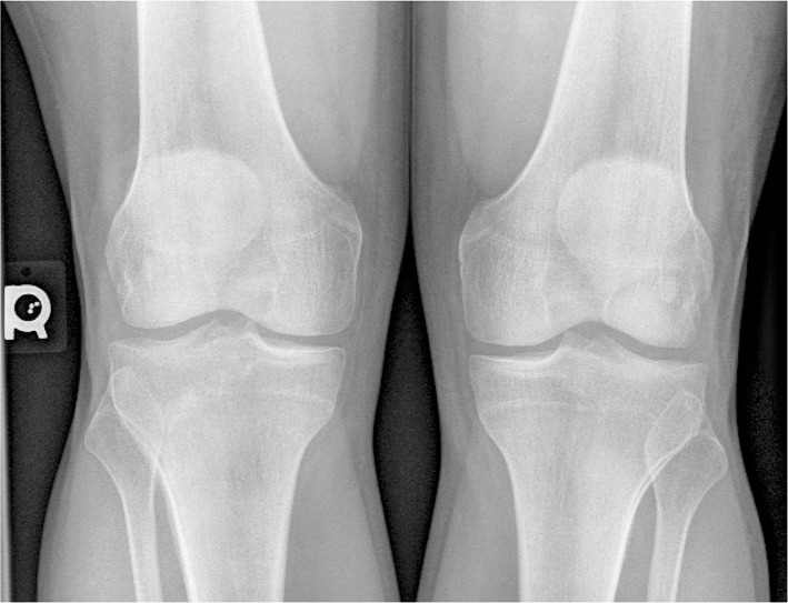
A plain-film radiographic series for the left ankle (Figures 1 and 2) was taken in addition to a bilateral AP knee radiograph (Figure 3). There was adequate bone density with no osteochondral defects detected at the tibial plafond and talar dome. All bony joints appeared unremarkable, however blurring of Kager’s fat pad was visualized. Radiographic findings included apparent chondrocalcinosis with joint effusion in the talotibial joint, which is highly suggestive of pseudogout associated with CPPD. As well, there was subtle chondrocalcinosis of the menisci bilaterally visualized on the bilateral AP knee radiographs (Figure 3).
Figure 1.
Left medial oblique ankle (left) and left AP ankle (right).
Figure 2.

Left lateral ankle.
Figure 3.
Bilateral AP knee – subtle chondrocalcinosis is visualized in the menisci bilaterally.
Diagnosis, Treatment and Referral
The patient was diagnosed, by plain-film radiography, with CPPD in the left ankle. At the time of writing, the patient did not receive any chiropractic treatment and was referred to a rheumatologist for further assessment. The patient declined conservative care, and was advised to treat the acute symptoms of CPPD at home and monitor his progress.
Discussion
Direct research evidence to support treatment recommendations are lacking therefore management strategies will vary according to the clinical presentation. Conservative treatment of CPPD is mainly symptomatic and often limited due to the etiology of the disease.1 Currently, there are no treatments that modify calcium pyrophosphate crystal formation or dissolution.1 High-quality evidence is limited for non-pharmacological treatment interventions for managing CPPD, however, expert opinion recommends the use of cryotherapy and temporary rest during acute attacks.7 Education is also an essential part of conservative care as it allows for patient involvement in the decision making process of their clinical management.7 Patient-centered care is optimized when the patient understands the characteristics of their diagnosis, available treatment options and associated benefits and risks.7
The medical management of CPPD is challenging due to the fact that the condition is often associated with other disorders. The course of treatment often varies widely across patients depending on their specific health conditions and their unique case of CPPD.5 Research shows the most common medications prescribed for CPPD are for symptomatic relief within the joint including conventional anti-inflammatory medications, such as NSAIDs.5
However it is important to note since CPPD prevails in older patients, additional caution and careful considerations should be taken when recommending NSAIDS due to drug interactions and harmful side effects. A safer alternative is joint aspiration or intra-articular injection of glucocoricosteroids.7 Glucocorticosteroids and joint aspiration have been shown to be a viable and effective option in the treatment of acute and painful CPPD attacks in addition to ice and rest.7 Dosage recommendations, however, are vague and often based on clinical expertise and research related to the management of gout.7
In addition to NSAID use, the management and treatment strategies should involve correcting the underlying metabolic abnormalities and treating the conditions.5 Newer therapies that require more evidence include substances targeting anti-crystal formation (such as probenecid) as well as anti-inflammatory medications (such as methotrexate) that target interleukin pathways to prevent recurrent attacks.5 At this time however, there is no definitive treatment available to dissolve the crystal deposits or prevent future crystal deposition.5 Due to the incurable nature of this condition, CPPD is classified as a chronic disorder with recurrent episodes. The prognosis is good, as long as symptoms are controlled and patients are monitored.8
Numerous clinical presentations of CPPD make diagnosing and treating this condition challenging. In this case specifically, CPPD targeted the menisci of the knees and the talotibial joint. A common location for CPPD includes, but is not limited to, the menisci of the knee and the patellofemoral joint, only one of which was seen in this case. It is apparent that there is irregular joint involvement, seeing that CPPD was also present in the ankle, which is a less common location for this condition. Although CPPD is often associated with osteoarthritis, it is important to understand the difference in clinical presentation between the two conditions. A large proportion of patients with CPPD follow a progressive course of articular degeneration in an irregular distribution pattern.5 Typically, around 50% of these individuals will experience acute attacks of pseudogout superimposed on their underlying osteoarthritis, whereas the remaining present with classical osteoarthritis.5 Features that distinguish CPPD from osteoarthritis include atypical joint distribution, presence of contractures and valgus knee deformities.5 Clinically, CPPD has the potential to mimic several forms of inflammatory arthritic conditions resulting in a wide array of clinical manifestations.5 These presentations include asymptomatic or lanthanic chondrocalcinosis, acute pseudogout, pseudo osteoarthritis (with or without acute attacks), pseudo rheumatoid arthritis, pseudo-polymyalgia rheumatica and pseudo-neuropathic arthropathy.5 It is possible for CPPD to co-exist with other arthritic conditions, further complicating diagnosis and management strategies. Further research on diagnosing, managing and reducing calcium pyrophosphate crystal deposition is essential seeing that CPPD and the associated CPPD-related arthropathies are likely to increase in prevalence due to the current aging population.5
Summary
This case report highlights a 36-year-old male patient who presented to a chiropractic clinic with an uncommon presentation of a common arthritic condition. Clinicians should take note of this unique presentation, as CPPD is prevalent in older, Caucasian populations and can often present concurrently with osteoarthritis.1,7 It is imperative to understand which joints can be targeted by CPPD and what treatment options are available. As well, it is clinically important to note the patient in this case report also had subtle findings of chondrocalcinosis in the menisci of the knees. Thus, inferring that if chondrocalcinosis is found in one joint in the body, there may be other joints in the body targeted as well. Further research is necessary to investigate the benefits of chiropractic care for the treatment and management of patients presenting with acute and chronic forms of CPPD, specifically with respect to providing pain relief and improving joint mobility.
Footnotes
The patient has provided written consent to have this case report published.
References
- 1.Abhishek A. Calcium pyrophosphate deposition. Br J Hosp Med. 2014;75(4):C61–C64. doi: 10.12968/hmed.2014.75.sup4.c61. [DOI] [PubMed] [Google Scholar]
- 2.Yochum T, Rowe L. Essentials of skeletal radiology. Baltimore: Williams & Wilkins; 1996. [Google Scholar]
- 3.Lindenmeyer C, Sobel A, Nazarian L, Mandel S, Raikin S. A case study of pseudoneuropathic pseudogout. Med Forum. 2013;14(26) [Google Scholar]
- 4.Netter P, Bardin T, Bianchi A, Richette P, Loeuille D. The ANKH gene and familial calcium pyrophosphate dihydrate deposition disease. J Bone Spine. 2004;71(5):365–368. doi: 10.1016/j.jbspin.2004.01.011. [DOI] [PubMed] [Google Scholar]
- 5.MacMullan P, McCarthy G. Treatment and management of pseudogout: insights for the clinician. Ther Adv Musculoskeletal Dis. 2012;4(2):121–131. doi: 10.1177/1759720X11432559. [DOI] [PMC free article] [PubMed] [Google Scholar]
- 6.Gamon E, Mouterde G, Barnetche T, Combe B. SAT0319 Diagnostic value of ultrasound in calcium pyrophosphate deposition disease: a systematic review and meta-analysis. Ann Rheum Dis. 2015;74(Suppl2):774.2–774. doi: 10.1136/rmdopen-2015-000118. [DOI] [PMC free article] [PubMed] [Google Scholar]
- 7.Zhang W, Doherty M, Pascual E, Barskova V, Guerne P, Jansen T, et al. EULAR recommendations for calcium pyrophosphate deposition. Part II: Management. Ann Rheum Dis. 2011;70(4):571–575. doi: 10.1136/ard.2010.139360. [DOI] [PubMed] [Google Scholar]
- 8.Schumacher H, Hasselbacher P. CPPD Deposition Disease [Internet] UW Orthopaedics and Sports Medicine, Seattle. 2012. [cited 2 January 2016]. Available from: http://www.orthop.washington.edu/?q=patient-care/articles/arthritis/cppd-deposition-disease.html.


