Abstract
A 10-month-old female Japanese black heifer presenting with sudden loss of appetite was diagnosed with extreme extension of the gallbladder. Laparotomy reaching from the right part of the 10th rib to the right flank showed an extended gallbladder greater than 50 cm in diameter. Cholecystectomy was performed as follows: 1) complete removal of the gallbladder distally from the base; 2) flushing via a catheter inserted into the common bile duct; and 3) covering of the hole opened in the common bile duct with a double-suturing method using the mucous membrane and muscular layers of the remaining gallbladder structures. Serum levels of total bilirubin gradually decreased from 7.5 mg/dl preoperatively to 4.7 mg/dl, 1.6 mg/dl and 0.6 mg/dl at 3, 8 and 34 days postoperatively, respectively. The heifer showed 1 month of clinical improvements, grew normally and finally became pregnant. To the best of our knowledge, this represents the first clinical report to describe cholecystectomy in cattle.
Keywords: bile duct, bilirubin, cholecystectomy, heifer, gallbladder
Cholecystectasia (extension of the gallbladder) in bovines is sometimes found in fields [1, 3, 5, 6, 9]. Such lesion is comparatively easily discovered by the specific clinical findings including jaundice and hyperbilirubinemia, and by clinical uses of ultrasonography to abdomen [1, 2]. In bovines, cholecystectasia is mainly associated with obstruction of the common bile duct and needs to be treated surgically in majority of the severe affected animals [1, 3, 6, 9]. Two types of the surgical techniques were previously reported; 1) cholecystoduodenostomy, which means that a new outlet for the bile is created by anastomosing the fundus of the gallbladder to the duodenum [1, 3, 4]; and 2) cholelithotripsy, which means that manually crushed stones of the gallbladder via the wall of the gallbladder are removed through the duodenum by massage [3, 9]. Cholecystectomy has never been performed, although this technique has also been considered as a surgical option for cattle [1].
A 10-month-old female Japanese black heifer showed sudden loss of appetite. Body temperature was recorded as 38.6–39.6°C during a month of clinical observation. The animal was voiding small amounts of tight, black feces or dark-green diarrhea. Abdominal pain and abdominal distension were not evident before admission. Clinical sign was not entirely ameliorated on injections of hepato-tonic drug (Urso-injection; ursodeoxycholic acid, DS Pharma Animal Health Co., Tokyo, Japan) and continuous fluid therapy. Jaundice was evident in the visible membranes of the vagina on day 20 after presentation. On day 26, hematological examination revealed normal results for both red blood cell count (995 × 104/µl) and white blood cell count (6,500/µl). Serum enzyme levels were high (aspartate aminotransferase [AST], 320 U/l [reference range, 30–104 U/l]; and γ-glutamyl transferase [GGT], 579 U/l [reference range, 9.5–39.0 U/l]. Hyperbilirubinemia was detected (total bilirubin, 7.5 mg/dl; reference range, <0.82 mg/dl) [1]. Eggs of Fasciola hepatica were not observed in feces. A ping sound was detected in the left 11th to 13th intercostal spaces. Ultrasonographic examination of the abdomen revealed an extreme large and anechoic structure in the right flank using an ultrasonographic device (HS-101V, Honda Electronics Co., Ltd., Toyohashi, Japan). The hepatic structure was almost invisible to be masked by the enlarged anechoic structure within the ultrasonographic view. The cholecystectasia diagnosed based on such finding should be treated surgically, because of the predictive poor therapeutic effect due to medical treatments.
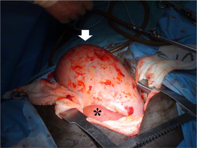
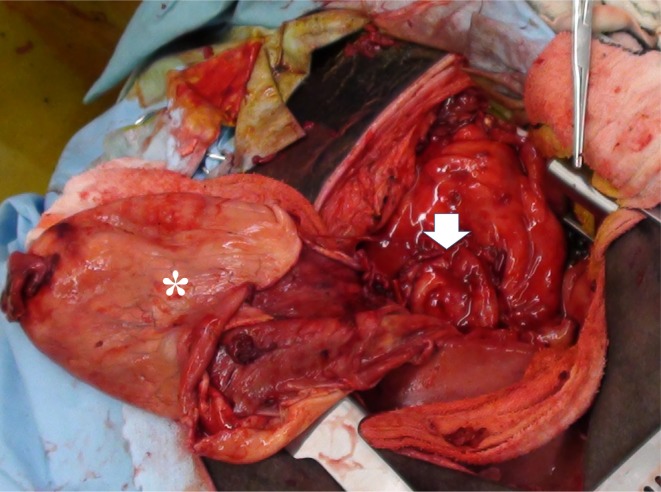
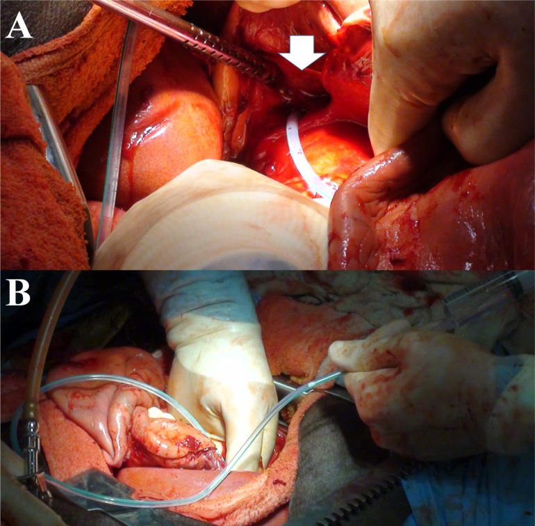
Laparotomy was performed under general anesthesia with 0.2–0.3% isoflurane via an inserted tracheal tube, after sedation with intravenous injection of xylazine hydrochloride (0.2 mg/kg). The animal was positioned in a left recumbent position. The skin was incised along the ribs in the right intercostal space between the 10th and 11th ribs. The intercostal muscles were separated by blunt dissection, and the peritoneum was incised. Swollen liver was partially observed from the opened intercostal space. To extend the intra-abdominal view, the costal bone-cartilage junctions of the 11th, 12th and 13th ribs were cut using a surgical saw. The incision line was extended caudally on the right flank due to incision across the obliquus externus abdominis muscle. Within the intra-abdominal view reaching from the right part of the 10th rib to the right flank, the gallbladder was observed to be severely extended greater than 50 cm within the distal region of the liver (Fig. 1). The gallbladder felt tight with bile fluid filling the cavity. Serous membranes of the gallbladder and liver structure were initially separated around the whole circumference of the gallbladder. The muscular layer and mucous membrane were completely incised within the stalk-like base of the gallbladder close to the common bile duct, after the removal of bile fluid by vacuuming (Fig. 2). Some bile fluid was then dropped into the abdominal cavity. After removal of the gallbladder, a catheter (Gastric tube [inner diameter: 2.5 mm and outer diameter: 5.0 mm], Fuji systems Co., Tokyo, Japan) was inserted into the common bile duct through the opened stalk of the gallbladder (Fig. 3A). Smooth flow of flushing sterilized saline via the catheter into the duodenum revealed no stenotic lesions and no foreign materials in the common bile duct (Fig. 3B). The partially remaining mucous membrane of the gallbladder was sutured with absorbable monofilament sutures (Maxon 2-0, Sherwood-Davis and Geck, St. Louis, MO, U.S.A.), so that the opened part of the common bile duct was covered. A final suture using the remaining mucous membrane of the gallbladder was performed after removal of the catheter into the common bile duct. Double-layered sutures were finally performed by following another continuous suture using the remained muscular layers of the gallbladder. The intra-abdominal drainage tube was set before abdominal closure. The costal bone-cartilage junctions were fixed with metal wires through holes opened into the proximal and distal edges of the cut lines. The peritoneum and muscles were sutured with absorbable monofilament sutures from the right part of the 10th rib to the right flank. The skin was sutured using non-absorbable sutures (Suprylon, Vömel, Gronberg, Germany). In total, cholecystectomy took 3 hr. Swelling was grossly evident without necrotic changes within the walls of the gallbladder. Papillary hyperplasia was observed on the mucous membrane of the gallbladder. Histological examination using hematoxylin and eosin staining revealed hyperplasia of the gallbladder mucosa and moderate infiltration of lymphocytes into the lamina propria of the mucous membrane and submucous tissues. The structure comprised necrotic and degenerative foci. These histological findings indicated chronic cholecystitis due to cholestasis. Bacterial examination revealed no growth, and eggs of Fasciola hepatica were not found in the bile fluid.
Fig. 1.
The abdominal cavity during laparotomy. Extreme extension of the gallbladder greater than 50 cm (arrow) is evident within the distal region of the liver (asterisk).
Fig. 2.
Intraoperative view during surgical resection of the gallbladder. The opening hole to the common bile duct (asterisk) is evident within the base of the gallbladder in surgical resection halfway around the wall of the gallbladder (arrow).
Fig. 3.
Intraoperative view during surgical resection of the gallbladder. (A) A tube is inserted through the opening hole into the common bile duct and toward the duodenum. (B) Flushing is performed via the catheter tube for lavage inside the common bile duct.
The present case was treated by 7 continuous days of antibiotic injections. Small amounts of exudate were removed by suction via the intra-abdominal drainage tube for 5 days after surgery, and the tube was then removed. The heifer showed no appetite and stiff gait by 3 days after surgery. Jaundice disappeared within a few days after surgery. Appetite and activity gradually increased over 7 days after surgery, and normal condition was acquired by 1 month postoperatively. Serum enzyme activities were dramatically improved in blood examinations at 3, 8 and 34 days after surgery as follows: AST, 104 U/l, 201 U/l and 74 U/l; GGT, 872 U/l, 999 U/l and 180 U/l; and total bilirubin, 4.7 mg/dl, 1.6 mg/dl and 0.6 mg/dl, respectively. As of the time of writing, the heifer was still growing and had achieved normal weight, and was delivered of two calves.
Obstruction of the common bile duct as main cause of cholecystectasia could be associated with cholangitis, abscess, cholelithiasis or neoplasm in cattle [6]. Cholangitis is a comparatively common lesion, with an occurrence rate of 7.2% [5]. Primary liver tumors of hepatocellular origin are a rare lesion, identified in around 0.09% of slaughtered cattle [9]. Rare bovine cases have also been reported with growth of liver abscesses resulting in obstruction of bile flow, although liver abscesses are identified in around 5.8% of slaughter cattle [9]. The present case may show a comparatively slow progression for the extreme expansion of the gallbladder, following downward dislocation of the gallbladder, and resulting in difficulty of bile flow due to torsion or physical pressure in the common bile duct, because stenosis or constriction was not evident within the common bile duct. The main treatment for these diseases is removal of the cause of obstruction in the common bile duct. This has typically been accomplished using various surgical techniques. A previous report described a surgical technique for a case of cholelithiasis in which the gallstone was crushed manually via the wall of the gallbladder without incising the gallbladder after laparotomy [3, 9]. The crushed pieces of gallstone were removed through the common bile duct and into the duodenum by massage [3, 9]. This surgical technique was termed cholelithotripsy [3, 9]. Manual repositioning of the gallbladder has been performed in a previous case with dislocation of the gallbladder; this surgical technique was called cholecystohepatopexy [1]. Another surgical technique called cholecystoduodenostomy creates a new outlet for the bile by anastomosing the fundus of the gallbladder to the duodenum [4]. These techniques allow rapid and dramatic improvements [1, 3, 4]. Cholecystectomy has also been considered as a surgical option for cattle [1]. However, to the best of our knowledge, cholecystectomy has never been performed in bovine cases for the following reasons: lack of information on postoperative nutritional management; the long time required for surgical procedures resulting in a greater risk to the animal; difficulty in exteriorizing the gallbladder; and risk of bile peritonitis [1, 3]. Cholecystectomy is commonly the first-choice surgical technique for gallbladder diseases in human medicine and small animal practice [7]. This convinced us that cholecystectomy can also become a common surgical technique for cattle. Advantages of cholecystopathy in cattle are as follows. First, cholecystopathy seems applicable for various types of cholecystopathy in cattle. Previous case reports have shown that application of a surgical technique was specialized to a specific type of cholecystopathy (e.g., cholelithotripsy for cholelithiasis and cholecystohepatopexy for dislocation of the gallbladder) [1, 3, 4]. Second, cholecystectomy is applicable in bovine cases with extreme extension of the gallbladder. This condition resembles atony of the gallbladder, which is a disease predisposing to acalculous cholecystitis in humans [8]. Preservation of an extended gallbladder may be inadequate with surgery, because the gallbladder may have loss of storage and excretion functions. Third, direct recognition of the causes of obstruction is possible with insertion of a catheter into the common bile duct. Gallstones present in the common bile duct can be removed to the duodenum by either flushing flow via the catheter or pushing the stones using the catheter itself.
In the present case, cholecystectomy achieved decreased blood bilirubin levels below 3.8 mg/dl (relative to exhibition of jaundice) for 8 days and gradual improvement of clinical signs over the course of 1 month [6]. This treatment also allowed the heifer to achieve normal growth following pregnancy. However, our results suggested a lengthened postoperative time required for clinical improvement due to cholecystectomy, compared with several days for the improvements in previous cases treated using other surgical techniques [1, 3, 4]. Normalization of total bilirubin levels was also recorded as very rapid in those cases, occurring in 24–48 hr [1, 3, 4]. Disadvantages of cholecystectomy in cattle are given as follows. First, diapedesis of some amount of bile fluid into the abdominal cavity occurs following the incision and removal of the gallbladder wall and vacuum removal of accumulated bile fluid. Fortunately, no postoperative complications were identified, because the bile fluid was sterile in the present case. Sterility of the biliary tract is maintained by the continual production and flow of bile into the intestine [9]. However, partial or complete obstruction of bile flow predisposes the individual to ascending infection by intestinal microorganisms in the biliary tract [9]. The risk of bile peritonitis will thus be important to consider in the application of cholecystectomy [1, 3, 7]. Postoperative use of antibiotics is indispensable, and intra-abdominal drainage is recommended. Second, cholecystectomy needs to be performed by right flank laparotomy under a left recumbent position. This will not be suitable for surgical procedures performed in the field. Cholecystohepatopexy, cholelithotripsy and cholecystoduodenostomy can be performed via a routine standing right flank laparotomy [1, 3, 4]. Third, an extended operative wound is needed for cholecystectomy, because of the need to exteriorize the gallbladder. This can result in considerable operative stress on the animal and lengthens operative time. Wound healing and improvement of clinical signs may thus be delayed. A point of improvement for further application of cholecystectomy to bovine practice may be to decrease the size of the gallbladder by vacuum suction of inclusions before making a complete surgical opening. The present animal showed normal growth postoperatively and became pregnant after surgical removal of the gallbladder, despite not receiving any specialized postoperative nutritional management. The present data indicate cholecystectomy as a meaningful surgical technique for postoperative maintenance or advancement of bovine productivity.
REFERENCES
- 1.Boerboom D., Mulon P. Y., Desrochers A.2003. Duodenal obstruction caused by malposition of the gallbladder in a heifer. J. Am. Vet. Med. Assoc. 223: 1475–1477, 1435. doi: 10.2460/javma.2003.223.1475 [DOI] [PubMed] [Google Scholar]
- 2.Braun U.2009. Ultrasonography of the liver in cattle. Vet. Clin. North Am. Food Anim. Pract. 25: 591–609. doi: 10.1016/j.cvfa.2009.07.003 [DOI] [PubMed] [Google Scholar]
- 3.Cable C. S., Rebhun W. C., Fortier L. A.1997. Cholelithiasis and cholecystitis in a dairy cow. J. Am. Vet. Med. Assoc. 211: 899–900. [PubMed] [Google Scholar]
- 4.Hofmeyr C. F. B.1955. Cholecystoduodenostomy in the cow. J. Am. Vet. Med. Assoc. 126: 459–462. [PubMed] [Google Scholar]
- 5.Kato M., Murakami Y., Shimizu M., Yamamoto H., Yonemoto Y., Ishii K., Kira S.2005. Survey of cattle fascioliasis in Tsuyama Abattoir. Environ. Health Prev. Med. 10: 162–165. doi: 10.1007/BF02900810 [DOI] [PMC free article] [PubMed] [Google Scholar]
- 6.McSherry B. J., Lumsden J. H., Valli V. E., Baird J. D.1984. Hyperbilirubinemia in sick cattle. Can. J. Comp. Med. 48: 237–240. [PMC free article] [PubMed] [Google Scholar]
- 7.Mehler S. J.2011. Complications of the extrahepatic biliary surgery in companion animals. Vet. Clin. North Am. Small Anim. Pract. 41: 949–967, vi. doi: 10.1016/j.cvsm.2011.05.009 [DOI] [PubMed] [Google Scholar]
- 8.Nies C., Zielke A., Hasse C., Klotter H. J., Rothmund M.1994. Atony of the gallbladder as a risk factor for acalculous cholecystitis. What is the effect of intensive care? Zentralbl. Chir. 119: 75–80 (in German). [PubMed] [Google Scholar]
- 9.Van Metre D. C., Tennant B. C., Whitlock R. H.2008. Infectious diseases of the gastrointestinal tract. pp. 200–294. In: Rebhun’s Diseases of Dairy Cattle, 3rd ed. (Peek, S. and Divers, T. eds.), W. B. Saunders Co., Philadelphia. [Google Scholar]


