
Extended Data Fig. 5. GMP clusters are clonal.
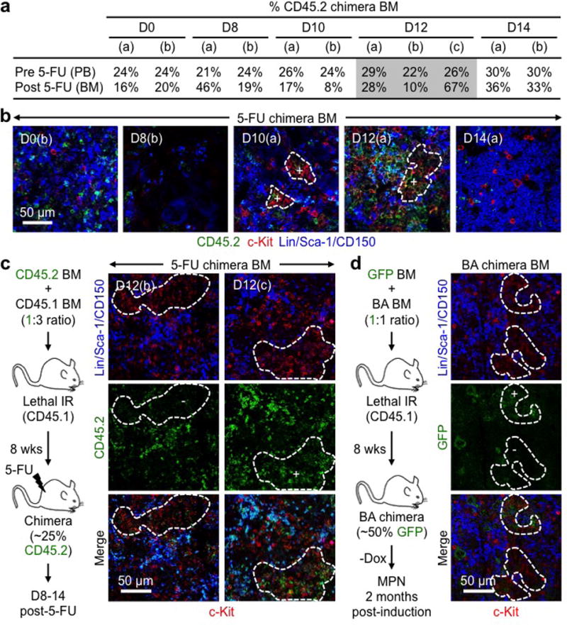
a–c, Clonality of regenerative GMP clusters: (a) Percent CD45.2+ cells in the peripheral blood (PB) pre- and BM post-5-FU treatment for each of the chimera mice used at the indicated days post-treatment (n = 2–3 mice/group); (b) Representative IF staining showing MPs (red) and CD45.2 (green) expression in 5-FU-treated chimera BM at the indicated days post-treatment; and (c) experimental scheme and representative IF staining showing MPs (red) and CD45.2 (green) expression separately in two independent d12 5-FU-treated chimera BM. Positive (+) clusters have ≥ 75% CD45.2+ cells and negative (−) clusters ≤ 5% CD45.2+ cells. Wks: weeks. d, Clonality of leukemic GMP clusters with experimental scheme and representative IF staining showing MPs (red) and GFP (green) expression from β-actin-Gfp cells in diseased BA chimera BM. Positive (+) clusters have ≥ 75% GFP+ cells and negative (-) clusters ≤ 5% GFP+ cells. Dotted lines indicate cGMPs.