Fig. 5.
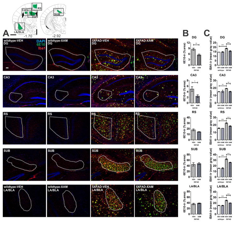
Xamoterol reduces microglia/macrophage (Iba1) gliosis and amyloid beta (6E10) immunoreactivity (-ir). Chronic dosing with the selective partial ADRB1 agonist, xamoterol, attenuates Iba1-ir in all regions in 5XFAD transgenic mice and reduces 6E10-ir in DG and CA3 regions of the hippocampus. A) Atlas plates (top left) indicate one of multiple levels from which the regions of interest (green) were selected for analyses [1]. Black boxes indicate image capture frame. Numbers below atlas plates indicate mm bregma; black scale bar, 1 mm. Photomicrographs (5X magnification) illustrate representative immunostaining (DAPI, blue; 6E10, green; Iba1, red) from each region of interest in wildtype and 5XFAD vehicle- (VEH) and xamoterol-treated (XAM) mice, white scale bar, 100 μm. Dashed white lines indicate region of quantification. B) Quantitative immunohistochemistry revealed decreases in 6E10-ir (% area) in DG and CA3. C) Increases in Iba1-ir (mean intensity) were detected in all regions analyzed in VEH-treated 5XFAD transgenic mice relative to VEH-treated wildtype littermates. These increases were attenuated in each region in transgenic mice chronically dosed with XAM. Sample size, n=4–5 per group, with exception of wildtype-XAM (n=3 for DG/CA3/SUB and n=2 for RS). Data represent mean ± SEM. *p < .05, **p < .01, ***p < .001, Bonferroni’s test for multiple comparisons following one-way ANOVA. RS, retrosplenial cortex; LA/BLA, lateral/basolateral amygdala; SUB, subiculum; CA3, CA3 region of the hippocampus; DG, dentate gyrus.

