Abstract
It has been shown that the L289A-mutated Newcastle disease virus (NDV) fusion (F) protein gains the ability to promote fusion of Cos-7 cells independent of the viral hemagglutinin-neuraminidase (HN) protein and exhibits a 50% enhancement in HN-dependent fusion over wild-type (wt) F protein. Here, we show that HN-independent fusion by L289A-F is not exhibited in BHK cells or in several other cell lines. However, similar to the results in Cos-7 cells, the mutated protein plus HN does promote 50 to 70% more fusion above wt levels in all of the cell lines tested. L289A-F protein exhibits the same specificity as the wt F protein for the homologous HN protein, as well as NDV-human parainfluenza virus 3 HN chimeras. The mutated F protein promotes fusion more effectively than the wt when it is coexpressed with either the chimeras or HN proteins deficient in receptor recognition activity. In addition, its fusogenic activity is significantly more resistant to removal of sialic acid on target cells. These findings are consistent with the demonstration that L289A-F interacts more efficiently with wt and mutated HN proteins than does wt F by a cell surface coimmunoprecipitation assay. Taken together, these findings indicate that L289A-F promotes fusion by a mechanism analogous to that of the wt protein with respect to the HN-F interaction but is less dependent on the attachment activity of HN. The phenotype of the mutated F protein correlates with a conformational change in the protein detectable by two different monoclonal antibodies. This conformational change may reflect a destabilization of F structure induced by the L289A substitution, which may in turn indicate a lower energy requirement for fusion activation.
Newcastle disease virus (NDV) is a member of the Avulavirus genus of the Paramyxoviridae family of viruses. Virion and infected cell surfaces contain two types of spikes, the hemagglutinin-neuraminidase (HN) and fusion (F) glycoproteins (25). The tetrameric HN protein spike mediates receptor recognition and also possesses neuraminidase (NA) activity, the ability to cleave a component of those receptors, sialic acid (20). The F protein directly mediates virus-cell and cell-cell fusion (20).
The paramyxovirus F spike is a trimeric, type I membrane glycoprotein; thus, it has the opposite orientation from that of HN. The three-dimensional structure of the NDV F protein has been solved (5). The F protein is produced as an inactive precursor, Fo, which must be cleaved by a cellular protease to produce an active, disulfide-linked F1-F2 complex. Cleavage creates a new C terminus of F1, a well-conserved fusion peptide, which is directly inserted into the membrane to initiate fusion (Fig. 1) (20).
FIG. 1.
Diagram of the structure of the NDV F glycoprotein. The F protein is proteolytically cleaved to produce the two disulfide-linked polypeptides, F1 and F2. Cleavage generates the hydrophobic fusion peptide (FP). There are three HRs in NDV F: HR1, adjacent to the FP; HR2, just outside the transmembrane (TM); and, HR3, located between the other two at residues 268 to 289.
For most paramyxoviruses, the promotion of fusion by the F protein is dependent on a contribution from HN, in addition to its recognition of receptors (14). This contribution is virus specific, in that most heterologous combinations of HN and F do not result in fusion (20). The role that HN plays in F-mediated fusion involves an interaction between the two proteins at the cell surface (7, 21, 33). Through the construction and evaluation of chimeric HN proteins composed of domains from proteins derived from heterologous viruses, it has been shown that the stalk region of the HN spike determines its specificity for the homologous F protein (8, 29, 31, 32).
Three heptad repeat (HR) domains have been identified in F1 (Fig. 1). HR1 is located adjacent to the fusion peptide at the N terminus of F1 (4). HR2 is located within the first 40 residues of the stalk just outside the membrane (3). These two HRs have been shown to refold into a six-helix bundle structure that is an intermediate in the fusion process (24). Another HR (HR3) is situated between HR1 and HR2 at residues 268 to 289 and may act during the conversion of F to the active form and/or in destabilizing the target membrane (12). The role of this HR in the structure and function of the NDV F protein was examined by the introduction of individual alanine substitutions for the heptadic leucines (26). One of these substitutions (L289A) resulted in a protein capable of promoting 70% of the amount of fusion promoted by wild-type (wt) HN and F in Cos-7 cell monolayers in the absence of HN. The substitution also resulted in more than 50% enhancement in fusion when L289A-F was coexpressed with HN compared to that achieved with the two wt proteins. Thus, a single amino acid substitution in F alters the requirement for HN in the promotion of fusion by NDV F.
Here, we have further characterized the relationship between receptor recognition and the fusion promotion activity of L289A-mutated NDV F. First, we have shown that the quite extensive HN-independent fusion exhibited by this mutated protein in Cos-7 cells cannot be duplicated in any of several other cell lines, including BHK, HeLa, 293T, CV-1, Hep-2, and CHO cells. However, enhanced HN-dependent fusion above wt levels is observed in all cell types tested. In addition, L289A-F is identical to the wt protein with respect to the requirement that the complementing HN protein be from the same virus and to the specificity for NDV-human parainfluenza virus 3 (hPIV3) HN chimeras. L289A-F also exhibits a pattern similar to that of the wt protein with respect to its interaction with HN and mutants derived from it, although the mutated F protein does interact more efficiently. Most significantly, the fusion-promoting activity of L289A-F is much more resistant to diminished receptor recognition by the coexpressed HN protein, resulting from either site-directed mutagenesis of HN or treatment of target cells with exogenous NA. Finally, two monoclonal antibodies (MAbs) can detect a conformational difference between L289A-F and the wt F protein. These findings support the idea that the L289A substitution converts F to a form that is less dependent on an interaction with HN for conversion to the fusion-active form and which may already be in an intermediate fusogenic conformation.
MATERIALS AND METHODS
Recombinant expression vectors.
Construction of the NDV-Australia-Victoria HN and F recombinant pBluescript SK(+) (Stratagene Cloning Systems, La Jolla, Calif.) and hPIV3 HN and F expression vectors (8, 22), as well as the pCAGGS expression vector containing the NDV-Australia-Victoria HN and F genes (21), has been described previously. Construction of the HN chimeras having NDV-derived N-terminal segments has also been previously described (8).
Site-directed mutagenesis.
Introduction of the L289A substitution into NDV F was accomplished by site-directed mutagenesis (6), using a mutagenic primer (5′-GTATACAGGTAACTGCCCCTTCAGTAGGAAACCTAAATAATATG-3′) (Integrated DNA Technologies, Inc., Coralville, Iowa). The codon for the alanine substitution is in boldface type. Screening for mutants was facilitated by the introduction of a unique EcoNI site (underlined in the sequence above). The presence of the desired mutation was verified by sequencing of double-stranded DNA, with the Sequenase Plasmid Sequencing Kit (United States Biochemical, Cleveland, Ohio), according to protocols provided by the company. Multiple clones were characterized. The preparation of site-directed HN mutants carrying a substitution of I175E, D198R, F220A, F220N, S222K, S222T, K236R, Y526L, or E547Q has been described previously (6, 7, 17), as has the preparation and characterization of the NDV-hPIV3 HN chimeras (8).
Cell lines and transient expression systems.
BHK-21, Cos-7, Hep-2, and CV-1 cells were obtained from the American Type Culture Collection (Manassas, Va.). CHO cells and sialic acid-deficient cells (Lec2) derived from them, as well as 293T cells, were gifts from Anne Moscona (Mount Sinai School of Medicine, New York, N.Y.). HeLa cells were obtained from the University of Massachusetts Medical Center Tissue Culture Facility. BHK-21 cells were maintained in Dulbecco's modified Eagle medium (high glucose), supplemented with 5% fetal calf serum, 20 mM l-glutamine, and antibiotics. All other cell lines were maintained in the same medium except that the medium was supplemented with 10% fetal calf serum and 20 mM nonessential amino acids.
All cells were seeded in six-well plates a day earlier at 4 × 10⁵ cells/well, except 293T cells, which were seeded at 6 × 10⁵ cells/well. All experiments, except for those on the effect of cell line and expression system on fusion promotion, were done with BHK-21 cells (American Type Culture Collection), with the recombinant vaccinia virus-T7 RNA polymerase expression system (11) by a previously described transfection protocol (22). The HN and F proteins were also expressed in HeLa and Hep-2 cells with the vaccinia virus-T7 RNA polymerase system. The viral glycoproteins were also coexpressed in these cell lines, as well as Cos-7, 293T, and CHO cells, with the pCAGGS expression vector. Plasmid DNA (1.5 μg/well) was transfected into the cells with PolyFect transfection agent (QIAGEN, Inc., Valencia, Calif.), according to protocols provided by the company and optimized for transfection of each specific cell line. Assays were performed 18 h posttransfection.
Hemadsorption assay.
Hemadsorption (HAd) activity of HN proteins was assayed by the ability of expressed proteins to adsorb guinea pig erythrocytes (Crane Laboratories, Syracuse, N.Y.). Monolayers were incubated for 30 min at either 4 or 37°C with a 2% suspension of erythrocytes in phosphate-buffered saline supplemented with 1% CaCl2 and MgCl2. After being extensively washed, adsorbed erythrocytes were lysed in 50 mM NH4Cl, and the lysate was clarified by centrifugation. HAd activity was quantitated by measuring the absorbance at 540 nm minus the background obtained with vector-expressing cells.
Production of MAbs.
Hybridomas producing MAbs Fa and Fb were prepared as described previously (16), following immunization of BALB/C mice with UV-inactivated virus and infected chicken embryo cells, respectively. Hybridomas were screened by an enzyme-linked immunosorbent assay and neutralization assays. Hybridoma supernatants were used in all experiments.
Cell surface expression and MAb reactivity.
The cell surface expression and MAb reactivity of L289A-F were compared to those of the wt F protein and its cleavage site mutant (CSM) by immunoprecipitation (IP) of the labeled proteins chased to the cell surface, as described previously for the HN protein (32). Briefly, mutant F-expressing cells were labeled for 3 h and chased with medium for 90 min, prior to IP by the individual MAbs or a rabbit antiserum prepared against the whole virus. Western blots for the detection of actin were used as a loading control. Diminished ability of the individual MAbs to recognize the mutated F protein was also confirmed by flow cytometric analysis.
Content-mixing assay for fusion.
The fusion promotion activity of the viral glycoproteins expressed using the vaccinia virus-T7 RNA polymerase expression system was quantitated with a content-mixing assay (6). Briefly, cell monolayers were infected with the vTF7-3 vaccinia virus recombinant as described above and cotransfected with the desired HN gene and either CSM F or L289A-CSM F, both of which carry a pair of cleavage site mutations which render the F protein nonfusogenic without exposure to trypsin. Another set of monolayers was infected with wt vaccinia virus (multiplicity of infection [MOI] of 10) and transfected with 1 μg of the plasmid pGINT7β-gal. Fusion of these two populations of cells resulted in expression of β-galactosidase, which was quantitated spectrophotometrically after a 5-h incubation period.
Fusion induced in the pCAGGS expression system was quantitated with a variation of this assay. The viral glycoproteins in pCAGGS vectors were cotransfected in one population of cells, along with pGINT7β-gal. These monolayers were trypsinized and mixed with cells previously infected with the vTF7-3 vaccinia virus recombinant (MOI of 10). The fusion of these two populations of cells resulted in expression of β-galactosidase, which was again quantitated spectrophotometrically.
Removal of receptors on target cells by exogenous NA treatment.
Target BHK cells transfected with pGINT7β-gal were treated for 1 h at 37°C with 50 mU of Vibrio cholerae NA (VCNA)/ml in Dulbecco's modified Eagle medium. After being extensively washed with Dulbecco's modified Eagle medium, the target cells were mixed with BHK cells expressing wt HN and either wt CSM F or L289A-mutated CSM F, and the extent of fusion was determined in the content-mixing fusion assay.
IP assay.
The IP protocol has been described previously (17). Briefly, at 22 h posttransfection, BHK cells were starved for 1 h at 37°C in medium lacking cysteine and methionine. Cells were then labeled with 1 ml of medium containing 100 μCi of Expre35S35S labeling mix (Perkin-Elmer Life and Analytical Sciences, Boston, Mass.) for 3 h at 37°C and chased for 90 min with medium. The cells were lysed, and F was immunoprecipitated with the appropriate antibody. Immune complexes were collected with Ultralink-Immobilized Protein A Plus (Pierce, Rockford, Ill.) and then analyzed by sodium dodecyl sulfate-polyacrylamide gel electrophoresis.
Co-IP.
The ability of HN to interact with F and L289A-F at the surface of transfected BHK cells was assayed at 16 h posttransfection, using a co-IP assay described previously (21). Briefly, monolayers consisting of equal numbers of cells were starved and labeled, prior to biotinylation of cell surface proteins. An interaction between HN and F at the surface was assayed by the ability of the two proteins to coimmunoprecipate from dodecylmaltoside cell lysates with a MAb to the F protein.
RESULTS
The HN-independent fusion activity of L289A-mutated F in Cos-7 cells is not exhibited in BHK cells.
A previous study showed that an L289A substitution in the NDV F protein eliminates the requirement for the HN protein in the promotion of fusion (26). In that study, fusion was assayed in Cos-7 cells with expression of the mutated F protein from the pSVL expression vector driven by the simian virus 40 promoter. Expression of L289A-F alone gave 70% of the fusion obtained with wt HN and F. Thus, in Cos-7 cells, the L289A substitution in F almost completely eliminated the requirement for HN in fusion promotion. However, coexpression of the mutated protein with HN did result in a 50% enhancement of fusion above the level achieved with the two wt proteins.
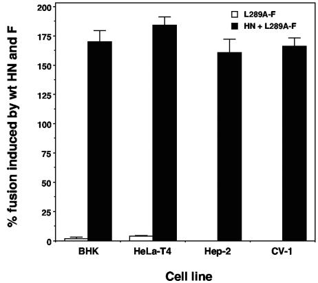
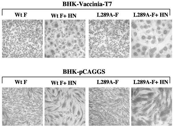
As part of a goal of exploring the relationship between receptor recognition by HN and fusion by F, we set out to determine the ability of L289A-F to promote fusion in BHK cells, using the vTF7-3 RNA polymerase expression system. Unlike the results obtained with the simian virus 40-Cos-7 system, expression of the mutated F protein in the vTF7-3-BHK cell system in the absence of HN failed to result in significant syncytium formation, inducing only 1.9% of the fusion obtained when wt F was coexpressed with wt HN (Fig. 2). Unfortunately, Cos-7 cells do not tolerate vaccinia virus well, and these cells cannot be assayed with this expression system. As shown in Fig. 3, BHK monolayers expressing the mutated F protein alone were indistinguishable from those expressing the wt F protein alone, which is nonfusogenic in the absence of HN. However, the enhanced fusion over wt levels achieved in monolayers coexpressing L289A-F and wt HN was also observed with BHK cells. This combination resulted in 170% of wt fusion (Fig. 2 and 3).
FIG. 2.
The promotion of fusion by L289A-F expressed alone and with wt HN in different cell lines. The mutated F protein is expressed both with and without HN in BHK, HeLa-T4, Hep-2, and CV-1 cells from the pBSK vector driven by the vaccinia virus-T7 RNA polymerase expression system. The extent of fusion is quantitated with the content-mixing fusion assay and expressed as the percentage of that obtained in the same cell line with HN and wt F. Error bars show the standard deviation from at least four independent determinations.
FIG. 3.
Syncytium formation in BHK cells induced by wt F and L289A-F with and without HN expressed by two different transient expression systems. In the top row, the indicated proteins are expressed from the pBSK vector driven by the vaccinia virus-T7 RNA polymerase expression system. In the bottom row, the indicated proteins are expressed from the pCAGGS vector driven by the chicken β-actin promoter. The monolayers were fixed with methanol and stained with Giemsa stain at either 22 h (vaccinia virus-T7) or 18 h (pCAGGS) posttransfection.
L289A-F also failed to promote HN-independent fusion in several other cell lines and with another expression system.
The failure to observe HN-independent fusion in BHK cells expressing L289A-F could be a phenomenon specific to these cells. To address this possibility, we determined the fusion-promoting activity of L289A-F with the vaccinia virus-T7 RNA polymerase expression system in three additional cell lines: HeLa-T4, Hep-2, and CV-1. In each case, expression of L289A-F with wt HN resulted in 50 to 75% more fusion than the two wt proteins, but barely detectable or undetectable fusion when the mutated F protein was expressed in the absence of HN (Fig. 2). Thus, the failure to detect HN-independent fusion in BHK cells was not specific for that cell line.
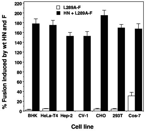
Another possibility for the lack of detectable HN-independent fusion in our system is the use of vaccinia virus. To address this possibility, the L289A-F protein was expressed in the absence of HN in the same cell lines as above, as well as two additional cell lines, CHO and 293T, with expression driven by the chicken β-actin promoter from the pCAGGS vector. Similar results were obtained with this expression system in all six cell lines. At 18 h posttransfection, L289A-F expressed in the absence of HN promoted less than 5% of wt HN- and F-induced fusion in each cell line (Fig. 4). Similar to results with the vaccinia virus-T7 RNA polymerase system, BHK cell monolayers expressing only the mutated F protein from pCAGGS were indistinguishable from wt F-expressing monolayers (Fig. 3). Even at 40 h posttransfection, both the vaccinia virus-T7 RNA polymerase and pCAGGS expression systems promoted significantly less than 10% of the control amount of fusion (data not shown).
FIG. 4.
The promotion of fusion by L289A-F expressed alone and with HN in different cell lines from the pCAGGS expression vector. The extent of fusion is quantitated with the content-mixing fusion assay and expressed as the percentage of that obtained in the same cell line with HN and wt F. Error bars show the standard deviation from at least four independent determinations.
To ensure that the failure to detect significant fusion in BHK cells is not the result of a defect in the L289A-F protein used in these studies, it was expressed without HN in Cos-7 cells from the pCAGGS vector. Similar to the earlier work, the mutated F protein promoted a significant level of HN-independent fusion in these cells, although in our hands, only 30% of the wt HN-F control (Fig. 4). This was approximately one-half of the previously reported level (26), although it should be noted that in the earlier report fusion was scored microscopically. However, coexpression of L289A-F with HN in Cos-7 cells led to almost 170% of wt fusion activity. Thus, the enhanced fusion obtained with HN and L289A-F was observed in all cell lines tested. But HN-independent fusion appeared to be specific to Cos-7 cells, at least among the cell lines tested here.
A heterologous attachment protein cannot substitute for NDV HN in L289A-F-mediated HN-dependent fusion.
Using vaccinia virus-T7 RNA polymerase-driven expression of HN and L289A-F in BHK cells, we set out to examine the relationship between HN′s recognition of receptors and the HN-dependent mode of fusion promoted by the mutated F protein. First, we wanted to know whether a heterologous attachment protein could replace NDV HN in the HN-dependent mode of fusion promoted by the L289A-F protein. Coexpression of the mutated NDV F protein with hPIV3 HN resulted in only 4% of the fusion obtained with the two wt NDV proteins, an extent of fusion not significantly different from that promoted by the L289A-F protein in the absence of HN (1.9% of wt). This was negligible compared to the 170% of the wt amount observed when the mutated F protein was coexpressed with NDV HN in this expression system (Fig. 2). Thus, a heterologous attachment protein could not significantly enhance the fusion ability of the L289A-F protein, indicating that the homologous HN protein is still required for efficient L289A-F-mediated fusion.
The homologous stalk domain of HN determines the fusion promotion ability of the L289A-F protein.
By evaluation of the ability of HN chimeras composed of domains derived from the NDV and hPIV3 HN proteins to complement either NDV F or hPIV3 F in the promotion of fusion, we have previously shown that the stalk domain of either HN protein determines its fusion specificity for the homologous F protein (8). A chimera (CH3) having only 141 N-terminal residues derived from NDV HN and the rest of the molecule, including the entire globular head, derived from hPIV3 HN, promoted more than 50% of wt NDV HN and F fusion. Similarly, a chimera with the entire NDV-derived ectodomain and a cytoplasmic tail and transmembrane anchor derived from the hPIV3 HN protein (CH6), as well as a chimera with only the stalk and transmembrane derived from NDV HN (CH11), promoted 45.5 and 39.5% of wt NDV HN and F fusion, respectively (8).
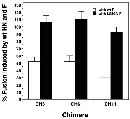
To determine whether L289A-F-mediated fusion exhibits the same dependence on the stalk region of the homologous HN protein, we evaluated the ability of NDV-hPIV3 chimeras with this region of the protein derived from NDV HN to complement the mutated NDV F protein in fusion. L289A-F expressed with chimeras CH3, CH6, and CH11 promoted 105.8, 110.3, and 92.0%, respectively, of wt fusion. This is compared to 52.2, 52.0, and 29.5%, respectively, of wt HN- and F-mediated fusion when the chimeras were expressed with wt F (Fig. 5). The latter values were very similar to those originally reported for these chimeras (8). Thus, in each case, the introduction of the L289A substitution resulted in at least a twofold increase relative to the extent of fusion promoted by wt F with the same chimera. Therefore, the promotion of fusion by L289A-F shows the same dependence on a contribution from the stalk domain of the homologous HN protein as the wt NDV F protein but again is more fusogenic.
FIG. 5.
Comparison of the extent of fusion promoted by wt F and L289A-F expressed with different NDV-hPIV3 HN chimeras. Proteins are expressed in BHK cells driven by the vaccinia virus-T7 RNA polymerase expression system. The extent of fusion is quantitated with the content-mixing fusion assay and expressed as the percentage of that obtained with wt HN and wt F. Error bars show the standard deviation from at least four independent determinations.
HN dimer interface mutants deficient in attachment and fusion at 37°C efficiently promote fusion with L289A-F.
We have previously shown that substitutions for residues F220 and S222 at the dimer interface of HN resulted in a destabilization of its interaction with receptors (6). These mutants exhibited near wt HAd at 4°C but were severely deficient in this activity at 37°C. This destabilization of attachment results in sharply diminished fusion activity, to less than 20% of the wt.
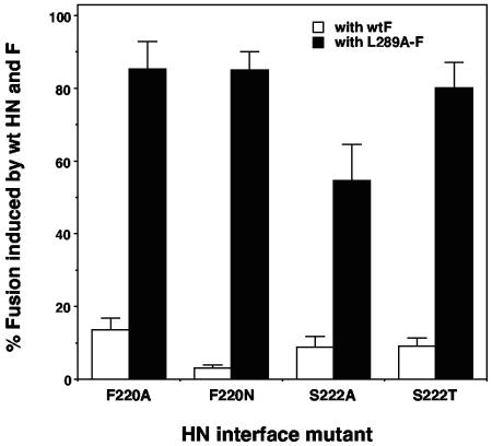
Figure 6 compares the ability of HN proteins carrying substitutions for these residues to complement wt NDV F and the L289A-F protein in the promotion of fusion. Consistent with earlier data (6), HN proteins carrying substitutions of F220A, F220N, S222A, or S222T severely impaired the ability of HN to complement wt F in the promotion of fusion, inducing amounts varying from 3.0 to 13.5% of the wt. However, in each case, expression with F carrying an L289A substitution resulted in a highly significant increase in fusion relative to that achieved with the wt F protein. Expression of L289A-F with HN carrying either an F220A or F220N substitution promoted approximately 85% of wt fusion activity (Fig. 6) while promoting only 13.5 and 3% of activity, respectively, with wt F. Similarly, HN carrying a substitution of S222A or S222T induced 54.7 or 80.3%, respectively, of wt fusion with the mutated F protein, while they promoted only about 9% wt fusion with the wt F protein. Thus, HN proteins carrying substitutions at two residues in the dimer interface, previously demonstrated to decrease receptor binding and in turn to decrease fusion with wt F, are capable of promoting extensive fusion with L289A-mutated F.
FIG. 6.
Comparison of the extent of fusion promoted by wt F and L289A-F expressed with HN proteins carrying mutations for residues F220 and S222 at the membrane-proximal end of the dimer interface. Proteins are expressed in BHK cells driven by the vaccinia virus-T7 RNA polymerase expression system. The extent of fusion is quantitated with the content-mixing fusion assay and expressed as the percentage of that obtained with wt HN and wt F. Error bars show the standard deviation from at least four independent determinations.
One possibility that could account for these findings is a stabilization of the attachment activity of the mutated HN proteins by coexpression of L289A-F. To address this possibility, HAd was compared in the cold and at 37°C for each of the above mutants expressed alone and in the presence of the CSM form of L289A-F. No difference in the attachment activity of any mutated HN protein was detected in the presence of the mutated F protein (data not shown).
NA-deficient HN proteins, which fail to bind receptors or promote fusion with wt NDV F, promote a significant level of fusion with L289A-F.
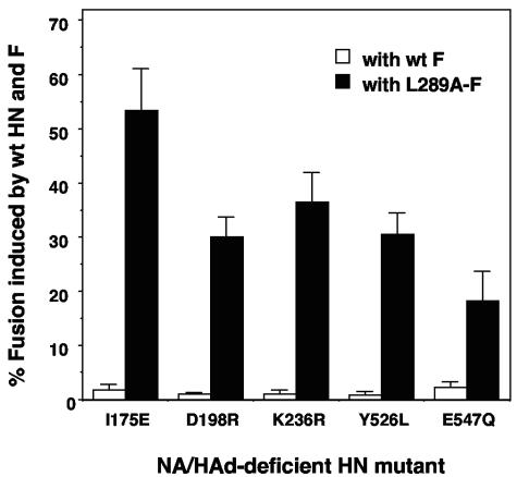
We have previously shown that several substitutions in the NA active site abolish the NA, HAd, and fusion promotion activities of HN, despite efficient expression at the cell surface (17, 21). These substitutions include I175E, D198R, K236R, Y526L, and E547Q. Based on the ability of L289A-F to fuse in the presence of dimer interface mutants, we assayed the fusogenic activity of the mutated F protein when it is coexpressed with each of these functionally deficient HN proteins (Fig. 7). Consistent with our earlier findings, none of the mutated HN proteins complemented wt F in the promotion of fusion at more than 3% of the wt level. This extremely low level of fusion may be due to a minimal amount of attachment activity that is undetectable by the HAd assay. More importantly, expression of each mutated HN protein with L289A-F did result in a significant amount of fusion, ranging from a low value of 18.2% for E547Q-HN to a high of 53.3% for I175E-HN. Thus, L289A-F is able to promote fusion when expressed with HN proteins that lack detectable receptor recognition activity.
FIG. 7.
Comparison of the extent of fusion promoted by wt F and L289A-F expressed with HN proteins carrying mutations in the NA active site that render the protein deficient in attachment function. Proteins are expressed in BHK cells driven by the vaccinia virus-T7 RNA polymerase expression system. The extent of fusion was quantitated with the content-mixing fusion assay and expressed as the percentage of that obtained with wt HN and wt F. Error bars show the standard deviation from at least four independent determinations.
Again, one possible explanation for the ability of these mutated HN proteins to promote fusion with L289A-F, but not the wt protein, is that L289A-F somehow rescues their receptor recognition activity. To test this possibility, the HAd assay was performed both at 4 and 37°C on monolayers in which the mutated HN proteins were coexpressed with the CSM form of L289A-F. None of the mutated HN proteins exhibited HAd activity significantly differently from that obtained when they were expressed in the absence of F (data not shown). Thus, the ability of these HN mutants to promote fusion with L289A-F is not related to a restoration of their receptor binding function.
The L289A substitution does not affect the ability of F to interact with HN at the cell surface.
An interaction between the NDV HN and F proteins at the surface of cells coexpressing the two proteins can be detected in a co-IP assay in which HN complexed with F is precipitated by an antibody to the latter protein (21). The ability of L289A-F to interact with HN, as well as with the NA-HAd-deficient mutants, was tested in this assay. In this experiment, lysates from equal numbers of cells are immunoprecipitated with either a mixture of MAbs to HN and an anti-F MAb or only the anti-F MAb. The percentage of the total amount of each HN protein at the cell surface that coimmunoprecipitates with F was determined.
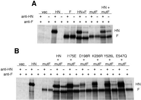
Figure 8A compares the amount of HN coimmunoprecipitated with F and the L289A-mutated form of the protein. In this experiment, both F proteins carry a pair of cleavage site mutations that render the protein dependent on protease treatment for fusion activation (7). This was done to eliminate the difference in the efficiency of IP of the F protein from fusing and nonfusing monolayers (7).
FIG. 8.
Co-IP of wt and mutated HN proteins with L289A-F. Equal numbers of cells were transfected as indicated. Both wt and L289A-mutated F proteins carry mutations in the cleavage site that render them uncleaved and nonfusogenic. After 16 h, the cells were starved and labeled. The cell surface proteins were biotinylated, and the cells were lysed. The lysate was divided into two aliquots and immunoprecipitated with an anti-F MAb and a mixture of anti-HN MAbs (15, 16) (the first lane in each pair) or with the anti-F MAb alone (the second lane in each pair). (A) Comparison of the co-IP of wt HN by the anti-F MAb through its interaction with wt F and L289A-F (indicated as mutF); (B) comparison of the co-IP of wt HN and several NA-HAd-deficient HN mutants by the anti-F MAb through their interaction with L289A-F (mutF).
The HN protein is coimmunoprecipitated by the anti-F MAb from the surface of cells in which it is expressed with either F protein. We previously demonstrated that the coimmunoprecipitated protein is authentic HN by sodium dodecyl sulfate-polyacrylamide gel electrophoresis under nonreducing conditions (21). In previous experiments, approximately one-third of the wt HN at the cell surface was brought down by the antibody to F in this assay (21). In this experiment, 21.5% of HN coimmunoprecipitated with wt F. However, 42% of HN coimmunoprecipitated with L289A-F. Thus, the L289A substitution may increase the ability of the F protein to interact with HN at the cell surface.
Figure 8B also shows the co-IP of HN mutants carrying substitutions of I175E, D198R, K236R, Y526L, or E547Q with L289A-F. Co-IP of these HN mutants is quite analogous to our previous findings with the wt F protein. The D198R- and K236R-mutated HN proteins were weakly coimmunoprecipitated in detectable amounts with the mutated F protein, though they were not with wt F. Y526L- and E547Q-mutated HN proteins, which are not precipitated in significant amounts with the wt F protein, were with L289A-F. Quantitation of the percentage of total HN at the cell surface that was precipitated with L289A-F revealed that 18.5 and 6.4% of the total cell surface of Y526L-HN and E547Q-HN, respectively, coimmnuoprecipitated with the mutated F protein. This may be enough of an interaction to account for the level of fusion obtained with these mutated HN proteins and L289A-F (Fig. 7). Most importantly, I175E-HN, which has been shown to coimmunoprecipitate with wt F, despite its lack of receptor recognition activity, also interacted with L289A-F. Of the total amount at the cell surface, 22.4% could be captured in a complex with the mutated F protein. This was slightly greater than the almost 15% of the protein that coimmunoprecipitated with wt F (21). Thus, L289A-F appears to interact at the cell surface more efficiently than the wt F protein with wt HN and several mutated HN proteins. Note that the difference in migration rate of F is dependent on the NA activity of the coexpressed HN protein and is due to the presence or absence of sialic acid.
Dependence of L289A-F-mediated fusion on sialic acid receptors.
Based on the ability of L289A-mutated F to promote a significant amount of fusion with HN proteins that are either deficient in receptor recognition or bind receptors with diminished stability, the possibility exists that the mutated F protein is capable of promoting fusion using a receptor other than sialic acid.
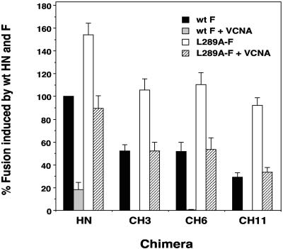
To test this possibility, we took two approaches. First, target cells in the content-mixing fusion assay were treated with exogenous VCNA prior to being mixed with cells coexpressing HN and either wt F or L289A-F. As shown in Fig. 9, the promotion of fusion by wt HN-F-expressing cells was reduced by more than 80% by treatment of the target cells with VCNA. However, the fusion-promoting activity of HN-L289A-F-expressing monolayers was significantly more resistant to removal of sialic acid receptors. They retained more than 50% of their original activity, which corresponded to nearly 90% of the activity exhibited by wt HN-F-expressing monolayers with untreated target cells. The fusion activity of wt HN-F-expressing cells that is resistant to exogenous NA treatment of target cells may be due to the inability of the VCNA treatment to completely deplete the cells of sialic acid. Even treatment with higher concentrations of VCNA did not completely eliminate wt HN-F-mediated fusion, suggesting that the amount of enzyme used is not limiting (data not shown). Thus, it seems more likely that, over the course of the 5-h incubation in the content-mixing assay, receptors are being replenished at the surface of the target cells.
FIG. 9.
Comparison of the effect of VCNA treatment of target cells on the promotion of fusion by wt F or L289A-F. Wt F and L289A-F were coexpressed with either wt HN or different NDV-hPIV3 HN chimeras. Target cells in the content-mixing assay for fusion were either untreated or treated for 1 h at 37°C with 50 mU of VCNA/ml. The extent of fusion induced by the two wt proteins was set at 100%, and fusion promotion by other combinations is expressed as a percentage of this amount. Error bars show the standard deviation from at least four independent determinations.
Thus, we used the same approach with the less fusogenic NDV-hPIV3 chimeras CH3, CH6, and CH11. The fusion-promoting activity of wt F complemented with each of these chimeras was completely eliminated by VCNA treatment (Fig. 9). These chimeras and wt F promoted 30 to 50% of wt fusion, but this fusion was completely eliminated by the VCNA treatment. Yet, fusion promoted by cells coexpressing CH3, CH6, or CH11 with L289A-F exhibited significant resistance to VCNA-mediated removal of sialic acid on target cells. The three chimeras normally promote fusion with L289A-F approximately as effectively as wt HN does with wt F. Even after VCNA treatment of target cells, chimeras CH3 and CH6 still retained one-half, and chimera CH11 retained one-third, of the fusion they exhibited with L289A-F (Fig. 9). Thus, the promotion of fusion by L289A-F is partially resistant to the depletion of sialic acid receptors.
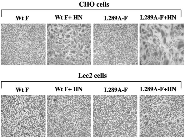
As a second approach to understanding the role of sialic acid receptors in L289A-F-mediated fusion, we used a sialic acid-deficient cell line. Lec2 cells are CHO-derived cells that are defective in the expression of sialic acid due to a block in the translocation of CMP-sialic acid to the lumen of the Golgi apparatus (9). CHO cells served as a positive control in this experiment. HN and either wt F or L289A-F were coexpressed in the two cell lines with the pCAGGS expression system. As shown in Fig. 3, coexpression of HN and L289A-F in CHO cells resulted in almost a twofold increase in the amount of fusion achieved with the two wt proteins in these cells. Figure 10 shows CHO and Lec2 monolayers transfected with wt F or the L289A-mutated protein in both the presence and absence of HN. Coexpression of HN and F in the sialic acid-deficient Lec2 cells failed to promote detectable amounts of fusion. However, coexpression of L289A-F and HN still resulted in a minimal, but detectable, amount of fusion in Lec2 cells. These cells still retained approximately 5 to 10% of the sialic acid present in the control cells (9). It is likely that this minimal amount of sialic acid was sufficient for the L289A-F protein to promote fusion, whereas there was not enough receptor binding activity to complement the wt F protein.
FIG. 10.
Syncytium formation in CHO and sialic acid-deficient Lec2 cells induced by wt F and L289A-F with and without HN. In all monolayers, the indicated proteins are expressed from the pCAGGS expression vector. In the top row, the indicated proteins are expressed in CHO cells. In the bottom row, the indicated proteins are expressed in Lec2 cells. The monolayers were fixed with methanol and stained with Giemsa stain at 18 h posttransfection.
MAbs detect a difference between L289A-mutated F and the wt protein.
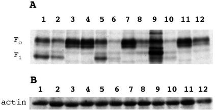
Cells expressing wt F and L289A-F, as well as the CSM forms of each protein, were individually immunoprecipitated with two MAbs specific for the NDV F protein. Equivalent amounts of cells were radiolabeled, and the proteins were chased to the surface. The F proteins were immunoprecipitated with one of two MAbs, Fa or Fb (Fig. 11A), or a polyclonal rabbit anti-NDV serum. The latter served as a control. The two MAbs recognized different conformation-dependent epitopes, based on their failure to bind to wt F in Western blots (data not shown). Wt F and L289A-F migrated as two bands, the uncleaved (Fo) and cleaved (F1) forms. However, as expected, F1 was not detectable in the IP of the CSM form of either the wt or the mutated F protein.
FIG. 11.
MAbs detect a conformational difference between L289A-F and the wt F protein. (A) Cells were transfected as follows: wt F, lanes 1, 5, and 9; L289A-F, lanes 2, 6, and 10; CSM F, lanes 3, 7, and 11; and CSM L289A-F, lanes 4, 8, and 12. Proteins were expressed in BHK cells driven by the vaccinia virus-T7 RNA polymerase expression system. Cells were starved for 1 h, labeled for 3 h, and chased for 90 min. Cells were lysed in Triton-deoxycholate. (A) Lanes 1 to 4, the F protein was immunoprecipitated with polyclonal rabbit anti-NDV serum; lanes 5 to 8, the F protein was immunoprecipitated with MAb Fa; lanes 9 to 12, the F protein was immunoprecipitated with MAb Fb. (B) An aliquot removed from each lysate before the addition of antibody was assayed by Western blotting to quantify cellular actin as a gel loading control.
Both MAbs immunoprecipitated significantly less of both L289A-mutated F proteins than the corresponding wt or CSM F protein (Fig. 11A, compare lanes 5 and 6, 7 and 8, 9 and 10, and 11 and 12). Densitometric quantitation of CSM F and CSM L289A-F immunoprecipitated by the two MAbs reveal L289A-F to wt F ratios of 0.59 and 0.54, respectively, for MAbs Fa and Fb. These results are supported by flow cytometric analyses, which showed that L289A-F is recognized 64.5% ± 6.2% and 55.7% ± 11.1% as well as the wt protein by MAb Fa and Fb, respectively. Unlike the two MAbs, the antiserum immunoprecipitated comparable amounts of CSM F and L289A-CSM F (Fig. 11A, lanes 3 and 4). Densitometric quantitation of lanes 3 and 4 in Fig. 11A gave a ratio of 1.07 for the CSM forms of L289A-F to that of the wt F protein. This indicates that the difference detected by the two MAbs was not due simply to a difference in cell surface expression.
The results with the cleaved form of the protein were similar (Fig. 11A). Both MAbs also immunoprecipitated L289A-F less efficiently than the wt protein, with ratios of L289A-F to wt F of 0.27 and 0.25 for Fa and Fb, respectively (Fig. 11A, compare lanes 5 and 6 and 9 and 10). Though the polyclonal serum also immunoprecipitated L289A-F less efficiently than the wt protein (ratio of 0.58) (Fig. 11A, lanes 1 and 2), this still meant that both L289A-mutated forms of the F protein were recognized about one-half as well as the corresponding wt form of the protein. Thus, two F-specific MAbs detected a conformational change in the F protein resulting from the L289A substitution. Figure 11B shows a Western blot loading control in which an aliquot from each lysate in panel A was probed with antibody to actin.
DISCUSSION
Similar to the activity of most paramyxoviruses, the fusion promotion activity of the wt NDV F protein is completely dependent on contributions from the homologous HN protein. One of the functions of HN in fusion is its ability to recognize sialic acid receptors. Despite the conservation of this activity among members of the paramyxovirus genus, heterologous HN proteins cannot substitute for NDV HN in complementing NDV F in the promotion of fusion. This is consistent with the existence of a virus-specific interaction between the two NDV glycoproteins demonstrable at the surface of both infected and transfected cells (7, 21). The specificity of NDV HN-F-mediated fusion is determined by the stalk region of the HN ectodomain (8, 32).
It has previously been demonstrated that a single amino acid substitution, L289A, in the NDV F protein alters the requirement for HN in the promotion of fusion (26). NDV F, carrying this substitution, is capable of promoting a significant level of syncytium formation in Cos-7 cells in the absence of HN. Here, we show that this HN-independent mode of fusion is apparently specific to this cell line. In several other cell types, L289A-F promotes less than 3% of wt fusion in the absence of HN.
The fact that HN-independent fusion is exhibited only in Cos-7 cells can potentially be explained in several ways. The possibility exists that only Cos-7 cells display an unidentified receptor molecule on their surfaces that is recognized by L289A-F. Alternatively, the unknown molecule may be present on the surfaces of all of the cells tested but may be present in significantly greater amounts on Cos-7 cells. Finally, HN-independent fusion may simply be a reflection of an inherently easier fusogenicity of Cos-7 cells.
We attempted to discriminate between these possibilities by performing the content-mixing fusion assay with HN and F coexpressed in one cell type (BHK or Cos-7) with the other cell type (BHK or Cos-7) serving as receptor-bearing target cells. We have been unable to detect significant fusion with either combination of cells. However, interpretation of these experiments is limited by the fact that Cos-7 cells do not tolerate the vaccinia virus well, necessitating the use of lower MOIs.
The combination of HN and L289A-mutated F consistently promotes a significantly greater amount of fusion than that promoted by the two wt proteins in all of the cell lines tested. Thus, we compared several parameters of the HN-dependent mode of fusion of this mutant F protein to that of wt F. In many ways, the mutated F protein is identical to the wt F protein. As we have shown previously for the wt F protein (8), L289A-F-induced fusion is not complemented by the heterologous hPIV3 HN protein. Thus, the contribution of receptor recognition activity, even by a heterologous HN protein, is not sufficient for L289A-F to induce fusion. Related to this finding, the mutated F protein shows the same fusion specificity exhibited by the wt protein with respect to NDV-hPIV3 HN chimeras. The stalk region of the HN spike determines the fusion specificity of L289A-F, just as it does the wt F protein.
Finally, L289A-mutated F appears to interact more efficiently with HN than does the wt F protein. We have previously shown that wt HN and F can be coimmunoprecipitated from the surface of cells coexpressing the two proteins with an antibody to the F protein (21). Approximately 42% of the HN present at the cell surface is coimmunoprecipitated through its interaction with L289A-F. This compares to the 21.5% of HN brought down with the wt F protein in the same experiment. This means that the changes induced in the F protein by the L289A substitution not only fail to interfere with its ability to interact with the HN protein at the cell surface, but rather may slightly enhance the interaction. The mutated F protein also displays a slightly different phenotype with respect to its ability to interact with a panel of NA-HAd- and fusion-deficient HN proteins. D198R- and K236R-mutated HN proteins are weakly coimmunoprecipitated with the L289A-F protein, though they are not with wt F. Also, Y526L- and E547Q-mutated HN proteins, which are not precipitated in significant amounts with the wt F protein, are precipitated in minimal amounts with L289A-F. Most importantly, I175E-HN, approximately 15% of which has been shown to coimmunoprecipitate with wt F despite its lack of receptor recognition activity, interacts more efficiently with L289A-F (approximately 23% of the total amount). Thus, not only does L289A-F interact with HN at the cell surface, but it does so more efficiently than the wt F protein.
However, L289A-F differs from the wt protein in one very important way. Although it requires the receptor recognition activity of the homologous HN protein in order to promote fusion (except in Cos-7 cells), it is much less dependent on this activity than the wt protein. This is demonstrated in several ways. First, the mutated F protein is capable of promoting significant levels of fusion with the I175E, D198R, K236R, Y526L, and E547Q HN mutants. Most notably, the combination of I175E-HN and L289A-F results in more than 50% of the level of fusion achieved with the two wt proteins. Fusion obtained with the other HN mutants and L289A-F ranges between 18 to 36% of that obtained with wt HN and F. This is in contrast to the negligible amount of fusion obtained by coexpression of these mutated HN proteins with wt F. The slight interaction detectable between these HN proteins and L289A-F does not appear to be sufficient to account for the significant level of fusion observed. Thus, L289A-F-induced fusion is still triggered under conditions where the HN-receptor interaction is undetectable by the HAd assay.
Second, L289A-F gives markedly enhanced levels of fusion with HN proteins carrying substitutions for residues F220 and S222. For example, coexpression of either F220N-HN or S222T-HN with wt F results in more than 80% of wt fusion. This is a dramatic increase over the less than 10% of wt fusion obtained when these mutated HN proteins are coexpressed with the wt F protein. The diminished fusogenic activity of these mutated HN proteins correlates with an unstable interaction between HN and receptors on target cells. Evidently, this interaction, although apparently too transient to enable the wt F protein to promote fusion, is strong enough to trigger fusion by L289A-F. The mutated F protein is able to promote fusion even with a weaker HN-receptor interaction.
Third, fusion induced by L289A-F is significantly more resistant to receptor removal than that induced by the wt F protein. Target cells were treated with exogenous NA in an attempt to deplete them of sialic acid receptors. This treatment did not completely eliminate fusion by wt HN and F; 18% of wt fusion survives this treatment. The remaining fusion is likely due to a very low level of receptors replenished during the incubation period of the content-mixing fusion assay. Still, the VCNA-treated monolayers coexpressing L289A-F and wt HN promote a level of fusion that is not significantly different from that obtained with untreated monolayers coexpressing the two wt proteins. Similarly, although fusion induced by wt F and NDV-hPIV3 HN chimeras CH3, CH6, and CH11 is completely blocked by VCNA treatment; monolayers coexpressing these chimeras and L289A-F also promote a level of fusion comparable to that exhibited by untreated monolayers coexpressing the respective chimera and the wt F protein. Again, these findings are consistent with a decreased dependence of L289A-F induced fusion on receptor binding by HN. In fact, the mutated F protein is capable of promoting fusion, albeit to a relatively minimal extent, in sialic acid-deficient Lec2 cells. This minimal amount of fusion is likely due to the fact that Lec2 cells still retain 5 to 10% of the wt amount of sialic acid (9). Although this amount of sialic acid is insufficient for the wt F protein to promote fusion, it is enough for the L289A-mutated protein.
There are several examples of membrane fusion induced by expression of only the fusion protein. Most notably, the F protein of the W3A strain of another paramyxovirus, simian virus 5, is capable of promoting HN-independent fusion. Based on sequence differences with the F protein of the highly homologous WR strain, which requires HN for fusion, it was shown that two amino acids, proline 22 and serine 443, are responsible for the HN-independent phenotype (23, 30). Other examples of attachment protein-independent fusion include the F proteins of several respiratory syncytial viruses in the Pneumovirus genus (19) and that of peste-des-petits-ruminants virus, a morbillivirus (27). However, it should be noted that the fusion promotion activity of each these attachment protein-independent F proteins is significantly enhanced by coexpression of the homologous receptor binding protein, as it is for L289A-mutated NDV F.
Attachment protein-independent fusion has also been induced by site-directed amino acid substitutions in important domains of various F proteins. In this way, it was shown that residues E132 in HR1 and A290 in HR3 also contribute to the HN-independent fusion activity of simian virus 5 F (18). Similarly, mutations in the cytoplasmic domain of the SER virus F protein rescue syncytium formation and eliminate the HN protein requirement for membrane fusion (28).
The mechanism of attachment protein independent fusion is poorly understood. Perhaps, the best-characterized system is that of the respiratory syncytial virus F protein. This protein can bind cellular heparin and heparan sulfate receptors and trigger a conformational change in the F protein, leading to extensive fusion in the absence of the viral attachment protein (1, 10). Recently, it has been shown that heparan sulfate also acts as a receptor for two other paramyxoviruses, Sendai virus and hPIV3 (2). Consistent with this, the F proteins of these viruses have several highly basic heparan sufate binding motifs (2, 10). However, it seems unlikely that heparan sulfate serves as a receptor for NDV F. The only such motif in the protein is the F cleavage site itself, which is not conserved in avirulent strains of the virus (13). Also, depletion of heparan sulfate from the cell surface with heparinase I has no significant effect on the extent of fusion induced by either wt F or L289A-F (data not shown).
The structural importance of HR3, of which L289 is a part, is most likely related to the fact that it is situated between HR1 and HR2. The L289A substitution may weaken the helical structure of HR3 and thereby lower the energy needed for the conformational change that results in HR1 and HR2 coming together in the fusion-active form of F. In addition, the reduced energy of activation of the L289A-mutated NDV F protein correlates with a conformational change in the protein detectable by conformation-specific antibodies.
Acknowledgments
We gratefully acknowledge Judith Alamares, Elizabeth Corey, and Paul Mahon for critical reading of the manuscript and helpful discussions. We also thank Robert Lamb and Trudy Morrison for the NDV F and HN genes, respectively, and Bernard Moss for the recombinant vaccinia virus.
This work was made possible by grant AI-49268 from the National Institutes of Health.
REFERENCES
- 1.Barretto, N., L. K. Hallak, and M. E. Peeples. 2003. Neuraminidase treatment of respiratory syncytial virus-infected cells or virions, but not target cells, enhances cell-cell fusion and infection. Virology 313:33-43. [DOI] [PubMed] [Google Scholar]
- 2.Bose, S., and A. K. Banerjee. 2002. Role of heparan sulfate in human parainfluenza virus type 3 infection. Virology 298:73-83. [DOI] [PubMed] [Google Scholar]
- 3.Buckland, R., E. Malvoisin, P. Beauverger, and F. Wild. 1992. A leucine zipper structure present in the measles virus fusion protein is not required for its tetramerization but is essential for fusion. J. Gen. Virol. 73:1703-1707. [DOI] [PubMed] [Google Scholar]
- 4.Chambers, P., C. R. Pringle, and J. J. Easton. 1990. Heptad repeat sequences are located adjacent to hydrophobic regions in several types of virus fusion glycoproteins. J. Gen. Virol. 71:3075-3080. [DOI] [PubMed] [Google Scholar]
- 5.Chen, L., J. J. Gorman, J. McKimm-Breschkin, L. J. Lawrence, P. A. Tulloch, B. J. Smith, P. M. Colman, and M. C. Lawrence. 2001. The structure of the fusion glycoprotein of Newcastle disease virus suggests a novel paradigm for the molecular mechanism of membrane fusion. Structure 9:255-266. [DOI] [PubMed] [Google Scholar]
- 6.Corey, E. A., A. M. Mirza, E. Levandowsky, and R. M. Iorio. 2003. Fusion deficiency induced by mutations at the dimer interface in the Newcastle disease virus hemagglutinin-neuraminidase is due to a temperature-dependent defect in receptor binding. J. Virol. 77:6913-6922. [DOI] [PMC free article] [PubMed] [Google Scholar]
- 7.Deng, R., Z. Wang, P. J. Mahon, M. Marinello, A. M. Mirza, and R. M. Iorio. 1999. Mutations in the NDV HN protein that interfere with its ability to interact with the homologous F protein in the promotion of fusion. Virology 253:43-54. [DOI] [PubMed] [Google Scholar]
- 8.Deng, R., Z. Wang, A. M. Mirza, and R. M. Iorio. 1995. Localization of a domain on the paramyxovirus attachment protein required for the promotion of cellular fusion by its homologous fusion protein spike. Virology 209:457-469. [DOI] [PubMed] [Google Scholar]
- 9.Eckhardt, M., B. Gotza, and R. Gerardy-Schahn. 1998. Mutants of the CMP-sialic acid transporter causing the Lec2 phenotype. J. Biol. Chem. 273:20189-20195. [DOI] [PubMed] [Google Scholar]
- 10.Feldman, S. A., S. A. Audet, and J. A. Beeler. 2000. The fusion glycoprotein of human respiratory syncytial virus facilitates virus attachment and infectivity via an interaction with cellular heparan sulfate. J. Virol. 74:6442-6447. [DOI] [PMC free article] [PubMed] [Google Scholar]
- 11.Fuerst, T. R., E. G. Niles, F. W. Studier, and B. Moss. 1986. Eucaryotic transient expression system based on recombinant vaccinia virus that synthesizes bacteriophage T7 RNA polymerase. Proc. Natl. Acad. Sci. USA 83:8122-8126. [DOI] [PMC free article] [PubMed] [Google Scholar]
- 12.Ghosh, J. K., M. Ovadia, and Y. Shai. 1997. A leucine zipper motif in the ectodomain of Sendai virus fusion protein assembles in solution and in membranes and specifically binds biologically-active peptides and the virus. Biochemistry 36:15451-15462. [DOI] [PubMed] [Google Scholar]
- 13.Glickman, R. L., R. J. Syddall, R. M. Iorio, J. P. Sheehan, and M. A. Bratt. 1988. Quantitative basic residue requirements in the cleavage-activation site of the fusion glycoprotein as a determinant of virulence for Newcastle disease virus. J. Virol. 62:354-356. [DOI] [PMC free article] [PubMed] [Google Scholar]
- 14.Hu, X., R. Ray, and R. W. Compans. 1992. Functional interactions between the fusion protein and hemagglutinin-neuraminidase of human parainfluenza viruses. J. Virol. 66:1528-1534. [DOI] [PMC free article] [PubMed] [Google Scholar]
- 15.Iorio, R. M., J. B. Borgman, R. L. Glickman, and M. A. Bratt. 1986. Genetic variation within a neutralizing domain on the haemagglutinin-neuraminidase glycoprotein of Newcastle disease virus. J. Gen. Virol. 67:1393-1403. [DOI] [PubMed] [Google Scholar]
- 16.Iorio, R. M., and M. A. Bratt. 1983. Monoclonal antibodies to Newcastle disease virus: delineation of four epitopes on the HN glycoprotein. J. Virol. 48:440-450. [DOI] [PMC free article] [PubMed] [Google Scholar]
- 17.Iorio, R. M., G. M. Field, J. M. Sauvron, A. M. Mirza, R. Deng, P. J. Mahon, and J. Langedijk. 2001. Structural and functional relationship between the receptor recognition and neuraminidase activities of the Newcastle disease virus hemagglutinin-neuraminidase protein: receptor recognition is dependent on neuraminidase activity. J. Virol. 75:1918-1927. [DOI] [PMC free article] [PubMed] [Google Scholar]
- 18.Ito, M., M. Nishio, H. Komoda, Y. Ito, and M. Tsurudome. 2000. An amino acid in the heptad repeat 1 domain is important for the haemagglutinin-neuraminidase-independent fusing activity of simian virus 5 fusion protein. J. Gen. Virol. 81:719-727. [DOI] [PubMed] [Google Scholar]
- 19.Kahn, J. S., M. J. Schnell, L. Buonocore, and J. K. Rose. 1999. Recombinant vesicular stomatitis virus expressing respiratory syncytial virus (RSV) glycoproteins: RSV fusion protein can mediate infection and cell fusion. Virology 254:81-91. [DOI] [PubMed] [Google Scholar]
- 20.Lamb, R. A., and D. Kolakofsky. 2001. Paramyxoviridae: the viruses and their replication, p. 689-724. In D. M. Knipe and P. M. Howley (ed.), Fields virology, 4th ed., Lippincott, Williams & Wilkins, Philadelphia, Pa.
- 21.Li, J., E. Quinlan, A. Mirza, and R. M. Iorio. 2004. Mutated form of the Newcastle disease virus hemagglutinin-neuraminidase interacts with the homologous fusion protein despite deficiencies in both receptor recognition and fusion promotion. J. Virol. 78:5299-5310. [DOI] [PMC free article] [PubMed] [Google Scholar]
- 22.Mirza, A. M., R. Deng, and R. M. Iorio. 1994. Site-directed mutagenesis of a conserved hexapeptide in the paramyxovirus hemagglutinin-neuraminidase glycoprotein: effects on antigenic structure and function. J. Virol. 68:5093-5099. [DOI] [PMC free article] [PubMed] [Google Scholar]
- 23.Paterson, R. G., C. J. Russell, and R. A. Lamb. 2000. Fusion protein of the paramyxovirus SV5: destabilizing and stabilizing mutants of fusion activation. Virology 270:17-30. [DOI] [PubMed] [Google Scholar]
- 24.Russell, C. J., T. S. Jardetzky, and R. A. Lamb. 2001. Membrane fusion machines of paramyxoviruses: capture of intermediates of fusion. EMBO J. 20:4024-4034. [DOI] [PMC free article] [PubMed] [Google Scholar]
- 25.Scheid, A., and P. W. Choppin. 1973. Isolation and purification of the envelope proteins of Newcastle disease virus. J. Virol. 11:263-271. [DOI] [PMC free article] [PubMed] [Google Scholar]
- 26.Sergel, T. A., L. W. McGinnes, and T. G. Morrison. 2000. A single amino acid change in the Newcastle disease virus fusion protein alters the requirement for HN protein in fusion. J. Virol. 74:5101-5107. [DOI] [PMC free article] [PubMed] [Google Scholar]
- 27.Seth, S., and M. S. Shaila. 2001. The fusion protein of peste des petits ruminants virus mediates biological fusion in the absence of hemagglutinin-neuraminidase protein. Virology 289:86-94. [DOI] [PubMed] [Google Scholar]
- 28.Seth, S., A. Vincent, and R. W. Compans. 2003. Mutations in the cytoplasmic domain of a paramyxovirus fusion glycoprotein rescue syncytium formation and eliminate the hemagglutinin-neuraminidase protein requirement for membrane fusion. J. Virol. 77:167-178. [DOI] [PMC free article] [PubMed] [Google Scholar]
- 29.Tanabayashi, K., and R. W. Compans. 1996. Functional interaction of paramyxovirus glycoproteins: identification of a domain in Sendai virus HN which promotes cell fusion. J. Virol. 70:6112-6118. [DOI] [PMC free article] [PubMed] [Google Scholar]
- 30.Tsurudome, M., M. Ito, M. Nishio, M. Kawana, H. Komada, and Y. Ito. 2001. Hemagglutinin-neuraminidase-independent fusion activity of simian virus 5 fusion (F) protein: difference in conformation between fusogenic and nonfusogenic F proteins on the cell surface. J. Virol. 75:8999-9009. [DOI] [PMC free article] [PubMed] [Google Scholar]
- 31.Tsurodome, M., M. Kawano, T. Yuasa, N. Tabata, M. Nishio, H. Komada, and Y. Ito. 1995. Identification of regions of the hemagglutinin-neuramindase protein of human parainfluenza virus type 2 important for promoting cell fusion. Virology 171:38-48. [DOI] [PubMed] [Google Scholar]
- 32.Wang, Z., A. M. Mirza, J. Li, P. J. Mahon, and R. M. Iorio. 2004. An oligosaccharide at the C-terminus of the F-specific domain in the stalk of the human parainfluenza virus 3 hemagglutinin-neuraminidase modulates fusion. Virus Res. 99:177-185. [DOI] [PubMed] [Google Scholar]
- 33.Yao, Q., X. Hu, and R. W. Compans. 1997. Association of the parainfluenza virus fusion and hemagglutinin-neuraminidase glycoproteins on cell surfaces. J. Virol. 71:650-656. [DOI] [PMC free article] [PubMed] [Google Scholar]


