Abstract
Background
Numerous procedures have been described for the treatment of chronic recurrent dislocation of the temporo-mandibular joint (TMJ), either in the form of enhancement or restriction of the condylar movement, with their obvious merits and demerits.
Materials and Method
We present a new technique of using U shaped iliac bone graft to restrict the condylar movement and its advantages over the conventional techniques.We have used this technique successfully in 8 cases where Dautrey’s procedure had failed with follow up period of 2 years.
Results
No patient complained of recurrent dislocation postoperatively.
Conclusion
This a very simple and effective technique where other procedures have failed.
Keywords: Chronic recurrent dislocation, Modified iliac bone graft, Temporo-mandibular joint
Introduction
Numerous procedures have been described for the treatment of chronic recurrent dislocation of the temporo-mandibular joint, either to enhance or restrict condylar movements with their obvious merits and demerits.
Procedures enhancing the path of condylar movement permit continued dislocation of condyle and may lead to articular damage. Procedures inhibiting the path of condylar movement may result in persistent medial dislocation of condyle, as cross-section of zygomatic arch is limited medio-laterally [1, 2]. Use of various alloplastic materials for restrictive procedures, pose threat of foreign body reaction [3].
Technique
We use preauricular approach to expose the articular eminence and zygomatic arch. A pocket of dimension 20 mm × 20 mm is created beneath the articular eminence and zygomatic arch. A bicortical graft of (L × H) 15 mm × 15 mm is harvested from tubercle of Ilium. The medullary portion of the graft is carved till the required depth, while maintaining the cortical height thus giving it a “U” shape (Fig. 1). The height of the graft can be varied as per need of the case. This “U” shaped graft is placed from inferior aspect at the root of zygoma to embrace it from three sides, inferior, lateral and medial within the created pocket beneath zygomatic arch. Two positional screws through a washer are placed to fix the graft and prevent fracture (Fig. 2). The screw passes through lateral cortex of graft, the zygoma and engages the medial cortex of the graft (Fig. 3). Thus, one cortex of the graft lays lateral to arch and the other lies on the medial aspect, while the arch lies between two cortices of the graft.
Fig. 1.

Harvested tubercle of ilium and shaped as U graft
Fig. 2.

Black solid short arrow showing zygomatic arch; black thin long arrow showing the graft fixation with positional screws through washer; white arrow with black border showing the condyle
Fig. 3.
Schematic representation of graft fixation to zygomatic arch as seen in coronal view
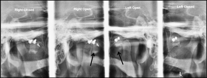
Intra-operatively condylar movements are checked for adequate restriction especially on medial aspect. Postoperatively soft diet is advised and radiographs are obtained to evaluate the efficacy of the procedure (Fig. 4).
Fig. 4.
Postoperative radiograph showing graft (black arrow) restricting movement of condyle
Total resolution of recurrent dislocation was achieved immediately after surgery in our patients. The width of the Ilium at the tubercle with optimal height of medial and lateral cortices of graft and wise removal of medullary bone provides perfect mechanical obstruction even for medial dislocation. We have used this technique successfully in 8 cases where Dautrey’s procedure had failed with minimal 2 years of follow-up. No patient complained of recurrent dislocation postoperatively.
Conclusion
Author`s technique is simple, effective and can be used in all age groups as gleno-temporal osteotomy is avoided. It provides superior restrictive capabilities due to cortical nature of graft and quadra-cortical fixation with lesser hardware making it cost effective with reduced possibility of foreign body reaction. This technique can successfully be used where Dautrey’s procedure has failed.
The only shortcoming of the procedure is need of second surgical site with associated morbidities.
Compliance with Ethical Standards
Conflict of interest
None.
Institutional Ethical Committee Approval
Obtained.
Informed Consent from All the Patients
Obtained.
References
- 1.Costas Lopez A, Monje Gil F, Fernandez Sanroman J, Goizueta Adame C, Castro Ruiz PC. Glenotemporal osteotomy as a definitive treatment for recurrent dislocation of the jaw. J Craniomaxillofac Surg. 1996;24:178–183. doi: 10.1016/S1010-5182(96)80053-9. [DOI] [PubMed] [Google Scholar]
- 2.Gadre K, Kaul D, Ramanojam S, Shah S. Dautrey’s procedure in treatment of recurrent dislocation of the mandible. J Oral Maxillofac Surg. 2010;68:2021–2024. doi: 10.1016/j.joms.2009.10.015. [DOI] [PubMed] [Google Scholar]
- 3.Medraa AM, Mahrous AM. Glenotemporal osteotomy and bone grafting in the management of chronic recurrent dislocation and hypermobility of the temporomandibular joint. Br J Oral Maxillofac Surg. 2008;46:119–122. doi: 10.1016/j.bjoms.2007.08.004. [DOI] [PubMed] [Google Scholar]


