Abstract
Translational initiation of most eukaryotic mRNAs occurs when a preinitiation complex binds to the 5′ cap, scans the mRNA, and selects a particular AUG codon as the initiation site. Selection of the correct initiation codon relies, in part, on its flanking residues; in mammalian cells, the core of the “Kozak” consensus is R−3CCAUGG+4 (R = purine; the A residue is designated position +1). The R−3 is considered the most important flanking residue, followed by G+4. Picornaviral mRNAs differ from most cellular mRNAs in several ways; they are uncapped, and they contain an internal ribosome entry site that allows the ribosome to bind near the initiation codon. The initiation codon of coxsackievirus B3 (CVB3) is flanked by both R−3 and G+4 (AAAATGG). Here, we report the construction of full-length CVB3 genomes that vary at these two positions, and we evaluate the effects of these variant sequences in vitro, in tissue culture cells, and in vivo. A virus with an A→C transversion at position −3 replicates as well as wild-type CVB3, both in tissue culture and in vivo. This virus is highly pathogenic, and its sequence is stable throughout the course of an in vivo infection. Furthermore, the in vitro translation products from this RNA are very similar to the wild type. Thus, R−3—thought to be the most functionally important component of the Kozak consensus—appears to be dispensable in CVB3. In contrast, a G-to-C transversion at G+4 is lethal; RNAs carrying this mutation fail to generate infectious virus either in tissue culture or in vivo. However, in vitro analysis indicates that G+4 has only a marginal effect on translational initiation, especially if R−3 is present; instead, the G+4 is required mainly because the second triplet of the polyprotein open reading frame must encode glycine, without which infectious virus production cannot proceed. In summary, our data indicate that CVB3 remains viable, even in vivo, in the absence of R−3, and we propose that the most important factor contributing to the high frequency of G+4—not only in CVB but also in other eukaryotic mRNAs, and thus in the consensus motif itself—may be the constraint upon the second amino acid rather than the requirements for translational initiation.
Translational initiation in eukaryotic cells usually requires that a 40S ribosomal subunit (along with other components, including a charged initiator  ) attaches to the m7GpppN cap at the 5′ end of an mRNA and moves along the molecule, in a process termed scanning, until it encounters the first potential translational initiation codon (AUG). At this point, the 60S subunit joins, and the resulting 80S ribosome begins protein synthesis. However, for some mRNAs, scanning is leaky; the preinitiation complex bypasses the first AUG, and translation is instead initiated at a downstream methionine codon. Sequences immediately flanking an AUG are thought to play a key part in the selection of the initiation codon by a scanning complex, and comparative sequence analysis of a variety of known vertebrate initiation codons (21) revealed an optimal consensus context of RYYAUGG (R = purine, Y = pyrimidine); subsequent molecular studies have slightly refined this, to RCCAUGG (23). By convention, the first A of the initiation codon is identified as base +1; with this nomenclature, the purine at position −3 (R−3) is the most highly conserved, being present in >90% of vertebrate mRNAs (22, 38), and is thought to be functionally the most important position (24). The G+4 also is highly conserved and contributes strongly, especially in the absence of an A at position −3 (23); however, the central CC in the consensus motif can vary without major consequences, as long as positions −3 and +4 conform (20).
Picornaviruses represent an exception to the general rules concerning ribosomal attachment. The mRNAs of these viruses do not have a 5′-terminal m7GpppN cap, and the AUG used for translational initiation lies far (usually ∼700 to ∼1,400 nucleotides) from the 5′ end of the RNA molecule. Strikingly, the intervening sequence—the 5′ untranslated region (5′UTR)—is by no means devoid of AUG codons; the number varies among different picornaviruses, but often is six to eight; and some of these codons are surrounded by a strong consensus motif. These observations raised two questions. First, how does the 40S subunit enter the mRNA, in the absence of a cap? Second, why does ribosomal scanning ignore several “good” AUGs present in the 5′UTR? Seminal studies showed that a portion of the 5′UTR could act as “landing pad” for ribosomes, thus enhancing translation of a downstream cistron (16, 34, 35). This structure, named the internal ribosomal entry site (IRES), can accept ribosomes even in the absence of a 5′ end (shown by IRES-dependent translation of a circularized mRNA molecule [5]). Further analysis of IRES function also suggested an answer to the second question: the IRES allows the 40S component to enter the mRNA downstream of the unused AUGs, so the preinitiation complex never encounters these potential translational initiation sites. Most picornaviral IRES elements belong to one of two groups, which differ in sequence, secondary structure, and the location of the translational initiation codon. Enteroviruses (e.g., polioviruses and coxsackieviruses) and rhinoviruses contain a type I IRES, and the initiation codon is usually ca. 50 to 150 nucleotides downstream from the 3′ end of the ribosome-binding site. In this case, ribosomal scanning seems likely, and there is evidence that it occurs (32). However, the importance of the AUG flanking sequences (in particular, the R−3 and G+4 residues) in IRES-driven translational initiation remains uncertain. Here, we investigate the requirement for R−3 and G+4 in the coxsackievirus B3 (CVB3) life cycle, by evaluating the viability of mutant viruses not only in tissue culture cells but also in vivo (a term which we reserve to denote experiments carried out in living animals). The latter approach distinguishes our studies from most previous analyses of AUG contexts, which relied solely on studies in vitro, or in transfected tissue culture cells (referred to by some workers as “in vivo”).
The goals of the present study were, therefore, (i) to determine whether the R−3 and/or the G+4 sequences of the wild-type (wt) CVB3 initiation codon (AAAAUGG) were required for viral viability and, if not, (ii) to measure the replicative capacity of the variant viruses in tissue culture, (iii) to evaluate the capacity of the variant viruses to replicate in vivo, (iv) to determine their pathogenicity and, finally, (v) to investigate the stability of variant sequences both in tissue culture and in vivo. All of these aims were achieved, and our data lead us to propose a novel explanation for the high frequency of G+4 in eukaryotic mRNAs.
MATERIALS AND METHODS
Cell lines, mice, and viruses.
HeLa cells, obtained from R. Wessely (formerly of the University of San Diego, San Diego, Calif.), were maintained in Dulbecco modified Eagle medium (DMEM; Invitrogen, Carlsbad, Calif.) supplemented with 10% fetal bovine serum (FBS), with 2 mM l-glutamine, penicillin (100 U/ml), and streptomycin (100 μg/ml) and were propagated at 37°C in 5% CO2. BALB/c mice were obtained from the breeding colony of The Scripps Research Institute and were utilized in accordance with National Institutes of Health animal care and use guidelines. Male mice were used at 8 to 10 weeks of age. The plasmid pH3, an infectious CVB3 clone (GenBank no. U57056), was used as the parental construct in these studies and was a generous gift from Kirk Knowlton (University of California, San Diego) (18).
Construction of recombinant plasmids.
To incorporate point mutations in the AUG region of the parental pH3 plasmid, the PCR was applied in the following cloning strategy. Briefly, four primers were used for each construct. Primers designated primer 1 (sense), which contained a NotI restriction enzyme site, and primer 4 (antisense), which contained an EcoRV restriction enzyme site, flanked the AUG region (spanning a total of 950 bp) and were the same for all three constructs. Primers 2 and 3, which differed for each construct, were complementary, and encoded the desired base pair changes around the AUG. Primers 1 and 3, and primers 2 and 4 were used to generate PCR fragments from the parental plasmid pH3, which encodes wt CVB3. The two fragments were combined and used as a template for another round of PCR, with primers 1 and 4. The 950-bp PCR product was shuttled into a TOPO cloning vector, PCRII-TOPO (TOPO TA Cloning; Invitrogen), and then the NotI/EcoRV fragment, carrying the mutated sequences, was cloned into pH3 that had been cut with the same enzymes.
In vitro transcription of RNA and transfection of HeLa cells.
mRNA was generated by using the mMessage mMachine kit (Ambion, Austin, Tex.) according to the manufacturer's directions. Recombinant plasmids were linearized by using the restriction enzyme ClaI, phenol-chloroform extracted, and ethanol precipitated. Then, 1 μg of linearized DNA was incubated with T7 RNA polymerase in a reaction buffer for 2 h at 37°C. After phenol-chloroform extraction and isopropanol precipitation, the RNA was quantified by spectrophotometry and stored at −80°C. For RNA transfection, 70% confluent monolayers of HeLa cells were established in a six-well plate. The transfection protocol combined 10 μg of Lipofectin (Invitrogen) with 3 μg of RNA in 1 ml of serum-free OptiMEM (Invitrogen) for a period of 4 h at 37°C. The transfection reagent was aspirated and replaced with 10% FBS-DMEM, and incubation continued for 20 h at 37°C. Supernatant and cells were collected and stored at −80°C prior to being tested for the presence of infectious materials.
Virus infections and in vivo RNA injections.
For one-step growth curves, 75% confluent monolayers of HeLa cells in six-well plates were infected at a multiplicity of infection (MOI) of 10; 1 h later, the inocula were removed, and the cell monolayers were washed twice with normal saline. After the addition of 10% FBS-DMEM, the monolayers were incubated (37°C, 5% CO2) and harvested at the time points indicated in the text. Samples were titrated as described below. For evaluation of the yield from stocks P1 to P3, HeLa cells were infected at an MOI of 0.1. After a 1-h incubation at 37°C, the inoculum was replaced with 10% FBS-DMEM, and incubation continued for an additional 23 h at 37°C, at which time the samples were harvested and titrated. For in vivo infections, 5 × 10³ PFU of Koz1 virus were injected intraperitoneally in BALB/c mice. In vivo inoculations of the Koz1, Koz4, and Koz1,4 RNAs were carried out by injecting the anterior tibial muscle of BALB/c mice with 50 μl of material containing either 15 or 0.75 μg of RNA dissolved in 1 N saline.
Virus harvesting and titration.
For infected cells, the supernatant and cells were collected and stored at −80°C. The materials were subjected to three freeze-thaw cycles before titration. Hearts and pancreata were collected and frozen in liquid nitrogen and then were thawed, weighed, and homogenized in 1 ml of DMEM by using an Ultra-Turrax T25 basic homogenizing apparatus (Ika Works, Inc., Wilmington, N.C.). For titrations, the viral supernatants or organ homogenates were serially diluted 1:10 in DMEM. Five dilutions per sample were plated, with 0.4 ml per well, on 95% confluent monolayers of HeLa cells in six-well plates. After a 1-h infection at 37°C the inoculum was removed, and monolayers were overlaid with a 5-ml agar plug (DMEM plus 0.6% agar). After a 42-h incubation at 37°C, the cells were fixed with 2 ml of methanol-acetic acid (3:1 [vol/vol]) for 10 min at room temperature; the agar plug was then removed, and the cells were stained with 1 ml of crystal violet solution (0.25% crystal violet made in 20% ethanol) for 1 h. After a tap water rinse, plaques were counted.
RNA isolation from tissue culture cells, supernatants, and tissues.
RNA was prepared from HeLa cells by using TRIzol LS reagent treatment as directed by the manufacturer (Invitrogen). Cell supernatants were frozen and thawed three times, and then RNA was isolated by mixing 250 μl of cell supernatant with 750 μl of TRIzol LS reagent at room temperature. After chloroform extraction and isopropanol precipitation, the RNA pellet was air dried and then redissolved in nuclease-free H2O (Ambion), quantified, and stored at −80°C. RNA was isolated from tissues (heart or pancreas) that had been frozen in liquid nitrogen. The tissues were homogenized in 1 ml of DMEM by using an Ultra-Turrax T25 basic homogenizing apparatus, and 250 μl of this homogenate was subjected to TRIzol LS treatment.
RT-PCR, cloning, and sequencing of viral RNA.
All studies were carried out in a designated PCR-clean area. RNA isolated from either HeLa cells or mouse tissue was subjected to reverse transcription by using Superscript II RNase H− reverse transcriptase (RT; Invitrogen). Primer 1, used to generate cDNA, was complementary to the genomic RNA between positions +191 and +166 with respect to the AUG. According to the manufacturer's directions, 50 μM primer was annealed to 1 to 5 μg of RNA at 70°C for 10 min. First-strand buffer, 100 mM dithiothreitol, and 10 mM nucleotide mix were added, and the mixture was prewarmed to 42°C. A total of 200 U of RT was added, and incubation at 42°C continued for 50 min, followed by 15 min at 70°C. PCR was carried out with primers 1 and 2; the latter corresponded to genomic sequence between positions −120 and −101 with respect to the AUG. The 311-bp PCR products were cloned into a TOPO cloning vector, PCRII-TOPO (TOPO TA Cloning; Invitrogen). After transformation, DNA was prepared from isolated bacterial colonies and was sequenced (Retrogen, San Diego, Calif.) by using the M13 reverse primer.
Histological analyses.
Hearts and pancreata were fixed overnight by immersion in 10% neutral-buffered formalin and then embedded in paraffin. Then, 5-μm sections were prepared and stained with hematoxylin-eosin or trichrome. Images were observed by using a Axiovert 200 inverted microscope (Carl Zeiss Light Microscopy, Gottingen, Germany) and acquired by using an AxioCam HR digital camera (Axiovert).
In vitro translation.
In vitro translation reactions were carried out as described previously (30). In brief, uncapped RNA was produced by using the MAXIscript kit (Ambion). For the translation, 65% of S10 extract from HeLa cells, 1× “all four” buffer (10× buffer composed of 300 μl of 1 M creatine phosphate; 300 μl of 2 M potassium acetate; 100 μl of creatine kinase [40 mg/ml in 10 mM HEPES-KOH; pH 7.4]; 155 μl of 1 M HEPES-KOH [pH 7.4]; 100 μl of ATP [0.1 M stock; Amersham Biosciences, Piscataway, N.J.]; and 25 μl each of CTP, GTP, and UTP [0.1 M stock; Amersham Biosciences]), 1.25 μg of RNA, and 50 μCi of [35S]methionine (>1,000 Ci/mmol; Amersham Biosciences) were mixed and used in 50-μl reactions. The translation was performed at 30°C for 4 h, after which 2× Laemmli sample buffer was added, and the reactions were heated to 90°C for 5 min. Then, 5-μl portions of the reactions were loaded and run on a sodium dodecyl sulfate-12.5% polyacrylamide gel. The gel was dehydrated with three washes with dimethyl sulfoxide for 30 min each time and finally impregnated with 20% PPO (2,5-diphenyloxazole) in dimethyl sulfoxide for 1 h. Following a 2-h wash in running water before the gel was dried at 80°C for 2.5 h and finally exposed to X-MR film (Kodak) for 16 h. The developed autoradiograph was scanned, and the image was analyzed by using the public domain software ImageJ, available at the NIH website (http://rsb.info.nih.gov/ij). This software was used to digitize individual lanes, yielding a two-dimensional plot for each lane, and these plots were further analyzed to measure the area under each peak (which is indicative of the quantity of labeled protein within the band).
RESULTS
Construction of variant CVB3 genomes that lack one or both of the critical Kozak residues.
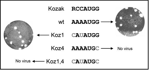
Three plasmid constructs were prepared, each encoding full-length infectious CVB3, but with R−3 and/or G+4 made nonconforming. The sequences around the AUG codons of wt CVB3 and of the three variants are indicated in Fig. 1. The first variant, termed Koz1, was prepared by constructing a site for the restriction enzyme NdeI; thus, it has an A→C transversion at position −3 and an A→U transition at position −1. The second variant, termed Koz4, has a G→C transversion at position +4. The third variant combines the first two, and so was termed Koz1,4. All plasmids contain a T7 RNA polymerase promoter immediately upstream of the viral genome and can be linearized at a unique ClaI site downstream of the 3′ end of the viral genome; this permits the production of potentially infectious RNA.
FIG. 1.
R−3 is not required for CVB3 production, but a transversion at G+4 is lethal. The optimal Kozak consensus (RCCAUGG) is shown, along with the equivalent sequences of wt CVB3 and the three mutant viruses, for which the mutated base(s) is shown in gray. The four full-length RNAs were transfected into HeLa cells and, 24 h later, the supernatants were tested for the presence of infectious virus. Despite multiple attempts, no virus was detected in the supernatant of cells transfected with the Koz4 or Koz1,4 RNAs. In contrast, the wt and Koz1 RNAs yielded viruses, which were titrated (plaque morphologies are shown) and named the P1 stocks.
Transfection of Koz1 RNA—but not of Koz4 or Koz1,4 RNA—generates infectious viruses with normal plaque morphologies, which replicate normally in tissue culture.
The three mutant RNAs (Koz1, Koz4, and Koz1,4) or RNA encoding wt CVB3 were transfected into HeLa cells, which are known to support productive CVB3 infection. Then, 24 h later, supernatants were harvested, and infectious virus titers were determined by a standard plaque assay. Supernatant from cells transfected with the Koz4 RNA (AAAAUGC) or the Koz1,4 RNA (CAUAUGC) yielded no plaques, suggesting that the G+4-to-C+4 transversion was lethal. However, infectious virus was detected in the Koz1 (CAUAUGG) and wt supernatants, and these viral stocks were termed P1. The plaque morphologies of the P1 viruses were of interest because coxsackieviruses that replicate and disseminate less efficiently than their wt counterparts—e.g., recombinant coxsackieviruses (10, 12, 13, 40)—often give rise to plaques that are smaller than those formed by wt virus. The plaques of the wt and Koz1 P1 stocks were similar in appearance (Fig. 1), suggesting that the viruses produced following Koz1 RNA transfection were not severely disabled in tissue culture.
The Koz1 virus lacking R-3 replicates as well as wt CVB3 in tissue culture and is stable after three serial passages.
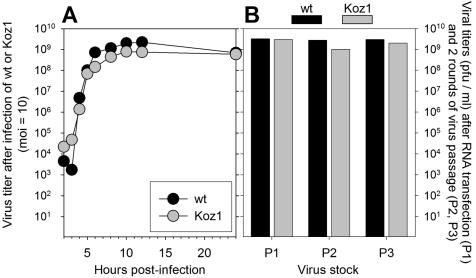
The Koz1 P1 virus stock was compared to wt virus by use of one-step growth curves and serial passage in tissue culture. For the one-step growth curve, stocks of wt or Koz1 virus, of similar titers, were used to infect HeLa cells at an MOI of 10, and samples were harvested at 2, 3, 4, 5, 6, 8, 10, 12, and 24 h postinfection and then titrated. As shown in Fig. 2A, the growth kinetics of the two viruses were very similar. For both viruses, the first substantial increase in titers was observed at 5 h postinfection. At this and most subsequent time points, the wt titer was very marginally higher (on average, ∼2-fold) than the Koz1 titer, but the endpoint titers were essentially identical (∼109 PFU/ml). Therefore, we conclude that CVB3 lacking the R−3 residue, thought to be the most important part of the Kozak consensus, replicates very similarly to wt virus in tissue culture.
FIG. 2.
One-step growth curves of wt and Koz1 viruses, and virus yields from the P1 to P3 stocks. (A) wt and Koz1 viruses were used to infect HeLa cells at an MOI of 10, and one-step growth curves were carried out. (B) Titers of the P1 stocks were determined, and the stocks were passaged in HeLa cells as described in the text to produce P2 and P3 stocks, whose titers were also were determined. The titers of all three stocks are shown. As described in the text, the viral RNA sequences of all stocks were determined, and all sequences were identical to that of the original input virus.
Next, serial passage studies were carried out to allow us to evaluate the viral yields over time and to assess the stability of the Koz1 sequence. The P1 stocks of wt and Koz1 viruses were used to infect HeLa cells at an MOI of 0.1. After 24 h, the supernatant was harvested, and this P2 stock was titrated and then used to infect HeLa cells (again at an MOI of 0.1), generating a P3 stock of virus, which was collected 24 h postinfection. As shown in Fig. 2B, all three stocks of wt and Koz1 viruses showed high and comparable titers. This suggested that either the Koz1 mutation might be relatively silent or that the virus had rapidly reverted to the wt sequence. To distinguish between these two possibilities, RNA was purified from the P1, P2, and P3 stocks of both wt and Koz1 viruses, and for each of the six resulting RNAs the region containing the initiation codon was amplified by RT-PCR. The six sets of PCR fragments were cloned, and four clones were sequenced from each set. All 12 sequences from the wt preparations contained AAAAUGG, as expected. Notably, all 12 sequences from the Koz1 preparations, representing three serial passages of this mutant virus, retained the input Koz1 sequence (CAUAUGG), indicating that this sequence is stable in tissue culture. These data confirm that R−3, thought to be the most important component of the Kozak consensus, is not absolutely required for CVB3 replication in HeLa cells because a virus lacking this sequence can replicate to levels similar to the wt virus and does not undergo rapid reversion toward the wt sequence.
The Koz1 (CAUAUGG) viral variant replicates in vivo, causes myocarditis and pancreatitis, and is stable.
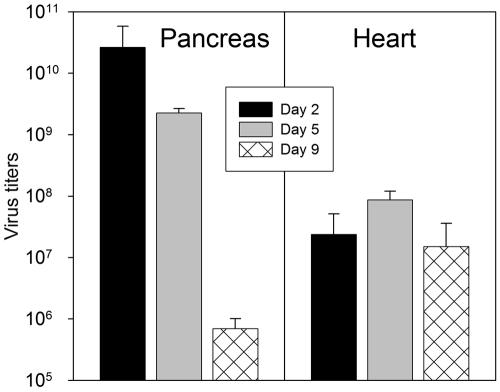
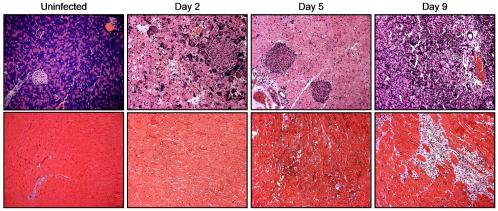
Having shown that CAUAUGG is stable through three passages in HeLa cells, we next evaluated the replicative capacity, pathogenicity, and sequence stability of the Koz1 virus in vivo. During wt CVB3 infection, the highest virus titers usually are found in the pancreas, with somewhat lower titers in the heart. The high titers are accompanied by pancreatitis (29, 37, 41) and myocarditis (9, 11, 15, 17, 42). Twelve BALB/c mice were infected intraperitoneally with 5 × 10³ PFU of the P1 stock of Koz1 virus. Three of the mice died within 2 days of infection, indicating that this mutant virus was highly pathogenic; this finding contrasts with the severe attenuation of most recombinant coxsackieviruses, in which doses as high as 107 PFU may be nonlethal (40). The hearts and pancreata of the nine surviving mice were harvested for analyses at 2, 5, and 9 days postinfection (four, three, and two mice, respectively). Immediately after harvest, each tissue was trisected, and the samples were processed, as described in Materials and Methods, for virus titration, histological analyses, and RNA extraction and subsequent sequencing. The mean pancreatic and myocardial virus titers at the three time points are shown in Fig. 3. The Koz1 virus replicated very efficiently in both tissues, reaching titers of >1010 PFU/g in the pancreas and >108 PFU/g in the heart. As might be expected, given the high titers of virus, the Koz1 variant also caused severe pathology in both organs, as shown in Fig. 4. Inflammatory cells were detectable in the pancreas as early as 2 days postinfection and became more numerous over the next week. Inflammatory changes were slower to develop in the myocardium (as occurs also during wt CVB3 infection) but, by 9 days postinfection, large inflammatory foci were visible. Thus, the Koz1 virus appears to be very similar to wt virus in its ability to replicate to high titers in the pancreas and heart and to cause disease.
FIG. 3.
The Koz1 virus replicates to high titers in the pancreata and hearts of BALB/c mice. Twelve BALB/c mice were infected with 5,000 PFU of the P1 stock of Koz1 virus (sequence CAUAUGG). Three mice died by 2 days postinfection; the hearts and pancreata of the remaining nine mice were harvested at 2, 5, or 9 days postinfection, and virus titers were determined. For each time point and in each tissue the means are shown, along with the standard deviation. As described in the text, RNA was isolated from all 18 organs, and sequence analyses of numerous clones from each showed that the Koz1 sequence remained stable in vivo, even as late as 9 days postinfection.
FIG. 4.
The Koz1 virus causes severe pancreatic and myocardial pathology. The pancreata and hearts of the Koz1 virus-infected mice, known to contain abundant virus (see Fig. 3), were evaluated for histopathologic changes. Paraffin sections (5 μm) were prepared and stained with hematoxylin and eosin (pancreata, top row) or trichrome (hearts, bottom row). Representative samples from both tissues are shown for the three time points indicated above each tissue pair and for uninfected tissues. A 20× objective lens was used.
One explanation for replicative and pathogenic similarities between wt and Koz1 viruses was that the Koz1 sequence had reverted to wt under selective pressure in vivo. To evaluate the in vivo stability of the Koz1 sequence, RNA was isolated from the hearts and pancreata of the nine surviving mice, and the 18 samples were subjected to RT-PCR, cloning, and sequencing as described in Materials and Methods. For all samples, multiple clones were sequenced, and all were identical to that of the input Koz1 virus (CAUAUGG), with a single exception; one clone from the pancreas of a day 2 mouse contained a U→C transition at position −1. Therefore, the Koz1 sequence CAUAUGG is stable in vivo for as long as 9 days postinfection; the −3 position, considered the most important feature of the Kozak consensus, does not revert to a purine.
Koz4 and Koz1,4 RNAs fail to yield infectious virus after in vivo injection.
The Koz4 and Koz1,4 mutations were severely restrictive in tissue culture, as judged by several unsuccessful attempts to generate infectious virus after RNA transfection of HeLa cells. However, picornavirus RNAs are infectious in vivo, and we considered it possible that the greater number and diversity of cells available in vivo might permit the generation of infectious (possibly revertant) virus after injection of Koz4 or Koz1,4 RNA. Therefore, these RNAs (or the Koz1 RNA, as a positive control) were prepared by in vitro transcription and were administered to BALB/c mice (four mice per RNA) by intramuscular injection (see Materials and Methods). At 9 days postinjection, the mice were sacrificed, and the pancreata and hearts were evaluated for virus titer and sequence and for histopathologic changes. Virus was present in all Koz1 RNA-injected mice and, at 9 days after Koz1 RNA injection, high titers were found, as well as marked pancreatic and myocardial pathology (data not shown). The viruses were sequenced, and all were Koz1, confirming the stability of this sequence in vivo. In contrast, neither the Koz4 RNA nor the Koz1,4 RNA yielded infectious progeny, and no histopathologic changes were noted 9 days postinjection (data not shown). These data indicate that the G+4 to C+4 change is lethal in vivo.
In vitro translation of wt RNA and of the three Koz mutant RNAs.
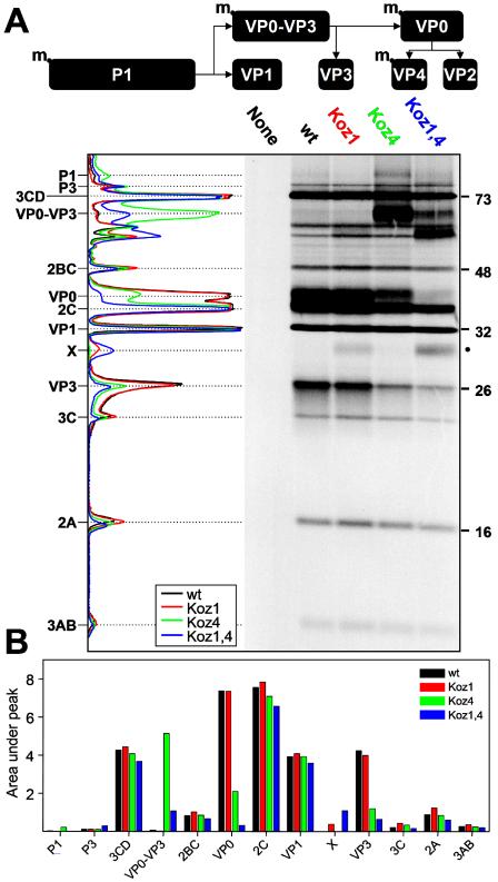
Next, we evaluated the ability of the three mutant RNAs to direct viral protein synthesis in vitro. The three Koz mutant RNAs and wt RNA were used as templates in an in vitro translation reaction as described in Materials and Methods; the protein products, separated on a polyacrylamide gel, are shown in Fig. 5A, together with digitized plots of each lane. To better quantitate the proteins within each band, the areas under the peaks were measured, and the results are shown in Fig. 5B. The sizes (Fig. 5A) and quantities (Fig. 5B) of viral proteins in the wt and Koz1 lanes (black and red plots, respectively) are essentially identical, indicating that the absence of R−3 from the CVB3 initiation site has little effect on translational initiation. Most significantly, the size and intensity of the VP0 bands are indistinguishable in the wt and Koz1 lanes; this protein represents the major N-terminal product of the coxsackievirus polyprotein, and its presence indicates that translation from the Koz1 RNA template began at the authentic AUG. These data confirm that the R−3 mutation is well tolerated by CVB3 and are consistent with the phenotype of the Koz1 virus, which behaves similarly to wt virus in tissue culture, as well as in vivo. Nevertheless, there is evidence that there is some leak-through at the Koz1 AUG. A new band of ∼29.7 kDa (here termed “X” and highlighted by a dot to the right of the autoradiograph in Fig. 5A) is present in the Koz1 lane. The size of this band is consistent with a protein that had initiated at the next in-frame AUG (sequence AUCAUGA) that lies 180 bases downstream of the authentic initiation codon. Thus, our in vitro translation data indicate that the Koz1 RNA shows some leak-through at the authentic codon, but this does not substantially alter the virus' ability to replicate in tissue culture and in vivo and to remain highly pathogenic. We cannot confidently attribute this leakiness to the change at R−3, since the Koz1 variant also contains a transition at the −1 position. However, we can conclude that R−3 is not required for the successful translation of CVB proteins.
FIG. 5.
In vitro translation of wt RNA, and the three mutant RNAs. In vitro translations were carried out as described in Materials and Methods, and the products were separated on a polyacrylamide gel and then visualized by autoradiography. (A) Diagram of normal P1 processing. m, myristoyl. The RNA templates are shown above the autoradiograph. The four lanes containing input RNA were digitized as described in Materials and Methods, and the four resulting plots are shown to the left of the autoradiograph. The viral proteins are indicated to the left side, and molecular masses are indicated to the right in kilodaltons. The novel band X with a molecular mass of ∼29.7 kDa is highlighted by a dot to the right of the autoradiograph. (B) To more precisely quantify the proteins generated by in vitro transcription, the four digitized plots shown in panel A were analyzed as described in Materials and Methods. The areas under the peaks were determined and are shown (in arbitrary units) for the indicated viral proteins.
Next, we evaluated in vitro translation from Koz4 RNA (green plot). The overall translation from the Koz4 template proceeds with an efficiency similar to that observed with wt template, as indicated by the near-identity, in size and intensity, of the 3CD, 2BC, 2C, VP1, 3C, 2A, and 3AB proteins. Furthermore, the novel ∼29.7 kDa protein X is absent, suggesting that mutation at G+4 alone does not result in leakiness. However, there are some marked differences in the Koz4 lane. First, the size of the VP0 protein is very slightly increased. This is expected, because the G+4-to-C+4 mutation alters the second amino acid of the polyprotein from glycine to the larger amino acid arginine. However, in addition, the intensities of the VP0 and VP3 bands are markedly reduced in the Koz4 lane. What might explain these observations? The processing cascade of the enteroviral P1 protein is shown diagrammatically in Fig. 5A. The first cleavage generates VP1 and VP0-VP3, and the latter molecule normally is rapidly processed to produce the VP0 and VP3 proteins. The Koz4 lane not only contains a normal amount of VP1 but also has a strong new band, of a size consistent with the VP0-VP3 precursor. The abundance of this band, together with the reduction in its cleavage products, VP0 and VP3, indicate that there is a defect in processing of the Koz4 VP0-VP3. To what might this defect be attributed? Early studies with poliovirus indicated that the true N terminus of the P1, VP0 and VP4 proteins was not methionine but instead was a blocked glycine (7), and subsequent analyses revealed that the material attached to the glycine was a myristoyl (C14H28O2) fatty acid side chain (6, 33). Myristoylation occurs only on a glycine residue (8, 28) and, when the glycine residue in poliovirus P1 was altered (for example, to arginine, as in Koz4), myristoylation was prevented, and no infectious poliovirus was produced (25-27, 31). The lethal effect of changing G+4 to C+4 could, in principle, be attributed to either the change in nucleotide (which could, for instance, alter the RNA structure) or to the resulting change in amino acid (Gly to Arg). These possibilities have been distinguished in poliovirus. Marc et al. changed the second codon from GGT (Gly) to GCT (Ala); despite the retention of the G+4, this mutation was lethal, indicating that the lethal effect is attributable to the absence of Gly and not to the absence of G+4 (26). Our data indicate that the same phenomena hold true for the closely related picornavirus CVB3. Some processing of the Koz4 VP0-VP3 does occur, since both VP0 and VP3 are detectable. Interestingly, the combined abundance of the VP0-VP3 precursor and of its products, together with the absence of protein X, indicate that translation initiation occurs efficiently at the authentic AUG in Koz4 (AAAAUGC). Therefore, as is true also for R−3, G+4 is not absolutely required for effective initiation at the authentic AUG codon in CVB3, but if G+4 is changed, the resulting loss of glycine is lethal.
The Koz1,4 lane combines the leaky initiation noted in the Koz1 lane, with the aberrant P1 processing identified in the Koz4 lane. The novel band of ∼29.7 kDa, indicative of leakiness, is present. The higher abundance of this band suggests that the addition of the G+4 mutation amplifies the effect of the R−3 mutation, increasing the leakiness of the authentic codon. This conclusion is supported by the reduced intensities of the VP0 and VP0-VP3 bands, and the presence of the latter demonstrates the abnormality in P1 processing.
DISCUSSION
The main goal of this study was to determine the importance of the Kozak consensus sequence R−3 and G+4 residues in translational initiation in CVB3. The R−3 in region 1 is well maintained in vertebrate mRNAs, being reported in ∼90% of cases (A exceeds G by a small margin, at a ratio of ca. 5:4 [36]), and it is thought to be the most important determinant in the flanking regions (22, 24); mutagenesis experiments indicate that its replacement by a pyrimidine can have consequences on translational initiation in vitro and in tissue culture, especially if G+4 is absent (19). Both of the purine samples also are more common than either of the pyrimidines at the +4 position, but in this case G outnumbers A, by ca. 5:2. In vitro toeprinting assays suggest that G+4, but not A+4, is beneficial for translational initiation (23), so the increased frequency of A+4 appears to be unrelated to translational initiation. Below, we suggest an explanation for the higher frequencies of both G and A at the +4 position.
Several reasons underpinned our selection of CVB3 as a model system in which to test the biological requirements for R−3 and G+4. As described in the introduction, ribosome binding to picornavirus mRNAs differs mechanistically from binding to the vast majority of eukaryotic messengers, so we wanted to determine whether the translational initiation consensus requirements also were different. More importantly, the availability of a full-length infectious clone allowed any changes around the AUG codon to be evaluated not only in vitro but also in vivo, and RNA viruses have a well-demonstrated capacity for rapid replication and evolution, permitting us to determine the stability of the variant sequences through many cycles of replication under stringent in vivo selective pressures. The data from the Koz1 mutation (CAUAUGG) show unequivocally that a virus lacking R−3 retains its ability to replicate in tissue culture. Plaque sizes are normal (Fig. 1A), and viral growth kinetics are comparable to that of wt virus (Fig. 2). The sequence of the Koz1 virus remains stable after three passages in tissue culture, suggesting that the sequence CAUAUGG serves as an acceptable initiation codon for this picornavirus. The in vivo data were even more striking: the Koz1 virus reached high titers in the heart and pancreas (Fig. 3) and caused profound histopathologic damage in these tissues (Fig. 4). Furthermore, even after 9 days of in vivo replication, the variant sequence was retained. The Koz1 RNA served as an effective template for in vitro translation, and the band sizes and intensities were essentially identical to those observed with the wt RNA as a template (Fig. 5). In particular, the VP0 bands were indistinguishable, indicating that the authentic AUG was used in Koz1. The Koz1 RNA did give rise to a band (X) whose size was consistent with a truncated VP0 product, suggesting that the Koz1 RNA was somewhat leaky. However, taken together, our data indicate that CVB3 does not have a strong requirement for R−3, at least when the G+4 is present.
Next, we evaluated the requirement for G+4 in CVB3 translational initiation. G+4 is present in >50% of eukaryotic mRNAs (36), and studies have shown that it is the only one of the four bases that confers an advantage at the +4 position (23). In contrast to our observations with Koz1, the biological consequences of a G+4 mutation were profound. Transfection of Koz4 RNA into HeLa cells, and Koz4 RNA injection into mice, failed to generate infectious virus, suggesting that the change was lethal. Despite this dramatic biological effect, in vitro translation studies showed that the majority of proteins synthesized from the Koz4 RNA template were remarkably similar in size and intensity to those produced from the wt and Koz1 templates (Fig. 5), and initiation at the authentic AUG codon was strong, as evidenced by the combined intensities of the VP0-VP3 precursor protein, and its products. Furthermore, no band was present at ∼29.7 kDa, indicating that the G+4→C+4 mutation did not cause leak-through at the authentic AUG codon. The Koz1,4 double mutation also was lethal but, in contrast to the Koz1 and Koz4 mutants, the Koz1,4 RNA showed a marked change in translational initiation, most likely due to substantial leakiness at the authentic AUG. Although some VP0-VP3 was detected (indicating initiation at the CAUAUGC sequence), most of the “VP0” was a truncated product X, of a size consistent with initiation at the next in-frame AUG. In summary, the authentic AUG codon in CVB3 is used with high efficiency, as long as either R−3 or G+4 is present; either of these positions can be mutated with little effect on translational initiation, so long as the other is retained. Most strikingly, a virus carrying C−3 is stable through many rounds of replication, in tissue culture and in vivo, suggesting that the evolutionary pressures favoring R−3 are subtle (or have not been encountered with the experimental manipulations used herein). In contrast to the minimal changes in translational initiation seen with the single mutants, removal of both R−3 and G+4 causes the authentic AUG to be frequently overlooked by the scanning preinitiation complex. This behavior is not atypical, and the Kozak rules do not demand that both R−3 and G+4 be present. For example, it is known that the effect of G+4 is greatest when R−3 is absent (23); this mirrors our observation that removal of G+4 from the wt sequence (to generate Koz4) has little effect on initiation, while removal of G+4 from Koz1 (to generate Koz1,4) markedly increases codon leakiness.
Our studies also provide an additional explanation for the high frequency of G at the +4 position of CVB and related picornaviruses. As noted in Results, the Koz4 and Koz1,4 P1 products are not processed correctly because the penultimate residue in the polyprotein, normally a glycine, has been changed to arginine, preventing myristoylation. Successful myristoylation requires at least two cotranslational modifications at the N terminus of the nascent wt viral polyprotein. First, the N-terminal methionine, specified by the authentic initiation codon, must be removed. This hydrolysis is carried out by a cellular enzyme, methionine (methionyl) aminopeptidase (MetAP), and the main criterion for MetAP activity is that the second amino acid should be small (glycine, alanine, serine, cysteine, threonine, proline, or valine); a large or highly charged penultimate residue prevents removal of the initiator methionine (1-3, 14, 39). The myristoyl chain is added by another cellular enzyme, N-myristoyltransferase, whose specificity is tighter still; this enzyme will attach the myristoyl moiety only to a glycine residue (8, 28). Thus, the production of infectious poliovirus and coxsackievirus will occur only if the polyprotein begins with Met-Gly; moreover, since all four Gly codons begin with a G residue, this provides a novel explanation—unrelated to the Kozak rules—for the G+4 requirement. Therefore, we propose that the primary driving force favoring G+4 in these picornaviruses is not translational initiation but instead is posttranslational modification of the encoded protein.
However, not all picornaviruses have G+4. For example, many isolates of foot and mouth disease virus, the prototype of the aphthovirus genus, have A+4 and the penultimate amino acids in these viral polyproteins can vary (e.g., Asn, Ser, and Lys); none of these residues can be myristoylated. Furthermore, even some picornaviruses with G+4 do not contain glycine as the second residue; the cardioviruses encephalomyocarditis virus and Theiler's murine encephalomyelitis virus, and the aphthovirus equine rhinovirus, all contain either alanine or valine at this position, and these residues cannot be myristoylated. The cardiovirus and aphthovirus genera of the picornavirus family achieve myristoylation via a different route; their polyprotein begins with a short protein (termed “leader” or L) which is removed, exposing an internal glycine that serves as the N terminus of the P1 product. The penultimate amino acids in these 14 viruses are, therefore, not constrained by myristoylation.
Similar constraints must apply to cellular proteins that undergo such N-terminal modifications; and, if large numbers of cellular proteins are so constrained, this would provide an alternative explanation for the high frequency of G+4 in the eukaryotic translational initiation consensus. Removal of the N-terminal methionine is remarkably common (2), suggesting that the seven residues named above must frequently occupy the penultimate position in a nascent eukaryotic protein. These seven amino acids can be encoded by 28 codons, and the ratio of G to A to T to C as the first base of the codon is 12:6:6:4. Other modifications can further skew the base representation at the +4 position in favor of a purine. Myristoylation, which, as noted above, requires a penultimate glycine (and hence, G+4), is important in eukaryotic biology, as demonstrated by the fact that mutations in N-myristoyltransferase can be lethal in yeast and Drosophila (reviewed in reference 8). The frequency of myristoylation in cellular proteins is relatively low (perhaps ∼1%) and thus is unlikely to greatly affect the overall frequency of G+4; however, other forms of acylation are extremely common. Some 70 to 80% of mammalian cytosolic proteins are acetylated (4), and the enzyme responsible for this modification, N α-acetyltransferase, preferentially targets one of two classes of N-terminal amino acid. The first class, termed GAST substrates, comprises glycine, alanine, serine, or threonine, all of which can be exposed by MetAP. The second class, M substrates, are acetylated on N-terminal methionine residues that are adjacent to a penultimate glutamate, aspartate, or asparagine. Thus, acetylation, which affects the majority of eukaryotic cytosolic proteins, can take place only when the penultimate residue is Gly, Ala, Ser, Thr, Asp, Asn, or Glu; these amino acids can be specified by 24 codons, in which the first base G/A/T/C ratio is 12:8:4:0. Therefore, the combined specificities of MetAP, N α-acetyltransferase, and N-myristoyltransferase predict that G would be the most common residue at the +4 position, followed by A. This is precisely what is observed; the +4 position is a purine in >70% of cases (G ∼53%, A ∼19%). Therefore, we propose that the high frequency of G and A at the +4 position may be driven by constraints on the nature of the second amino acid which, in turn, is driven by requirements for posttranslational processing. This idea not only provides an alternative explanation for the preference for G+4; it also explains the excess of A residues over either C or T, even though A does not improve the efficiency of translational initiation.
In summary, we show here that a coxsackievirus carrying a mutation in R−3 remains viable and is pathogenic and stable in vivo; R−3 may be dispensable in coxsackievirus. Furthermore, we show that the high frequency of G+4 in the picornavirus family is driven by posttranslational modifications which constrain the penultimate amino acid and, thereby, the bases that encode it. Since, for any amino acid, the first base of the codon is the least variable, it follows that constraint on the nature of the penultimate amino acid will most tightly constrain the nucleotide that lies at the +4 position with respect to the initiating AUG. In CVB3, the importance of the penultimate Gly results in an absolute requirement for G+4. We argue that similar considerations will apply to many, perhaps most, eukaryotic open reading frames, providing an alternative interpretation for the very high frequency of purine samples at the +4 position in the eukaryotic translational initiation consensus.
Acknowledgments
We are grateful to Ralph Feuer for helpful discussions and to Annette Lord for excellent secretarial support.
This study was supported by NIH grant R-01 AI-42314 to J.L.W. C.T.C. was supported by NIH training grant T32 AI007354.
Footnotes
Manuscript number 16179-NP from The Scripps Research Institute.
REFERENCES
- 1.Arfin, S. M., R. L. Kendall, L. Hall, L. H. Weaver, A. E. Stewart, B. W. Matthews, and R. A. Bradshaw. 1995. Eukaryotic methionyl aminopeptidases: two classes of cobalt-dependent enzymes. Proc. Natl. Acad. Sci. USA 92:7714-7718. [DOI] [PMC free article] [PubMed] [Google Scholar]
- 2.Boissel, J. P., T. J. Kasper, and H. F. Bunn. 1988. Cotranslational amino-terminal processing of cytosolic proteins: cell-free expression of site-directed mutants of human hemoglobin. J. Biol. Chem. 263:8443-8449. [PubMed] [Google Scholar]
- 3.Bradshaw, R. A., W. W. Brickey, and K. W. Walker. 1998. N-terminal processing: the methionine aminopeptidase and Nα-acetyltransferase families. Trends Biochem. Sci. 23:263-267. [DOI] [PubMed] [Google Scholar]
- 4.Brown, J. L., and W. K. Roberts. 1976. Evidence that approximately eighty per cent of the soluble proteins from Ehrlich ascites cells are Nα-acetylated. J. Biol. Chem. 251:1009-1014. [PubMed] [Google Scholar]
- 5.Chen, C. Y., and P. Sarnow. 1995. Initiation of protein synthesis by the eukaryotic translational apparatus on circular RNAs. Science 268:415-417. [DOI] [PubMed] [Google Scholar]
- 6.Chow, M., J. F. Newman, D. Filman, J. M. Hogle, D. J. Rowlands, and F. Brown. 1987. Myristoylation of picornavirus capsid protein VP4 and its structural significance. Nature 327:482-486. [DOI] [PubMed] [Google Scholar]
- 7.Dorner, A. J., L. F. Dorner, G. R. Larsen, E. Wimmer, and C. W. Anderson. 1982. Identification of the initiation site of poliovirus polyprotein synthesis. J. Virol. 42:1017-1028. [DOI] [PMC free article] [PubMed] [Google Scholar]
- 8.Farazi, T. A., G. Waksman, and J. I. Gordon. 2001. The biology and enzymology of protein N-myristoylation. J. Biol. Chem. 276:39501-39504. [DOI] [PubMed] [Google Scholar]
- 9.Gebhard, J. R., C. M. Perry, S. Harkins, T. Lane, I. Mena, V. C. Asensio, I. L. Campbell, and J. L. Whitton. 1998. Coxsackievirus B3-induced myocarditis: perforin exacerbates disease but plays no detectable role in virus clearance. Am. J. Pathol. 153:417-428. [DOI] [PMC free article] [PubMed] [Google Scholar]
- 10.Halim, S. S., S. E. Ostrowski, W. T. Lee, and A. I. Ramsingh. 2000. Immunogenicity of a foreign peptide expressed within a capsid protein of an attenuated coxsackievirus. Vaccine 19:958-965. [DOI] [PubMed] [Google Scholar]
- 11.Henke, A., S. A. Huber, A. Stelzner, and J. L. Whitton. 1995. The role of CD8+ T lymphocytes in coxsackievirus B3-induced myocarditis. J. Virol. 69:6720-6728. [DOI] [PMC free article] [PubMed] [Google Scholar]
- 12.Henke, A., R. Zell, G. Ehrlich, and A. Stelzner. 2001. Expression of immunoregulatory cytokines by recombinant coxsackievirus B3 variants confers protection against virus-caused myocarditis. J. Virol. 75:8187-8194. [DOI] [PMC free article] [PubMed] [Google Scholar]
- 13.Hofling, K., S. Tracy, N. Chapman, K. S. Kim, and L. J. Smith. 2000. Expression of an antigenic adenovirus epitope in a group B coxsackievirus. J. Virol. 74:4570-4578. [DOI] [PMC free article] [PubMed] [Google Scholar]
- 14.Huang, S., R. C. Elliott, P. S. Liu, R. K. Koduri, J. L. Weickmann, J. H. Lee, L. C. Blair, P. Ghosh-Dastidar, R. A. Bradshaw, K. M. Bryan, B. Einarson, R. L. Kendall, K. Kolacz, and K. Saito. 1987. Specificity of cotranslational amino-terminal processing of proteins in yeast. Biochemistry 26:8242-8246. [DOI] [PubMed] [Google Scholar]
- 15.Huber, S. A., and L. P. Job. 1983. Cellular immune mechanisms in coxsackievirus group B, type 3 induced myocarditis in BALB/c mice. Adv. Exp. Med. Biol. 161:491-508. [DOI] [PubMed] [Google Scholar]
- 16.Jang, S. K., H. G. Krausslich, M. J. Nicklin, G. M. Duke, A. C. Palmenberg, and E. Wimmer. 1988. A segment of the 5′ nontranslated region of encephalomyocarditis virus RNA directs internal entry of ribosomes during in vitro translation. J. Virol. 62:2636-2643. [DOI] [PMC free article] [PubMed] [Google Scholar]
- 17.Kim, K. S., G. Hufnagel, N. M. Chapman, and S. Tracy. 2001. The group B coxsackieviruses and myocarditis. Rev. Med. Virol. 11:355-368. [DOI] [PubMed] [Google Scholar]
- 18.Knowlton, K. U., E. S. Jeon, N. Berkley, R. Wessely, and S. A. Huber. 1996. A mutation in the puff region of VP2 attenuates the myocarditic phenotype of an infectious cDNA of the Woodruff variant of coxsackievirus B3. J. Virol. 70:7811-7818. [DOI] [PMC free article] [PubMed] [Google Scholar]
- 19.Kozak, M. 1986. Point mutations define a sequence flanking the AUG initiator codon that modulates translation by eukaryotic ribosomes. Cell 44:283-292. [DOI] [PubMed] [Google Scholar]
- 20.Kozak, M. 1987. At least six nucleotides preceding the AUG initiator codon enhance translation in mammalian cells. J. Mol. Biol. 196:947-950. [DOI] [PubMed] [Google Scholar]
- 21.Kozak, M. 1981. Possible role of flanking nucleotides in recognition of the AUG initiator codon by eukaryotic ribosomes. Nucleic Acids Res. 9:5233-5262. [DOI] [PMC free article] [PubMed] [Google Scholar]
- 22.Kozak, M. 1987. An analysis of 5′-noncoding sequences from 699 vertebrate messenger RNAs. Nucleic Acids Res. 15:8125-8148. [DOI] [PMC free article] [PubMed] [Google Scholar]
- 23.Kozak, M. 1997. Recognition of AUG and alternative initiator codons is augmented by G in position +4 but is not generally affected by the nucleotides in positions +5 and +6. EMBO J. 16:2482-2492. [DOI] [PMC free article] [PubMed] [Google Scholar]
- 24.Kozak, M. 2002. Pushing the limits of the scanning mechanism for initiation of translation. Gene 299:1-34. [DOI] [PMC free article] [PubMed] [Google Scholar]
- 25.Krausslich, H. G., C. Holscher, Q. Reuer, J. Harber, and E. Wimmer. 1990. Myristoylation of the poliovirus polyprotein is required for proteolytic processing of the capsid and for viral infectivity. J. Virol. 64:2433-2436. [DOI] [PMC free article] [PubMed] [Google Scholar]
- 26.Marc, D., G. Drugeon, A. L. Haenni, M. Girard, and S.van der Werf. 1989. Role of myristoylation of poliovirus capsid protein VP4 as determined by site-directed mutagenesis of its N-terminal sequence. EMBO J. 8:2661-2668. [DOI] [PMC free article] [PubMed] [Google Scholar]
- 27.Marc, D., G. Masson, M. Girard, and S. van der Werf. 1990. Lack of myristoylation of poliovirus capsid polypeptide VP0 prevents the formation of virions or results in the assembly of noninfectious virus particles. J. Virol. 64:4099-4107. [DOI] [PMC free article] [PubMed] [Google Scholar]
- 28.Maurer-Stroh, S., M. Gouda, M. Novatchkova, A. Schleiffer, G. Schneider, F. L. Sirota, M. Wildpaner, N. Hayashi, and F. Eisenhaber. 13February2004. MYRbase: analysis of genome-wide glycine myristoylation enlarges the functional spectrum of eukaryotic myristoylated proteins. Genome Biol. 5(3):R21. [Online.] http://genomebiology.com/2004/5/3/R21. [DOI] [PMC free article] [PubMed]
- 29.Mena, I., C. Fischer, J. R. Gebhard, C. M. Perry, S. Harkins, and J. L. Whitton. 2000. Coxsackievirus infection of the pancreas: evaluation of receptor expression, pathogenesis, and immunopathology. Virology 271:276-288. [DOI] [PubMed] [Google Scholar]
- 30.Molla, A., A. V. Paul, and E. Wimmer. 1991. Cell-free, de novo synthesis of poliovirus. Science 254:1647-1651. [DOI] [PubMed] [Google Scholar]
- 31.Moscufo, N., J. Simons, and M. Chow. 1991. Myristoylation is important at multiple stages in poliovirus assembly. J. Virol. 65:2372-2380. [DOI] [PMC free article] [PubMed] [Google Scholar]
- 32.Ohlmann, T., and R. J. Jackson. 1999. The properties of chimeric picornavirus IRESes show that discrimination between internal translation initiation sites is influenced by the identity of the IRES and not just the context of the AUG codon. RNA. 5:764-778. [DOI] [PMC free article] [PubMed] [Google Scholar]
- 33.Paul, A. V., A. Schultz, S. E. Pincus, S. Oroszlan, and E. Wimmer. 1987. Capsid protein VP4 of poliovirus is N-myristoylated. Proc. Natl. Acad. Sci. USA 84:7827-7831. [DOI] [PMC free article] [PubMed] [Google Scholar]
- 34.Pelletier, J., G. Kaplan, V. R. Racaniello, and N. Sonenberg. 1988. Cap-independent translation of poliovirus mRNA is conferred by sequence elements within the 5′ noncoding region. Mol. Cell. Biol. 8:1103-1112. [DOI] [PMC free article] [PubMed] [Google Scholar]
- 35.Pelletier, J., and N. Sonenberg. 1988. Internal initiation of translation of eukaryotic mRNA directed by a sequence derived from poliovirus RNA. Nature 334:320-325. [DOI] [PubMed] [Google Scholar]
- 36.Peri, S., and A. Pandey. 2001. A reassessment of the translation initiation codon in vertebrates. Trends Genet. 17:685-687. [DOI] [PubMed] [Google Scholar]
- 37.Ramsingh, A. I. 1997. Coxsackieviruses and pancreatitis. Front. Biosci. 2:e53-e62. [DOI] [PubMed] [Google Scholar]
- 38.Sakai, H., T. Washio, R. Saito, A. Shinagawa, M. Itoh, K. Shibata, P. Carninci, H. Konno, J. Kawai, Y. Hayashizaki, and M. Tomita. 2001. Correlation between sequence conservation of the 5′ untranslated region and codon usage bias in Mus musculus genes. Gene 276:101-105. [DOI] [PubMed] [Google Scholar]
- 39.Sherman, F., J. W. Stewart, and S. Tsunasawa. 1985. Methionine or not methionine at the beginning of a protein. Bioessays 3:27-31. [DOI] [PubMed] [Google Scholar]
- 40.Slifka, M. K., R. R. Pagarigan, I. Mena, R. Feuer, and J. L. Whitton. 2001. Using recombinant coxsackievirus B3 to evaluate the induction and protective efficacy of CD8+ T cells in controlling picornaviral infection. J. Virol. 75:2377-2387. [DOI] [PMC free article] [PubMed] [Google Scholar]
- 41.Tracy, S., K. Hofling, S. Pirruccello, P. H. Lane, S. M. Reyna, and C. J. Gauntt. 2000. Group B coxsackievirus myocarditis and pancreatitis: connection between viral virulence phenotypes in mice. J. Med. Virol. 62:70-81. [DOI] [PubMed] [Google Scholar]
- 42.Woodruff, J. F. 1980. Viral myocarditis: a review. Am. J. Pathol. 101:425-484. [PMC free article] [PubMed] [Google Scholar]


