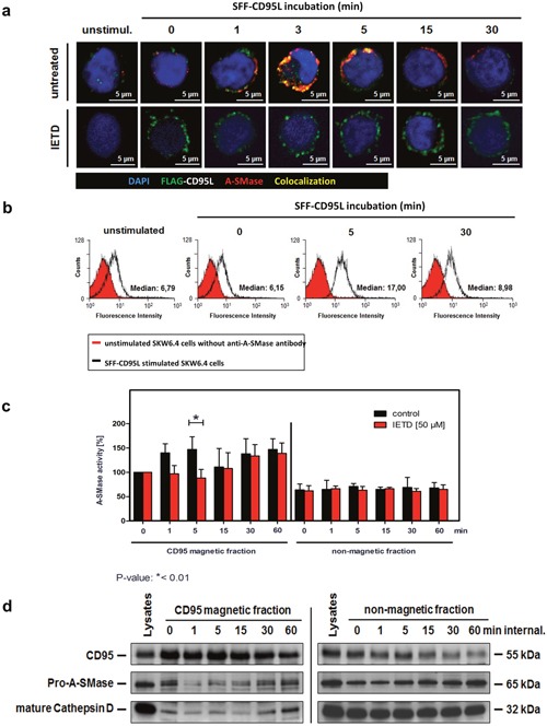
Figure 4. Characterization of the first A-SMase activation peak.

a. Translocation of A-SMase to the plasma membrane in non-permeabilized SKW6.4 cells. Stimulation with 100 ng/ml SFF-CD95L resulted in a rapid translocation of A-SMase to the cell surface (upper panel) which was abrogated after co-treatment with 50 μM caspases-8 inhibitor IETD (lower panel). b. Transient exposure of the A-SMase to the plasma membrane induced by SFF-CD95L. Non-permeabilized SKW6.4 cells were stimulated for indicated time points with 100 ng/ml SFF-CD95L and analyzed by flow cytometry. c. Comparison of A-SMase activation in CD95 magnetic and non-magnetic fractions from SKW6.4 cells treated with or without IETD (50 μM). In contrast to the control, IETD blocked the first activation peak of A-SMase in CD95 magnetic fractions. Represented are the mean values of four independently performed assays with control cells and three independent experiments with IETD treated cells. (2way Anova with Bonferroni post-test) d. Western blot analysis of CD95 magnetic and non-magnetic fractions isolated from IETD treated SKW6.4 cells with anti-CD95, anti-A-SMase and anti-Cathepsin D antibodies.
