. 2017 Mar 7;6:e23661. doi: 10.7554/eLife.23661

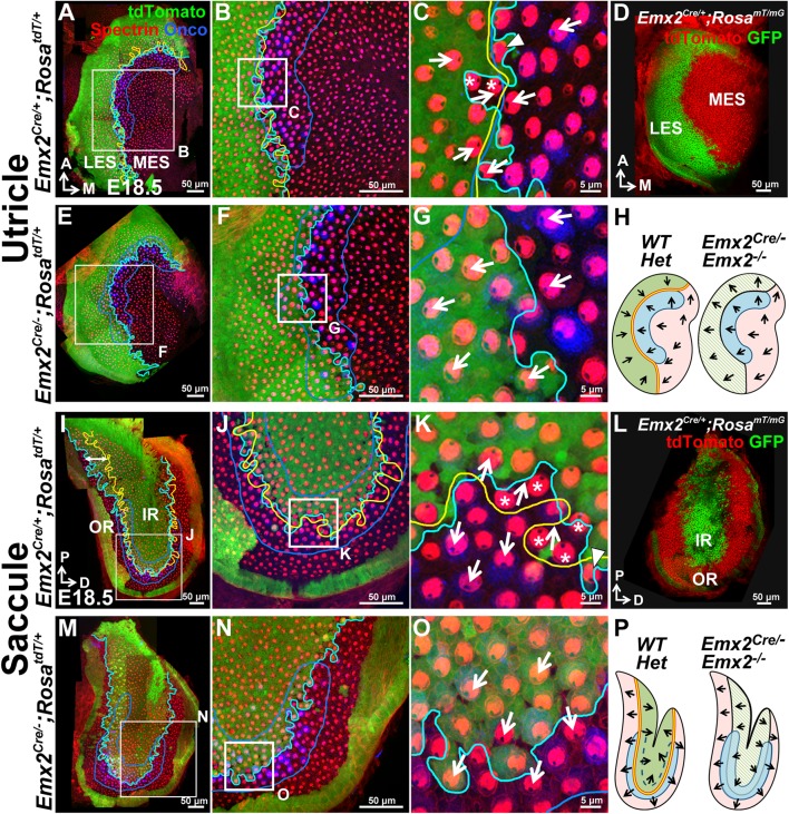
Figure 2. The Emx2 lineage domain is present in Emx2 functional null maculae.

(AC) and (IK) are the same specimens as (CE’) and (FH’) in Figure 1. (AC) In Emx2cre/+;RosatdT/+ utricles, the border of the Emx2 lineage domain (cyan line) coincides largely with LPR (yellow line), located at the lateral edge of the oncomodulin-positive striola (blue outlined; n=5). (C) HCs point toward each other (arrows) across the LPR. Asterisks label HCs that are negative for cre reporter signals but positive for Emx2 immunostaining (Figure 1E’; n=18), whereas arrowhead labels a HC that is cre-reporter positive but negative for Emx2 immunostaining (Figure 1E’; n=21). (D) A similar Emx2 lineage domain (GFP) is observed using a different Cre reporter, RosamT/mG. (EG) The Emx2 lineage domain (green) remained in Emx2Cre/-;RosatdT/+ utricles, but hair bundle polarity in this region is reversed (G; n=5) compared to controls (C; n=5). (IK) In Emx2Cre/+;RosatdT/+ control saccules, the border of the Emx2 lineage domain (cyan line) mostly coincides with the LPR except in the ventral-posterior region (I, double-headed arrow). (K) Across the LPR, HCs are pointing away from each other (arrows). Asterisks label HCs that are negative for cre reporter signals but positive for Emx2 immunostaining (Figure 1H’; n=60), whereas arrowhead labels a HC that is cre-reporter positive but negative for Emx2 immunostaining (Figure 1H’; n=105). (L) A similar lineage domain (GFP) is observed using the Cre reporter RosamT/mG. (MO) In Emx2Cre/-;RosatdT/+ mutant saccules, the border of the Emx2 lineage domain (cyan line) remained located in the middle of the striolar region (blue outline), but the hair bundles within the lineage domain are reversed (O; n=5), relative to controls (green region in K). (H) and (P) Schematics of respective utricles and saccules showing relationships among the Emx2 expression domain (green), hair bundle polarity pattern, LPR (yellow line) and striola (blue outlined) in controls and Emx2 mutants.

DOI: http://dx.doi.org/10.7554/eLife.23661.004

Figure 2—source data 1. Sensory area quantifications for Emx2 mutant maculae.
DOI: 10.7554/eLife.23661.005
Figure 2—source data 2. Total hair cell number in utricles of loss- and gain-of-function Emx2 mutants.
DOI: 10.7554/eLife.23661.006

Figure 2.

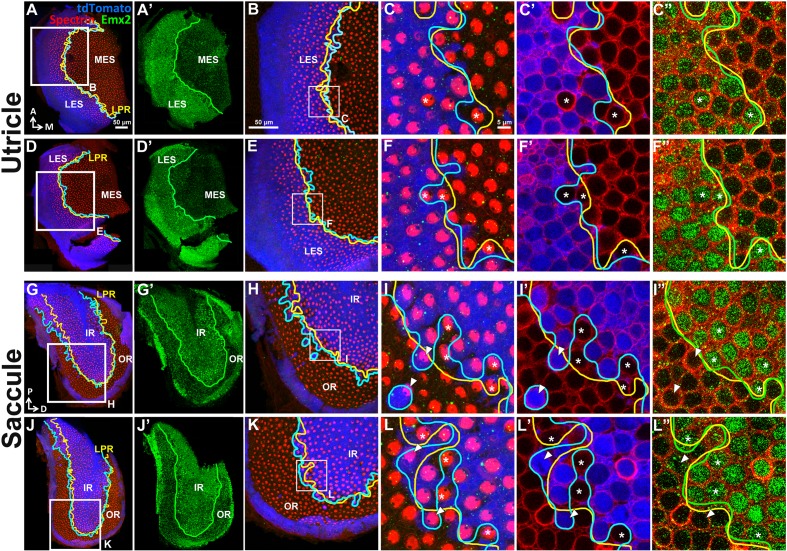
Figure 2—figure supplement 1. Emx2 immunostaining in the Emx2Cre/+ maculae correlates with the reversed hair bundle polarity.

Figure 2—figure supplement 1.

In the utricle (AF”) and saccule (GL”), the lineage (A,D,G,J, blue) and expression (A’,D’,G’,J’, green) domains of Emx2 are both restricted to the LES and IR, respectively. (C,F,I,L) show Emx2 lineage (blue) and anti-spectrin staining (red). (C’,F’,I’,L’) and (C”,F”,I”,L”) are same regions as (C,F,I,L) showing lineage staining (blue) and anti-Emx2 staining (green) at the nucleus level of HCs, respectively. The LPR (C”,F”,I”,L”, yellow line) correlates with the border of Emx2 immunostaining (green line) better than the border of Emx2-lineage domain (C’,F’,I’,L’, cyan line). Asterisks label the Emx2-lineage negative HCs with reversed bundle polarity that are positive for Emx2 staining, whereas arrowheads label Emx2-lineage HCs with default bundle polarity that are negative for Emx2 immunoreactivity.
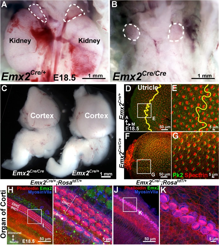
Figure 2—figure supplement 2. Mutant phenotypes of Emx2Cre/Cre and Emx2Cre/- embryos are similar to Emx2 -/- mutants.

Figure 2—figure supplement 2.

(AB) The kidneys below the adrenal glands (dotted lines) are absent in Emx2Cre/Cre (B) compared to Emx2Cre/+ embryos (A). (C) The size of the cortex is smaller in Emx2Cre/Cre than Emx2Cre/+ embryos. Both kidney and cortical phenotypes have been described in Emx2 -/- mutants (n=5; Pellegrini et al., 1996; Miyamoto et al., 1997). (DG) In contrast to controls (D-E; n=3), the LPR (yellow line) is absent in Emx2Cre/Cre utricles (FG; n=3) and hair bundle polarity defects are similar to those reported in Emx2-/- ears (Holley et al., 2010). The distribution of Prickle-like 2 (Pk2) immunostaining in Emx2Cre/Cre utricles is associated with the medial border of HCs (G), similar to control (E) and Emx2-/- utricles (Figure 6D–E). (HK) Compared to controls (HI; n=5), outer HCs are absent and two rows of poorly organized inner HCs with aberrant hair bundle polarity are evident in Emx2Cre/-;RosatdT/+ cochlea (J-K; n=5), similar to the phenotypes reported in Emx2 knockout mutants (Holley et al., 2010).