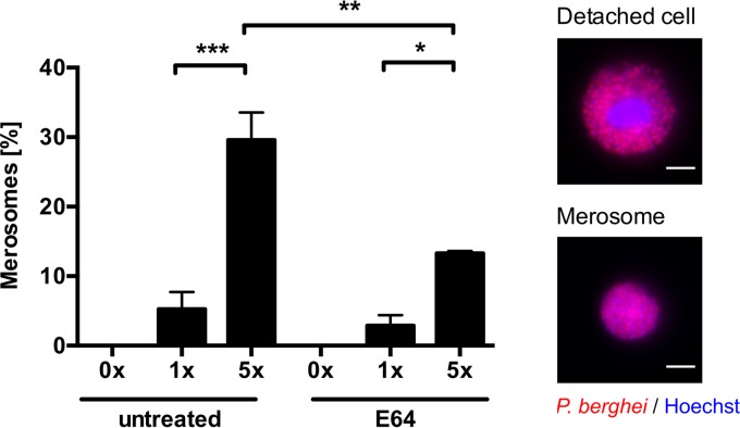
FIG 3 .
In vitro merosome formation is dependent on shear forces and is sensitive to E64. HeLa cells were infected with mCherry-expressing parasites, and at 55 hpi, cells were treated with 10 µg/ml E64 or left untreated. At 65 hpi, the detached-cell-containing cell culture supernatant was either directly analyzed without pipetting (0×) or pipetted up and down one or five times (1×, 5×) before analysis. Detached cells (containing a host cell nucleus) and merosomes (lacking a host cell nucleus) were discriminated by staining with Hoechst 33342. Representative images of detached cells and merosomes are shown on the right, where the parasite cytoplasm is displayed in red and nuclei are in blue. The relative percentage of merosome formation is shown as the mean and the standard error of the mean of three independent experiments in which a total of 1,134 untreated and 811 E64-treated detached cells and merosomes were analyzed. For statistical analysis, a one-way ANOVA, followed by a Holm-Sidak multiple-comparison test, was performed. Statistically significant differences are indicated by asterisks (*, P < 0.05; **, P < 0.01; ***, P < 0.001). All scale bars, 10 µm.

