Fluorescence in frogs

Fluorescence in H. punctatus.
Fluorescence is a fairly common phenomenon among aquatic vertebrates, such as sharks and ray-finned fishes, but rare among land-dwelling vertebrates, particularly amphibians, which count around 7,600 species among their number. Carlos Taboada et al. (pp. 3672–3677) report fluorescence in juveniles and adults of the South American tree frog Hypsiboas punctatus, specimens of which were collected in Argentina. The frog’s translucent skin emitted bright blue/green light when excited with UV-A blue light. Chemical analysis revealed that the frog’s eerie cyan glow under ultraviolet light largely originated in the lymph and glandular secretions of the skin, and could be attributed to a previously unreported group of compounds dubbed hyloins. Analysis revealed that hyloin-mediated fluorescence accounts for around 18% and 30% of the total light emerging from the frogs under moonlit and twilight conditions, respectively; H. punctatus is active from dusk to dawn. Skin color in most amphibian species stems from the chemical and structural features of cells called chromatophores. Hence, the authors suggest, hyloins might represent a supplemental source of skin coloration in frogs. According to the authors, fluorescence, often deemed superfluous to vertebrate life on land, might help individuals stand out in visually riotous terrestrial habitats. — P.N.
Mitigation of Alzheimer’s pathogenesis

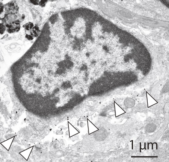
KS glycans on microglial cell surface in the Alzheimer’s brain.
The glycan molecule keratan sulfate (KS), which is found in microglia, has been implicated in amyotrophic lateral sclerosis, a neurodegenerative disorder that affects nerve cells in the brain and spinal cord. However, the functional roles of KS and the enzyme that synthesizes it, GlcNAc6ST1, in neurodegenerative disorders, such as Alzheimer’s disease (AD), remain unclear. Zui Zhang et al. (pp. E2947–E2954) found that levels of KS and GlcNAc6ST1 were upregulated in the brains of two transgenic mouse models, including J20 mice, a model of AD, and in postmortem brain samples from AD patients. Subsequently, the authors crossbred GlcNAc6ST1-deficient (GlcNAc6ST1−/−) mice with J20 mice. In primary microglial cells isolated from the resulting J20/GlcNAc6ST1−/− mice, the authors failed to find KS, but report correspondingly increased levels of amyloid β (Aβ) phagocytosis and a hyperactive response to IL-4, a potent antiinflammatory cytokine. Moreover, the J20/GlcNAc6ST1−/− mice exhibited reduced levels of cerebral Aβ plaques. The findings suggest that GlcNAc6ST1 might regulate microglial function. According to the authors, inhibition of KS synthesis by targeting GlcNAc6ST1 might provide a therapeutic approach for mitigating AD pathogenesis. — C.S.
Dead zones and coral reefs

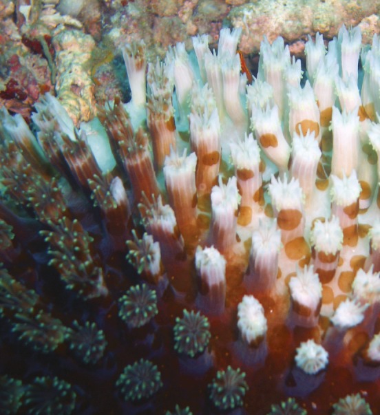
Bleached coral. Image courtesy of Flickr/Samuel Chow.
The potential impact of low oxygen “dead zones” in coastal waters on coral reefs is not well understood. Andrew Altieri et al. (pp. 3660–3665) documented the effects of a massive hypoxic event that occurred on the Caribbean coast of Panama in September 2010, in which more than 90% of corals died on some reefs. Both the fraction of live coral remaining and the diversity of live coral in 2012 correlated with dissolved oxygen levels measured during the hypoxic episode. Following the hypoxic episode, the dominant coral shifted from lettuce corals to star corals, the latter of which was found to be much more hypoxia-tolerant than the former. Analysis of a global database of all known dead zones showed that more than half of the dead zones in the tropics were associated with coral reefs, and that more than 10% of coral reefs worldwide are at increased risk of hypoxia due to anthropogenic and geographic factors. Documented dead zones are underrepresented in the tropics relative to temperate regions, given the length of coastline in each region, suggesting that the impact of dead zones on coral reefs may have been underreported by an order of magnitude. The predominance of research on dead zones by investigators from temperate regions may have contributed to the observed bias, according to the authors. — B.D.


