Ensuring high-fidelity DNA replication is essential for maintaining genome stability in organisms from Escherichia coli to humans. This task requires an intricate network of cellular components that carries out replication and postreplication DNA repair (1). In eukaryotes, replication is carried out by Pol ε and Pol δ that synthesize the leading and lagging strands, respectively, using short RNA–DNA primers synthesized by Pol α. Although replication gives rise to a low spontaneous mutation rate of ∼10−10 mutations per base pair per generation, errors in the nucleotide incorporation step occur about once every 104 to 105 insertions on average, although the frequency varies considerably depending on a number of parameters including the particular mismatch, chromosomal context, and the DNA polymerase. In addition to stringent selectivity at each step of nucleotide incorporation, high-fidelity DNA polymerases possess 3′-endonuclease activities that act as proofreaders, removing errant bases whose abnormal geometries act as speed bumps, allowing excision of the incorrect base and insertion of the correct one. Working closely with the replication machinery is a postreplication DNA repair pathway: DNA mismatch repair (MMR). MMR targets replication errors that have escaped proofreading by excising a region that contains the mismatched base(s) on the newly synthesized strand and giving a high-fidelity DNA polymerase a second chance. In cells whose MMR function is compromised by mutation or epigenetic silencing a hypermutator phenotype ensues. Loss of MMR results in inherited cancer susceptibility (e.g., Lynch syndrome), as well as an increased incidence of sporadic cancers (2). MMR has been reconstituted with a heteroduplex (mismatched) DNA substrate and purified proteins from E. coli and subsequently with yeast and human proteins (3–5). Until now, eukaryotic systems have all used Pol δ. In PNAS, Bowen and Kolodner report the first reconstitution with purified Saccharomyces cerevisiae proteins of 5′ and 3′ MMR that is dependent on Pol ε (6).
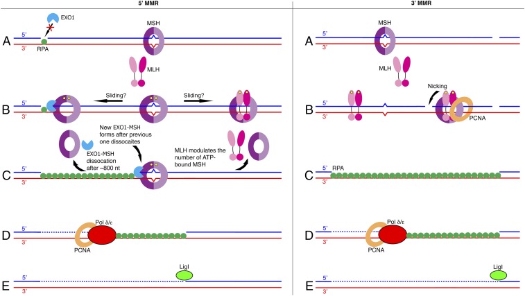
The general schemes of MMR seem deceptively simple (Fig. 1). The newly synthesized strand containing mismatches is targeted for excision followed by high-fidelity resynthesis and ligation. Nevertheless, any scheme must reconcile action at a distance (i.e., the mismatch and a strand-specific signal for excision can be far apart) (7–9). MMR in eukaryotes features two families of MMR proteins, heterodimeric homologs of bacterial MutS (MSH) or MutL (MLH). Early models of E. coli MMR invoke MMR protein-mediated DNA looping between a mismatch and a DNA-methylation excision signal or ATP-driven MutS translocation along the DNA. The nucleotide switch model posits the existence of multiple diffusing MSH–MLH clamps (8). Other studies suggest that a single MSH clamp at a mismatch can load multiple MLH clamps to license excision (1, 9). MutSα (MSH2-MSH6) and MutSβ (MSH2-MSH3) are related to the AAA+ family of ATPases and carry out mismatch recognition (10, 11). MutL homologs MutLα (yeast ScMlh1-Pms1 and human HsMLH1-PMS2), MutLβ (Mlh1-Mlh2), and MutLγ (Mlh1-Mlh3) have N- and C-terminal domains connected by a long helix (12). The N-terminal domain mediates interactions with MutS proteins and DNA and has a GHKL family ATP-binding motif. The C-terminal dimerization domain has a latent endonuclease activity in Pms1 (HsPMS2) and Mlh3 that is activated by proliferating cell nuclear antigen (PCNA), a replication processivity clamp. Mismatches belong to two classes, base–base mispairs (e.g., G opposite T) and unpaired insertion and deletion (indel) mismatches at repetitive sequences caused by strand slippage. Indels are recalcitrant to proofreading because they are often too far from the polymerase active site, leading to microsatellite instability, the contraction and expansion of mono- and dinucleotide repeats whose presence in MMR-deficient bacteria and yeast presaged the discovery of MMR defects in Lynch syndrome colorectal cancer (13). In vitro, 5′ MMR requires MutSα or β, EXO1, an obligate 5′ to 3′ exonuclease, RPA single-strand binding protein, RFC clamp loader PCNA, and Pol δ or ε, whereas 3′ MMR requires these proteins plus MLH (e.g., MutLα) (3–6).
Fig. 1.
Schemes for in vitro eukaryotic MMR. (Left) 5′ MMR. (A) The MutS homolog proteins (MSH, purple) MutSα (MSH2-MSH6), or MutSβ (MSH2-MSH3) recognize and bind a mismatch. RPA (green) bound to single-strand DNA prevents EXO1 (blue) from accessing and degrading DNA. (B) In the sliding clamp model, MutSα/β at a mismatch binds ATP (yellow) and undergoes nucleotide switch activation, becoming a sliding clamp that diffuses along the DNA. Multiple MSH clamps are loaded at a single mismatch. The interaction of EXO1 with MSH sliding clamps overcomes the RPA barrier and activates EXO1 for 5′ to 3′ excision from the 5′ nick. MutL homolog proteins (MLH, pink) (MutLα is ScMlh1-Pms1 or HsMLH1-PMS2) bind ATP and may interact with MSH sliding clamps, though MLH is not absolutely required in vitro for 5' MMR. In other models, MSH remains at the mismatch to authorize excision or can load multiple MLH clamps onto the DNA in the vicinity of the mismatch (not shown). (C) In the sliding clamp model, the EXO1/MSH complex dissociates after excising several hundred nucleotides. Iterative rounds of MSH-EXO1 excision create an excision tract coated with RPA that extends from the 5′ nick to just beyond the mismatch. MLH may limit excision by modulating the number of MSH clamps on DNA. (D) RFC (not shown) loads PCNA clamps (orange) with specific orientation at 3′ termini of strand breaks or gaps, and PCNA facilitates high-fidelity DNA synthesis by Pol δ or ε (red). (E) DNA ligase I (green) seals the nick. (Right) 3′ MMR. (A) MSH recognizes a mismatch. (B) In the sliding clamp model, ATP-dependent binding and nucleotide switching creates MSH sliding clamps that diffuse from the mismatch. The interaction of ATP-bound MLH heterodimers with MSH sliding clamps and PCNA oriented with respect to 3′ termini activates MLH strand-specific nicking. Alternatively, ATP-activated MSH may remain at the mismatch to load MLH and activate nicking (not shown). (C) Excision is EXO1-dependent or -independent, leading to an RPA-coated excision track. An EXO1-independent Pol δ strand-displacement pathway is not shown. (D) Pol δ or ε (red) with the aid of PCNA completes gap filling. (E) DNA ligase I (green) seals the nick.
The first stage of MMR is mismatch recognition by MutSα or MutSβ (Fig. 1A). Using related but distinct mismatch binding sites, MutSα repairs base–base mispairs and small indels of 1–2 nt, whereas MutSβ handles primarily indels of 1–15 bases. Mismatch provoked ADP → ATP exchange in the nucleotide binding sites converts MutSα and MutSβ into sliding clamps that diffuse along the DNA, facilitating the mismatch search and subsequent interaction with MutLα. PCNA interacts with several MMR components including MutS and MutL homologs, EXO1, Pol δ, and Pol ε. MSH3 and MSH6 interact with PCNA via PCNA-interaction-peptide (PIP) motifs in an N-terminal domain. The ability of PCNA to provide a link between replication and MMR and to facilitate the mismatch search is supported by the observation that colocalization of ScMutSα foci with replication centers is dependent on an intact PIP motif in Msh6 (14). Paradoxically, the mutator phenotype of Msh6 PIP mutants is relatively mild, suggesting an alternate pathway for mismatch recognition that does not require interaction with the replication machinery. ScMsh2, but not Msh6, has been implicated in MSH–MLH interactions, and a cocrystal structure of E. coli MutS cross-linked to the N-terminal domain of MutL indicates extensive conformational changes in the complex (9, 15). Defining the physical interaction between MSH and MLH proteins and PCNA and determining the dynamics of complex formation and dissociation remain important problems.
The next stage is excision, a carefully regulated step whose details are unresolved (Fig. 1 B and C). A central tenet of MMR is that DNA excision must be targeted to the newly synthesized strand containing the error rather than the parental or template strand. Almost uniquely, E. coli uses d(GATC) methylation to direct excision (8). In many prokaryotes and all eukaryotes strand discrimination must originate elsewhere. Currently, the leading candidate is PCNA. In vitro MMR reactions require a preexisting nick or single-strand gap in the heteroduplex DNA substrate that can reside on the 5′ (5′ MMR) or 3′ (3′ MMR) side of the mismatch. Importantly, this strand discontinuity serves to direct excision exclusively to the nicked strand. These nicks and gaps are potential PCNA loading sites.
In 5′ MMR reactions, excision is catalyzed by EXO1, a 5′ to 3′ exonuclease that is activated by MutSα in a mismatch-dependent manner (Fig. 1, Left). Although HsMLH1-PMS2 is not required for repair in vitro, its presence modulates the excision step such that excision tracts terminate just past the mismatch (5). Real-time single-molecule imaging studies of the excision step of 5′ MMR support a dynamic molecular switch/sliding clamp model in which multiple ATP-bound HsMutSα sliding clamps diffuse along the DNA following mismatch binding (16). Although HsRPA bound to a single-strand gap or nick inhibits EXO1, preventing runaway excision, an HsMutSα-EXO1 complex at the 5′ break can promote excision over a distance of ∼800 nt. In the case of a mismatch located some distance from the initiating strand break or gap, iterations of MutSα/EXO1-mediated excision starting with the MutSα clamp closest to the 5′ end could extend the excision tract to reach the mismatch, explaining the need for multiple sliding clamps. In this model, HsMutLα modulates excision indirectly by stochastically limiting the number of HsMutSα sliding clamps that are available to activate EXO1 excision. Whether HsMutLα also functions to limit loading of HsMutSα at a mismatch or renders an essential factor such as EXO1 limiting remains unknown. This stochastic sliding clamp model for MMR stands in contrast to models in which MMR proteins remain at the mismatch to license excision (1, 9). Another paradox centers on the requirement for EXO1 in in vitro MMR reactions but its seemingly minor role in vivo (9).
Excision in the case of 3′ MMR (Fig. 1, Right) in which a break or gap is located on the 3′ side of the mismatch is less well understood because EXO1 is, to date, the only exonuclease activity implicated in MMR. The endonuclease activity of MutLα is clearly essential in 3′ reconstitution studies in the presence or absence of EXO1, and it is activated by the interaction of MutLα with both MutSα/β and PCNA (5, 17). It has been proposed based on single-molecule imaging and other studies that multiple rounds of MLH incision between a 3′ break loaded with PCNA and the mismatch can effect strand excision by itself or in combination with EXO1 (8, 9). In an alternative EXO1-independent pathway, Pol δ mediates strand-displacement synthesis from a nick (9, 18). Visualization of fluorescently tagged MMR proteins in yeast and human cells reveals that MutSα and β localize in S phase to replication centers, highlighting ongoing debates of MSH clamps as diffusing or mismatch-bound (14, 19). Interestingly, ScMlh1-Pms1 is oftentimes found by itself even though ScMsh2-Msh6 is required for ScMlh1-Pms1 foci formation. What is the stoichiometry of MMR proteins on DNA, and can activated MutLα function independently of MSHs? How does PCNA confer strand discrimination of MLH nicking? RFC loads PCNA with a defined orientation at 3′ termini. One of PCNA’s two nonidentical faces is always oriented in the direction of synthesis and is the surface that interacts with PIP motifs. It is surmised that retention of this geometry when MutLα interacts with PCNA ensures that cleavage is directed to the newly synthesized strand (17). Gaps between Okazaki fragments in lagging strands or residual breaks in leading strands can also provide strand-discrimination signals in cells. Details regarding strand-discrimination signals and the role of PCNA in vivo remain unresolved, as do the roles of MutLβ and MutLγ.
In the third stage, error-free DNA gap filling by a high-fidelity polymerase and ligation by DNA ligase I restore a corrected and intact DNA duplex (Fig. 1 D and E). PCNA loaded by RFC at a 3′ terminus and replicative Pols δ and ε can use the RPA-coated gapped DNA to carry out synthesis. Several issues remain unresolved. For example, how does MMR differ on leading versus lagging strands, and do replicative polymerases exhibit a strand preference for correction? Is the choice of polymerase influenced by the type of mismatch? Perhaps most intriguing is how these polymerases are recruited to MMR-provoked gaps that are not positioned at origins of replication or part of the advancing replication fork. Bidirectional DNA replication in archaea and eukaryotes is, not surprisingly, exquisitely regulated (20). Replication is temporally restricted to S phase and spatially regulated to initiate only at DNA origins positioned throughout the genome. Strand separation by a DNA helicase must precede synthesis, necessitating the careful choreography of multiprotein complexes. However, MMR has no helicase requirement in vitro. A fully reconstituted system of replication and MMR is required to help address these questions.
The development of an in vitro MMR system in S. cerevisiae that allows interrogation of MMR in the presence of replicative DNA polymerases is a welcome and critical component of approaches that can eventually support a coupled DNA replication–MMR system. In particular, the ability to move seamlessly between in vivo and in vitro studies confers a tremendous advantage. In the future, the adaptation of reconstituted MMR systems holds great promise to examine other important cellular functions of MMR such as DNA repair and homologous recombination in somatic and meiotic cells (21), cellular responses to DNA damage (22), and triplet repeat expansion (23).
Acknowledgments
This work was supported by the Division of Intramural Research, National Institute of Diabetes and Digestive and Kidney Diseases, National Institutes of Health.
Footnotes
The authors declare no conflict of interest.
See companion article on page 3607.
References
- 1.Kunkel TA, Erie DA. Eukaryotic mismatch repair in relation to DNA replication. Annu Rev Genet. 2015;49:291–313. doi: 10.1146/annurev-genet-112414-054722. [DOI] [PMC free article] [PubMed] [Google Scholar]
- 2.Heinen CD. Mismatch repair defects and Lynch syndrome: The role of the basic scientist in the battle against cancer. DNA Repair (Amst) 2016;38:127–134. doi: 10.1016/j.dnarep.2015.11.025. [DOI] [PMC free article] [PubMed] [Google Scholar]
- 3.Bowen N, et al. Reconstitution of long and short patch mismatch repair reactions using Saccharomyces cerevisiae proteins. Proc Natl Acad Sci USA. 2013;110:18472–18477. doi: 10.1073/pnas.1318971110. [DOI] [PMC free article] [PubMed] [Google Scholar]
- 4.Constantin N, Dzantiev L, Kadyrov FA, Modrich P. Human mismatch repair: Reconstitution of a nick-directed bidirectional reaction. J Biol Chem. 2005;280:39752–39761. doi: 10.1074/jbc.M509701200. [DOI] [PMC free article] [PubMed] [Google Scholar]
- 5.Zhang Y, et al. Reconstitution of 5′-directed human mismatch repair in a purified system. Cell. 2005;122:693–705. doi: 10.1016/j.cell.2005.06.027. [DOI] [PubMed] [Google Scholar]
- 6.Bowen N, Kolodner RD. Reconstitution of Saccharomyces cerevisiae DNA polymerase ε-dependent mismatch repair with purified proteins. Proc Natl Acad Sci USA. 2017;114:3607–3612. doi: 10.1073/pnas.1701753114. [DOI] [PMC free article] [PubMed] [Google Scholar]
- 7.Jiricny J. Postreplicative mismatch repair. Cold Spring Harb Perspect Biol. 2013;5:a012633. doi: 10.1101/cshperspect.a012633. [DOI] [PMC free article] [PubMed] [Google Scholar]
- 8.Fishel R. Mismatch repair. J Biol Chem. 2015;290:26395–26403. doi: 10.1074/jbc.R115.660142. [DOI] [PMC free article] [PubMed] [Google Scholar]
- 9.Goellner EM, Putnam CD, Kolodner RD. Exonuclease 1-dependent and independent mismatch repair. DNA Repair (Amst) 2015;32:24–32. doi: 10.1016/j.dnarep.2015.04.010. [DOI] [PMC free article] [PubMed] [Google Scholar]
- 10.Warren JJ, et al. Structure of the human MutSalpha DNA lesion recognition complex. Mol Cell. 2007;26:579–592. doi: 10.1016/j.molcel.2007.04.018. [DOI] [PubMed] [Google Scholar]
- 11.Gupta S, Gellert M, Yang W. Mechanism of mismatch recognition revealed by human MutSβ bound to unpaired DNA loops. Nat Struct Mol Biol. 2011;19:72–78. doi: 10.1038/nsmb.2175. [DOI] [PMC free article] [PubMed] [Google Scholar]
- 12.Guarné A, Charbonnier JB. Insights from a decade of biophysical studies on MutL: Roles in strand discrimination and mismatch removal. Prog Biophys Mol Biol. 2015;117:149–156. doi: 10.1016/j.pbiomolbio.2015.02.002. [DOI] [PubMed] [Google Scholar]
- 13.Strand M, Prolla TA, Liskay RM, Petes TD. Destabilization of tracts of simple repetitive DNA in yeast by mutations affecting DNA mismatch repair. Nature. 1993;365:274–276. doi: 10.1038/365274a0. [DOI] [PubMed] [Google Scholar]
- 14.Hombauer H, Campbell CS, Smith CE, Desai A, Kolodner RD. Visualization of eukaryotic DNA mismatch repair reveals distinct recognition and repair intermediates. Cell. 2011;147:1040–1053. doi: 10.1016/j.cell.2011.10.025. [DOI] [PMC free article] [PubMed] [Google Scholar]
- 15.Groothuizen FS, Sixma TK. The conserved molecular machinery in DNA mismatch repair enzyme structures. DNA Repair (Amst) 2016;38:14–23. doi: 10.1016/j.dnarep.2015.11.012. [DOI] [PubMed] [Google Scholar]
- 16.Jeon Y, et al. Dynamic control of strand excision during human DNA mismatch repair. Proc Natl Acad Sci USA. 2016;113:3281–3286. doi: 10.1073/pnas.1523748113. [DOI] [PMC free article] [PubMed] [Google Scholar]
- 17.Pluciennik A, et al. PCNA function in the activation and strand direction of MutLα endonuclease in mismatch repair. Proc Natl Acad Sci USA. 2010;107:16066–16071. doi: 10.1073/pnas.1010662107. [DOI] [PMC free article] [PubMed] [Google Scholar]
- 18.Kadyrov FA, et al. A possible mechanism for exonuclease 1-independent eukaryotic mismatch repair. Proc Natl Acad Sci USA. 2009;106:8495–8500. doi: 10.1073/pnas.0903654106. [DOI] [PMC free article] [PubMed] [Google Scholar]
- 19.Kleczkowska HE, Marra G, Lettieri T, Jiricny J. hMSH3 and hMSH6 interact with PCNA and colocalize with it to replication foci. Genes Dev. 2001;15:724–736. doi: 10.1101/gad.191201. [DOI] [PMC free article] [PubMed] [Google Scholar]
- 20.Bleichert F, Botchan MR, Berger JM. Mechanisms for initiating cellular DNA replication. Science. 2017;355:eaah6317. doi: 10.1126/science.aah6317. [DOI] [PubMed] [Google Scholar]
- 21.Manhart CM, Alani E. Roles for mismatch repair family proteins in promoting meiotic crossing over. DNA Repair (Amst) 2016;38:84–93. doi: 10.1016/j.dnarep.2015.11.024. [DOI] [PMC free article] [PubMed] [Google Scholar]
- 22.Li Z, Pearlman AH, Hsieh P. DNA mismatch repair and the DNA damage response. DNA Repair (Amst) 2016;38:94–101. doi: 10.1016/j.dnarep.2015.11.019. [DOI] [PMC free article] [PubMed] [Google Scholar]
- 23.Iyer RR, Pluciennik A, Napierala M, Wells RD. DNA triplet repeat expansion and mismatch repair. Annu Rev Biochem. 2015;84:199–226. doi: 10.1146/annurev-biochem-060614-034010. [DOI] [PMC free article] [PubMed] [Google Scholar]


