Abstract
Homologous recombination between the circular chromosomes of bacteria can generate chromosome dimers. They are resolved by a recombination event at a specific site in the replication terminus of chromosomes, dif, by dedicated tyrosine recombinases. The reaction is under the control of a cell division protein, FtsK, which assembles into active DNA pumps at mid-cell during septum formation. Previous studies suggested that activation of Xer recombination at dif was restricted to chromosome dimers in Escherichia coli but not in Vibrio cholerae, suggesting that FtsK mainly acted on chromosome dimers in E. coli but frequently processed monomeric chromosomes in V. cholerae. However, recent microscopic studies suggested that E. coli FtsK served to release the MatP-mediated cohesion and/or cell division apparatus-interaction of sister copies of the dif region independently of chromosome dimer formation. Here, we show that these apparently paradoxical observations are not linked to any difference in the dimer resolution machineries of E. coli and V. cholerae but to differences in the timing of segregation of their chromosomes. V. cholerae harbours two circular chromosomes, chr1 and chr2. We found that whatever the growth conditions, sister copies of the V. cholerae chr1 dif region remain together at mid-cell until the onset of constriction, which permits their processing by FtsK and the activation of dif-recombination. Likewise, sister copies of the dif region of the E. coli chromosome only separate after the onset of constriction in slow growth conditions. However, under fast growth conditions the dif sites separate before constriction, which restricts XerCD-dif activity to resolving chromosome dimers.
Author summary
DNA synthesis, chromosome segregation and cell division must be coordinated to ensure the stable inheritance of the genetic material during proliferation. In eukaryotes, this is achieved by their temporal separation and the existence of checkpoint mechanisms that delay certain steps until others are completed. In contrast, replication, segregation and cell division are interconnected in bacteria. For instance, studies in slowly growing Escherichia coli cells revealed that sister copies of the replication terminus of its chromosome are tethered together at the division site by the binding of a protein, MatP, which interacts with the cell division machinery, to be orderly segregated by a cell division protein, FtsK, which assembles into oriented DNA pumps at the time of constriction. Here, we show using both genetic and fluorescent video microscopy that it is not the case when E. coli cells undergo multiple rounds of replication.
Introduction
DNA synthesis, chromosome segregation and cell division must be coordinated to ensure the stable inheritance of the genetic material during proliferation. In eukaryotes, this is achieved by coupling the assembly and activity of the cell division apparatus to the assembly and activity of the mitotic spindle, a subcellular structure that serves to separate chromosomes. Yeast and animal cells also evolved a checkpoint mechanism that delays cell scission when chromatin remains trapped in the division plane. No such checkpoint exists in bacteria. Instead, they rely on a highly conserved protein, FtsK, to transport any trapped DNA from one daughter cell compartment to another during septation [1].
FtsK is a bi-functional protein. It includes an integral domain at its amino-terminus (FtsKN), a low complexity ‘linker’ region that lacks any evolutionarily conserved feature (FtsKL) and a conserved RecA-type ATPase fold at its carboxyl-terminus (FtsKC) [2]. It was initially discovered because of its essential role in cell division in Escherichia coli [3]. However, only FtsKN and FtsKL are implicated in the cell division process [4]. FtsKC serves to transport DNA between daughter cell compartments before final scission [5,6]. It assembles into hexamers on double stranded DNA at the initiation of septation and uses the energy from binding/hydrolysis of ATP to translocate on it [7–10]. E. coli harbours a single circular chromosome with a single origin of replication, oriC. A winged helix domain at the extreme carboxyl-terminus of FtsK, FtsKγ, binds to specific 8-bp polar DNA motifs, the KOPS, which orientates its loading [11–13]. KOPS are over-represented on the E. coli chromosome. They point from oriC toward a specific 28 bp site, dif, within the replication terminus of the chromosome, ter. As a result, FtsKC motors are directed towards dif [12]. Daughter chromosomes segregate progressively as they are replicated. However, the delay between the time of replication and the time of separation of sister loci is variable [14]. In particular, it was observed that ter sister copies remained close together at mid-cell until the very end of the cell cycle in different growth conditions [14–17]. This is at least in part explained by the binding of MatP, a protein that interacts with the cell division machinery, to specific DNA motifs within ter [18,19]. Microscopic observations of the cellular arrangement of pairs of chromosome loci under slow growth conditions recently suggested that FtsK translocation served to release the MatP-mediated cohesion and/or cell division apparatus-interaction of ter sisters in a KOPS-oriented manner, placing it at the centre of the coordination between the E. coli replication/segregation and cell division cycles [20].
Prior to this observation, FtsK translocation was only considered as a safeguard against the formation of chromosome dimers [21]. Chromosome dimers are generated by homologous recombination events between chromatid sisters during or after replication. They physically impede the segregation of genetic information at cell division, which generates a substrate for FtsK translocation. They are resolved by the addition of a crossover at dif by a dedicated pair of chromosomally encoded tyrosine recombinases, XerC and XerD. Xer recombination-deficient E. coli strains were extensively characterised by microscopy [22,23], growth competition [23], direct measurements of recombination rates at dif using density label assays derived from the Meselson and Stahl experiment [24–26] and the excision of a DNA segment inserted between two dif sites in direct repetition (dif-cassette) at the dif locus [2,23,27]. These studies demonstrated that chromosome dimers were due to recA-dependent homologous recombination initiated by either the recF or recB pathways and independently of the role of recA in SOS. They also suggested that chromosome dimers formed at a generation rate of less than 20% whatever the growth conditions. However, it was observed that mutations decreasing the processivity of replication forks increased their formation in agreement with the multiple roles played by homologous recombination in replication fork progression [26,28,29]. Growth competition and dif-cassette excision experiments further indicated that dif only functioned within the ter region, at the zone of convergence of the KOPS motifs [12,23,30,31]. Finally, density label and dif-cassette excision assays showed that recombination at dif took place at a late stage of cell division, after the initiation of septum constriction [25,32].
FtsK plays two essential roles in chromosome dimer resolution. First, KOPS-oriented FtsK-dependent DNA transport ensures that the two dif sites of a chromosome dimer are brought together at mid-cell [2,12,33]. Second, FtsK activates the addition of a crossover by the Xer recombinases via a direct interaction between FtsKγ and XerD [6,34,35]. The roles played by FtsK in chromosome dimer resolution explains the spatial restriction of the activity of dif on the chromosome to the KOPS convergence zone [12,23,30,31] while the temporal restriction of Xer recombination at chromosomal dif sites suggests that the action of FtsKC is delayed compared to its recruitment to the septum [25,32]. Correspondingly, ectopic production of FtsKC was sufficient to activate dif recombination outside of the KOPS convergence zone, independently of cell division and of recA [2,27]. Taken together, these results suggested that FtsK normally only acted on chromosome dimers. The mild phenotype of FtsK translocation deficient mutants and their suppression by the inactivation of recA corroborated this hypothesis [33,36–38], in apparent contradiction with the general role of FtsK in the release of MatP-mediated cohesion and/or cell division apparatus-interaction of ter sisters [20].
The dimer resolution machinery is conserved in almost all bacteria [21]. This is notably the case in Vibrio cholerae, which harbours two distinct, non-homologous circular chromosomes, chr1 and chr2. The dimer resolution sites of chr1 and chr2, dif1 and dif2, respectively, differ in sequence. However, we previously showed that V. cholerae FtsK controlled the addition of a crossover by V. cholerae XerC and XerD at both sites [39]. As in E. coli, V. cholerae dif-cassette excision was restricted to the KOPS convergence zone within chr1 and chr2 ter regions and only took place after the initiation of septum constriction [40]. However, dif-cassette excision on both V. cholerae chromosomes was independent of recA [40]. In addition, it was too high to be solely explained by the estimated rate of chromosome dimer formation at each generation [39,40]. This observation prompted us to re-visit how the E. coli and V. cholerae chromosomes are managed at the time of cell division using a combination of careful dif-cassette excision assays and newly available fluorescence microscopy techniques.
Here, we show that the E. coli recA-dependency and V. cholerae recA-independency of dif-cassette excision are not determined by differences in the dimer resolution machineries of the two bacteria but by differences in the timing of segregation of their chromosomes: whatever the growth conditions, V. cholerae chr1 ter sister copies remain together at mid-cell until the onset of constriction, which increases the chances for FtsK to activate recombination at dif independently of recA. Likewise, we show that in slow growth conditions, E. coli ter sister copies separate after the onset of constriction and dif-recombination is independent of recA. In contrast, our results suggest that MatP does not prevent E. coli ter sister copies from separating away from each other and from mid-cell before constriction in fast growth conditions. We show that separation of ter sisters is independent of FtsK, which explains why recombination at dif becomes dependent on the formation of chromosome dimers by homologous recombination during fast growth.
Results
The recA-dependency of recombination at dif is species specific
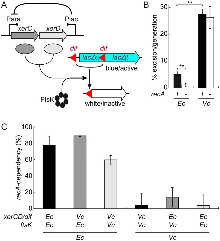
We first checked whether the respective recA-dependency and recA-independency of dif-cassette excision in E. coli and V. cholerae was not due to differences in the design of the assays that were previously used in the two species. To this end, we created dif- and dif1-excision cassettes by introducing a first copy of these sites in the coding region of the E. coli lacZ gene in such a manner that the produced peptide retained its β-galactosidase activity and a second copy of the sites ahead of the lacZ ORF (Fig 1A). Recombination between the two sites of the cassettes excises a third of the lacZ ORF, which abolishes β-galactosidase production. Plating cells on X-Gal gives the cassette recombination frequency for different time points in growing cultures. We inserted the cassettes at the dif locus in strains in which the endogenous lacZ, xerC and xerD genes were deleted. XerC and XerD were produced from a xerC-xerD operon under the control of the arabinose promoter. The E. coli lacZ promoter and the E. coli lacI repressor gene were added in anti-orientation at the end of the operon to help repress any leaky expression of the recombinases (Fig 1A). In the case of E. coli, the xerC-xerD operon was introduced on a pBAD vector. Due to the instability of the vector in V. cholerae, the xerC-xerD operon was integrated in place of the xerC gene locus in the V. cholerae strains.
Fig 1. Genetic determinants of Xer recombination at dif.
(A) Scheme of the dif-cassette excision genetic assay. (B) Influence of homologous recombination on the rate of dif-cassette excision in E. coli and V. cholerae cells grown in LB for 16 h and 3 h, respectively. Mean of 5 independent experiments. Ec: E. coli; Vc: V. cholerae; **: p<0.001 (t-test with a Two-tailed distribution). (C) recA-dependency of dif-cassette excision in E. coli and V. cholerae cells harbouring hybrid dimer resolution machineries. Mean of at least 3 independent experiments. Ec: E. coli cells grown for 16 h in LB; Vc: V. cholerae cells grown for 3 h in LB; recA-dependency: fraction of the dif-cassette excision rate that is linked to recA, 1-frecA-/frecA+.
In a dif-excision cassette experiment, the frequency of recombination per cell per generation (f) is deduced from the initial and final Ratios of the non-recombined cells to the total number of cells (Ri and Rf, respectively) and the number of divisions (n) that occurred during the course of the experiment with the following formula, f = 1-eln(Rf/Ri)/n, which can be simplified into f = 1-eln(Rf)/n because Ri normally equals 1. Rf is best monitored when in the 10–90% range. Large n minimises the possible error made on its estimation. Previous work showed that both conditions were reached with overnight cultures of E. coli strains in LB [23,27]. Using these conditions for our lacZ dif-cassette, we measured an excision rate in the order of 5% per generation (Fig 1B; n = 19.3±1.5, Rf = 36.4±5.4). The rate dropped to little more than 1% per generation in ΔrecA cells (Fig 1B; n = 19.9±1.1, Rf = 79.8±7.3).
Previous results indicated that in the case of V. cholerae strains, only 3 h of growth in LB had to be used because of the elevated number of Xer recombination events at both dif1 and dif2 [40]. This short incubation time was large enough for n to reach a 6 to 7 value due to the fast growth rate of V. cholerae. Under these conditions, the excision rate of the dif1-cassette was in the order of 27% (Fig 1B; n = 6.8±0.5, Rf = 10±1.8) and was not affected by the deletion of recA (Fig 1B; in the order of 26%, n = 6.5±1.1, Rf = 13.7±2.5).
We confirmed that the respective recA-dependency and recA-independency of dif-cassette excision in E. coli and dif1-cassette excision in V. cholerae were not due to a difference in the number of divisions that cells performed during the course of the experiment by monitoring E. coli dif-cassette excision after only 8 h, i.e. at a time when n reached a 7 to 8 value (S1 Fig). Together, these results confirmed the species specificity of the recA-dependency of dif recombination.
Dimer resolution machineries do not specify the recA-dependency of dif recombination
Cultures of E. coli and V. cholerae recA- strains displayed similar loss of colony forming units (S2A Fig) and produced anucleate cells at a similar rate (S2B Fig), suggesting that the E. coli recA-dependence and V. cholerae recA-independence of dif recombination were not linked to any species specific role of RecA in the two bacterial species.
On the contrary, V. cholerae XerC and XerD seemed remarkably different from their E. coli counterparts in that they were known to act on sites with highly divergent central regions [39,41–43]. To check if the recA-independent cassette excisions observed in V. cholerae were linked to this peculiarity, we swapped the arabinose-inducible xerC-xerD operon and the excision cassettes of the E. coli and V. cholerae reporter strains. We also swapped the C-terminal domains of E. coli and V. cholerae FtsK to create reporter strains harbouring fully heterospecific Xer recombination systems. Cassette excision remained dependent on recA in the two E. coli hybrids and independent from it in the two V. cholerae hybrids, indicating that the FtsK/XerCD/dif systems did not specify the recA-dependency of dif recombination (Fig 1C).
E. coli sister termini segregate ahead of septation in fast growth
As E. coli and V. cholerae dif-cassette excisions depend on the initiation of cell constriction [25,32,40] and are restricted to the ter regions [25,32,40], we wondered if differences in recA-dependency were linked to differences in the timing of segregation of the terminus of the E. coli and V. cholerae chromosomes with respect to the assembly of their cell division apparatus.
To test this hypothesis, we compared the position of ydeV, a locus 8 kb away from dif on the E. coli chromosome, and the position of the V. cholerae chr1 dif1 locus in cells under exponential growth in liquid. LB-background fluorescence prevented the use of the exact same growth conditions as those of the cassette excision assays. However, E. coli and V. cholerae dif-cassette excisions remained recA-dependent and independent, respectively, in M9 supplemented with 10% of LB, 0.1% casamino acids and 0.2% of glucose (S3 Fig, M9-Rich). V. cholerae cells had a generation time of 23 min in this medium, which is only slightly longer than their 22 min LB generation time, whereas the generation time of E. coli cells increased from 24 min in LB to 40 min in M9-Rich.
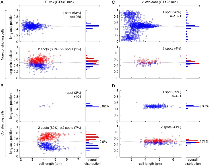
When grown in M9-Rich medium, 37% of E. coli cells displayed two or more ydeV sister loci before a constriction event could be detected (Fig 2A). The proportion of cells with two foci reached 89% when constriction was visible (Fig 2B, left panels). In addition, most of the foci were spatially separated with only 6% of them remaining close to mid-cell, i.e. at a distance from the cell centre of less than 5% of the cell length (Fig 2B, right panels). Thus, only ~8% of the foci, whether single or double, were in the immediate vicinity of the cell division apparatus in constricting E. coli cells with a 40 min generation time.
Fig 2. Fluorescence microscopy snapshot analysis of the position of the dif region in fast growing cells.
(A) Position of the ydeV locus of the E. coli chromosome in cells with no visible indentation. (B) Position of the ydeV locus of the E. coli chromosome in cells with visible indentation. (C) Position of the dif1 locus of V. cholerae chr1 in cells with no visible indentation. (D) Position of the dif1 locus of V. cholerae chr1 in cells with visible indentation. GT: generation time; n: number of cells analysed; left panels: relative long axis position of foci as a function of cell length; right panels: overall distribution of foci positions; upper panels: cells presenting a single focus; lower panels: cells presenting 2 foci. Cells were arbitrarily oriented.
In contrast, a single dif1 spot was observed in 96% of V. cholerae cells that were grown in M9-Rich medium and that presented no visible sign of constriction (Fig 2C). The spot was at mid-cell in the largest cells (Fig 2C). A single dif1 spot was also observed in 59% of the cells in which constriction was visible (Fig 2D, upper left panel). 89% of them were in the immediate vicinity of the cell division apparatus at mid-cell, i.e. at a distance from the cell centre of less than 5% of the cell length (Fig 2D, upper right panel). 71% of the foci observed in the constricting cells with two spots (Fig 2D, lower left panel) were also in close vicinity of mid-cell (Fig 2D, lower right panel). Thus, 82% of the foci, whether single or double, were in the immediate vicinity of the cell division apparatus in constricting V. cholerae cells with a 23 min generation time.
Finally, we observed that in V. cholerae cells with a single dif1 focus, the spot was located at a pole in the shortest cells, i.e. newborn cells (Fig 2C), whereas the ydeV focus of E. coli cells with a single spot was already positioned around mid-cell in the shortest cells (Fig 2A). Correspondingly, sister dif1 spots remained located at mid-cell in the longest V. cholerae cells, i.e. ready to divide cells (Fig 2D), whereas sister ydeV spots relocated toward the 1/4 and 3/4 positions in the longest E. coli cells (Fig 2B).
These observations were consistent with the idea that sister dif sites split and migrated away from mid-cell before the onset of constriction could be manually detected. Sister dif sites separation would then prevent FtsK-mediated Xer recombination activation in E. coli unless a dimer was present. On the contrary, late sister dif1 segregation would permit the action of FtsK on monomeric chromosomes in V. cholerae.
Growth conditions influence sister ter segregation
The results of Fig 2 seemed to contradict the idea that FtsK drove the orderly segregation of E. coli sister ter independently of chromosome dimer formation [20]. However, the latter phenomenon had been documented for cells under extreme slow growth, with a generation time of 210 min [20]. It led us to suspect that growth conditions influenced sister ter segregation, with fast growth conditions accelerating their segregation.
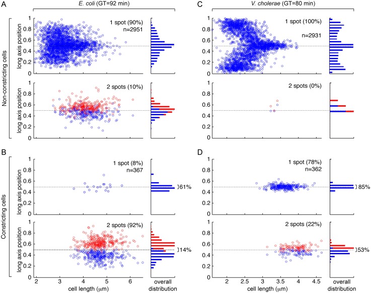
In order to verify this hypothesis, we inspected the localisation of the ydeV and dif1 loci, in snapshot images of cells grown in liquid in minimal medium supplemented with only 0.2% of fructose (M9). Under this condition, E. coli cells presented a generation time of 92 min whereas V. cholerae cells had a generation time of 80 min. In the case of E. coli, 90% of the cells with no visible sign of constriction contained a single ydeV spot, the localisation of which reached mid-cell in the largest cells (Fig 3A). Most of the E. coli constricting cells (92%) still presented two ydeV spots (Fig 3B, lower left panel). However, 14% of them remained in close proximity to the cell centre, i.e. at a distance of less than 5% of the cell length (Fig 3B, lower right panel). Thus, foci, in the order of 18%, whether single or double, remained in the immediate vicinity of the cell division apparatus in constricting E. coli cells with a 92 min generation time. Correspondingly, the single ydeV spot of the shortest cells often located off the cell centre, toward the cell poles (Fig 3B).
Fig 3. Fluorescence microscopy snapshot analysis of the position of the dif region in slow growing cells.
(A) Position of the ydeV locus of the E. coli chromosome in cells with no visible indentation. (B) Position of the ydeV locus of the E. coli chromosome in cells with visible indentation. (C) Position of the dif1 locus of V. cholerae chr1 in cells with no visible indentation. (D) Position of the dif1 locus of V. cholerae chr1 in cells with visible indentation. GT: generation time; n: number of cells analysed; left panels: relative long axis position of foci as a function of cell length; right panels: overall distribution of foci positions; upper panels: cells presenting a single focus; lower panels: cells presenting 2 foci. Cells were arbitrarily oriented.
A similar trend was observed for V. cholerae cells, with a single dif1 spot detected in almost 100% of the cells that presented no visible sign of constriction (Fig 3C) and in 78% of the cells in which constriction was visible (Fig 3D). In total, 78% of the foci, whether single or double, were in the immediate vicinity of the cell division apparatus in constricting V. cholerae cells with an 80 min generation time, i.e. at a distance from the cell centre of less than 5% of the cell length.
These results suggested that growth conditions influenced sister ter segregation in both E. coli and V. cholerae.
E. coli dif-cassette excision becomes recA-independent in slow growth conditions
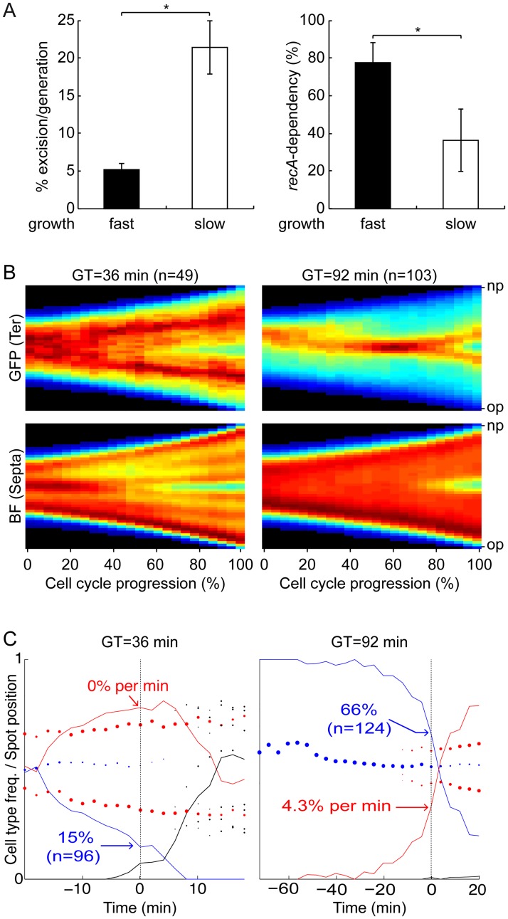
We next wondered if the delay in the separation of the E. coli ter sisters that we observed in cells growing with a generation time of 92 min was sufficient to influence dif-cassette excision. Indeed, the rate of dif-cassette excision, as measured in a 16 h experiment, increased from 5% in LB to 20% in M9 (Fig 4A, left panel). In addition, a significant proportion of the observed recombination events (>60%) were now independent of recA (Fig 4A, right panel). These results were confirmed with 8 h dif-cassette recombination assays (S4 Fig).
Fig 4. Initiation of multiple rounds of replication uncouples the final stages of chromosome segregation from cell division.
(A) Rate of dif-cassette (left panel) and recA-dependency (right panel) of dif-cassette excision measured over 16 h in fast (LB) and slow (M9) growing E. coli cells. Mean of 5 independent experiments. *: p <0.01 (t-test with a Two-tailed distribution). (B) Consensus images summarising the cell cycle choreography of the ydeV fluorescence marker (upper panels, GFP) and the cell shape (lower panels, BF) in fast (left panels) and slow (right panels) growing E. coli cells. Only cells that were followed from birth to division were taken into consideration. At each time point, the maximal and minimal intensities of the projections were set to 1 and 0, respectively. We created images describing the evolution of the fluorescence and cell shape in the cell cycle by plotting the different projections of individual cells as a function of time using a jet colour code. In the heat maps, black corresponds to the lowest and dark red to the highest intensities. In the GFP maps the red areas indicate the presence of the ydeV marker, in the BF maps the green line indicates the Septa appearance. Individual fluorescence and cell shape images were compiled into consensus images summarising the results. Y-axis: position along the cell length. X-axis: cell cycle. GT: generation time; n: number of complete cell cycles that were analysed; op: old pole; np: new pole. 0: birth; 1: division. (C) Frequency of cells displaying a single spot (blue line), two spots (red line) and 3 or more spots (black line) as a function of the time before or after septation was detected. The Y-axis serves for both the cell type frequency lines and to the relative spot positions, with 0 indicating 0% frequency or the old pole position and 1 indicating 100% frequency or the new pole position. Left panels: M9-Rich; right panels: M9. The percentage of cells with a single ydeV spot at the time when septation was detected is indicated in blue with the number of observed cells (n) between parentheses. The percentage of new ydeV duplication events 0% per min and 4.3% per min represented at the time when septation was detected is indicated in red. GT: generation time.
E. coli ter sister copies separate ahead of constriction in fast growth
As dif-cassette excisions depend on the initiation of cell constriction [25,32,40], the results of Fig 4A could be explained if separation of the E. coli chromosome ter sister copies was strictly connected to the onset of constriction under slow growth but that they separated ahead of constriction in fast growth, as suggested by the snapshot results of Figs 2 and 3. We therefore decided to follow the cell cycle choreography of sister ydeV loci by fluorescent video-microscopy to obtain direct evidence that their separation was strictly connected to the onset of constriction under slow growth but that it took ahead of it in fast growth.
In brief, a stack of 32 bright-field images below and above the focal plane of the cells and a single fluorescence image at the focal plane were taken at regular intervals. The bright-field stacks served to reconstruct a high definition image of the cell shapes. Cell genealogy was reconstructed, with cells arising from fully observed division events being oriented from their new pole (the pole originating from the division of the mother cell) to their old pole. Each of the individual cell lives were then analysed by two different methods.
First, we plotted the long cell axis projections of the fluorescence and reconstituted cell shape as a function of time for cells for which a complete cell cycle was recorded. At each time point, the maximal and minimal intensities of the projections were set to 1 and 0, respectively. We created images describing the evolution of the fluorescence and cell shape as a function of time by plotting the different projections of individual cells as a function of time using a jet colour code. Individual fluorescence and cell shape images were then compiled into consensus images summarising the results (Fig 4B). In the fluorescence consensus images (Fig 4B, upper panels, GFP), red colouring signals the position of the ydeV loci (ter). In the cell shape consensus images (Fig 4B, lower panels, BF), red colouring corresponds to regions of high bright-field signal with blue colouring at mid-cell signalling cell constriction (Septa). Use of the cell shape projections was as efficient as the use of a fluorescent derivative of the SPOR domain, which specifically labels nascent peptidoglycan at the septum, to detect constriction events (S5 Fig, [44]). Because projections were scaled from 0 to 1, each of the individual cell lives equally contributed to the consensus images. Consensus images indicated that in slowly growing cells, most sister termini separated and migrated away from mid-cell at the same time as constriction became visible, at approximately 80% of the cell cycle (Fig 4B, right panels). In addition, the ydeV fluorescence signals remained close to mid-cell up to the end of the cell cycle, and lagged at the new pole of newborn cells (Fig 4B), in agreement with the snapshot results of Fig 3. In contrast, in fast growing cells, sister termini already separated and located away from mid-cell at 40% of the cell cycle whereas constriction became visible only after 60% of the cell cycle had elapsed (Fig 4B, left panels). In addition, they already relocated to the 1/4 and 3/4 positions at the end of the cell cycle (Fig 4B).
Second, the position along the cell long axis of each fluorescence focus was manually determined as well as the position of any observed septation trace. This method could give a late estimate of the onset of constriction in cells. Note, however, that septation leads to the formation of a darker line across the cell section in our BF reconstructed images, which helps limit this bias (S6 Fig). We then aligned the cycles of cells for which the initiation of septation was monitored using as a reference the time when septation was first detected (Fig 4C, dashed vertical line at time 0). We computed the median positions of the fluorescence foci (Fig 4C, filled circles) and the frequency of cells with a single focus (Fig 4C, blue line and radius of blue circles), with two foci (Fig 4C, red line and radius of red circles) and with 3 or more foci (Fig 4C, black line and radius of black circles). It revealed that in fast growth conditions only 15% of ter sisters were not separated at the time when septation was first detected (Fig 4C, left panel, blue curve). Indeed, most ter sisters were already separated before the onset of constriction could be detected with our BF reconstruction method (Fig 4C, left panel, red curve), with the two foci having already moved away from mid-cell (Fig 4C, left panel, red spots). Finally, cells with 3 or 4 foci could be observed at late stages of the cell cycle, in agreement with the frequent birth of cells with duplicated ter sisters (Fig 4C, left panel, black spots and black line), which explained the decrease in the frequency of cells with two spots at these stages. In contrast, 100% of the slowly growing cells contained a single focus at birth and 66% of them still presented a single spot at the onset of constriction (Fig 4C, right panel, blue curve). In addition, the rate of duplication of sister termini was maximal at the onset of constriction, with 4.3% of new duplication events per min (Fig 4C, right panel, red curve).
Taken together, these results suggested that separation of sister copies of the E. coli chromosome ter region was connected to the onset of constriction under slow growth but that they separated ahead of constriction in fast growth.
It could somehow seem surprising that, in half of the cells grown in M9-Rich, 2 ydeV spots were visible at birth and 3–4 ydeV spots were visible at the time of division (Fig 4C) whereas the shortest cells of our snapshot image analysis results only had a single ydeV spot and the longest cells 2 ydeV spots (Fig 2). We cannot rule out the possibility that differences in the video-microscopy and snapshot image observations are due to differences in the growth condition of the cells in the two sets of experiments. Indeed, the median generation time of cells observed by video-microscopy was 10% shorter than the generation time of cells grown in liquid. However, we wish to emphasize that differences in the methods of analysis of snapshot images and time-lapse experiments are sufficient to explain the observed differences. Snapshot image analysis permits to observe a distorted cell cycle based on cell length instead of cell age because there is a considerable variation in the length of dividing and newborn cells, which is linked to the intrinsic randomness of growth and cell cycle regulation [16,45]. In addition, it is difficult to assess during the segmentation of snapshot images if joint cells correspond to (i) randomly juxtaposed cells, (ii) daughter cells from a recent cell scission event or (iii) the two halves of a cell in the process of constriction. It is essential to differentiate the first case from the two others, which implies the use of cell segmentation parameters that overestimate cell scission events, i.e. tend to separate the two halves of a cell in the process of constriction. As a result, some non-constricting cells shown in Fig 2 probably correspond to the two halves of a mother cell before scission. In contrast, time-lapse observations permit to unambiguously determine when cell scission has occurred because once separated the two daughter cells re-orientate and slide along each other. Correspondingly, in our video-microscopy experiments, the median length of cells just before cell scission was found to be in the order of 8 μm whereas the longest cell segments in snapshot images were in the order of 7 μm.
MatP does not maintain E. coli ter sister copies at the division site in fast growth
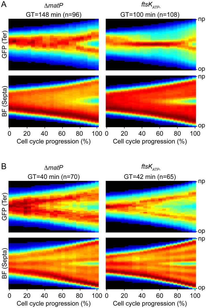
The ter domain of the E. coli chromosome harbours MatP-specific DNA binding motifs [19]. MatP interacts with ZapB, an early component of the cell division machinery [18]. As the MatP-ZapB interaction was shown to tether sister copies of ter loci at mid-cell and prevent their separation until the very end of cell division in cells grown on minimal media [18], we decided to check the action of MatP under our slow (M9) and fast (M9-Rich) growth conditions. When compared to the doubling time of matP+ E. coli cells, the generation time of ΔmatP cells increased by almost 60% in M9 and 10% in M9-Rich (Fig 5A and 5B, ΔmatP). Inspection of the cell contour and ydeV fluorescence consensus images further indicated that sister copies of the ydeV locus separated further ahead of the initiation of septation in the ΔmatP cells than they did in the matP+ cells: they separated at 70% of the cell cycle whereas constriction became visible at 80% of the cell cycle in M9 (Fig 5A, ΔmatP); most sister copies of the ydeV locus were already separated at 20% of the cell cycle whereas constriction was only visible at 60% of the cell cycle in M9-Rich (Fig 5B and S7 Fig). Taken together, these results indicated that MatP participated in chromosome organisation under both slow and fast growth conditions.
Fig 5. ter segregation in matP- and ftsKATP- cells.
(A) Slow growth conditions (M9). (B) Fast growth conditions (M9-Rich). Consensus images summarising the cell cycle choreography of the ydeV fluorescence marker (upper panels, GFP) and of the cell shape (lower panels, BF) in matP- (left panels) and ftsKATP- (right panels) E. coli cells. GT: generation time; n: number of complete cell cycles analysed by fluorescence video-microscopy.
However, whether in rich or poor growth conditions, the number of oriC to ter foci remained constant during the growth of filaments induced by the addition of cephalexin (S8 Fig and S1 and S2 Movies). Cephalexin drives the formation of smooth filaments by blocking the activity of FtsI without disassembling the cell division apparatus and without impeding new rounds of chromosome replication [46]. These results suggested that MatP maintained ter regions attached to the division machinery for only a limited period of time between each new round of replication.
FtsK translocation activity is not required for E. coli ter sister copies separation in fast growth
As FtsK translocation was shown to drive the orderly separation of sister copies of the terminus region of the E. coli chromosome under slow growth conditions [20], we next decided to check the role of FtsK in the separation of ydeV sister copies in M9 and M9-Rich conditions. To this end, we compared when ydeV sister copies separated with respect to the initiation of constriction in cells harbouring an ATPase deficient allele of ftsK (ftsKATP-). We observed a similar 17% increase in the doubling time of ftsKATP- cells in M9 and M9-Rich conditions (Fig 5A and 5B, ftsKATP-). In M9, sister copies of ydeV only separated at 90% of the cell cycle whereas constriction became visible at 70% of the cell cycle, thereby allowing the management of the terminus region by FtsK (Fig 5A, ftsKATP-). However, sister copies of ydeV separated in cephalexin treated cells, suggesting that FtsK translocation is not absolutely required for sister ter separation under slow growth conditions (S8 Fig and S1 and S2 Movies). In M9-Rich, sister copies of ydeV separated at 40% of the cell cycle whereas constriction became visible at 70% of the cell cycle (Fig 5B, ftsKATP-), demonstrating that ydeV sister copies separation did not require FtsK translocation in rich growth conditions. Careful inspection of individual cell lineages revealed frequent splitting of sister ydeV copies into 3–4 foci at the late stage of the cell cycle, further suggesting that FtsK translocation was not necessary to split the pairs of sister copies (S9 Fig). Indeed, ydeV sister copies seemed to separate 10% of the cell cycle time earlier in ftsKATP- cells than they did in FtsK+ cells in M9-Rich (Fig 5B, ftsKATP-). The phenomenon was not unexpected since cells that require FtsK translocation to complete chromosome segregation during constriction (such as cells harbouring a chromosome dimer) are necessarily excluded from the analysis because they fail to complete their cell cycle.
Discussion
dif-cassette excision rates report the processing of chromosomes by FtsK
The activity of FtsKC is the sole limiting factor for dif-recombination in WT cells, which suggested that dif-cassette excision could be used to monitor the processing of chromosomes by FtsK [2,6,39,40]. Indeed, restriction of the assembly of FtsK pumps to mid-cell at the onset of cell division [8] was reflected in the spatial and temporal restriction of dif-recombination to the ter region of chromosomes [2,23,40] and to the initiation of constriction [32,40]. Correspondingly, the low excision rate of dif-cassettes integrated at the E. coli dif locus and its dependence on recA were attributed to the low frequency with which the ter regions of monomeric chromosomes remained trapped in the septum at the onset of constriction [2,27]. However, this view was contradicted by recent microscopic observations, which suggested that FtsK promoted the orderly segregation of loci within the E. coli ter region, whether chromosome dimers were present or not [20], which raised the possibility that an as yet unknown mechanism restricted Xer recombination to chromosome dimers in E. coli.
The excision of dif-cassettes was not restricted to chromosome dimers in V. cholerae [40], further suggesting that the E. coli recA-dependence of dif-cassette excision might be due to a specific property of its dimer resolution system. The results we present demonstrate that it is not the case (Fig 1). Instead, they suggest that recA-dependency and recA-independency are due to the choreography of segregation adopted by the ter regions of chromosomes in the two species (Fig 2). In particular, the rate of dif-cassette excision increased and became less recA-dependent in conditions in which E. coli ter sister copies remained more frequently located at mid-cell at the onset of constriction (Figs 3 and 4). Taken together, these results suggest that differences in dif-cassette excision rates report the relative proximity of sister chromosomal regions at the time of constriction in fast and slow growing E. coli and V. cholerae cells.
Fast growth conditions uncouple ter segregation and cell division in E. coli
Sister copies of the ter region of chromosomes co-localize at mid-cell until the initiation of cell division in both E. coli and V. cholerae [47,48], at least in part because of the MatP/matS macrodomain organisation system [18,19,40]. This mode of segregation participates in the coordination between chromosome segregation and cell division. In particular, a nucleoid occlusion factor, SlmA, impedes the assembly of the cell division machinery until a time when the only genomic DNA left at mid-cell consists of the sister copies of the terminus region of chromosomes [49,50].
Previous reports suggested that the separation of ter sisters was mediated by FtsK translocation, which stripped MatP off DNA during constriction [20,40]. It led to the idea that MatP and FtsK served to coordinate the final stages of chromosome segregation with cell division in bacteria [51]. Our observations of the position of sister termini in V. cholerae cells under slow and fast growth (Figs 3 and 2, respectively) and in E. coli cells under slow growth (Figs 3 and 4) are fully consistent with this idea: sister termini separated at a late stage of the cell cycle, concomitantly with cell constriction (Figs 2, 3 and 4); correspondingly, the chromosomal terminus region lagged at one pole (the new pole) of newborn cells (Figs 2, 3 and 4). In fast growth conditions, however, we found that the sister termini of the E. coli chromosome separated prior to the initiation of septation (Fig 4). They even often reached the 1/4 and 3/4 positions before the onset of constriction could be detected, emphasizing that ter segregation was independent from cell division (Figs 2 and 4). Together, these results suggested that ter segregation was generally independent of FtsK in fast growth conditions, which we confirmed by analysing the choreography of segregation of ter in ftsKATP- cells (Fig 5).
MatP maintains E. coli ter sister copies at the division site for a limited amount of time
Our observations of ΔmatP cells suggested that MatP still participated in the organisation of the ter region in both slow and fast growth conditions (Fig 5). What mechanism could explain FtsK-independent sister ter separation in fast growing E. coli cells (Fig 5 and S8 Fig)? We are attracted to the idea that new rounds of replication/segregation reorganise chromosomal DNA and in particular separate sister ter copies by pulling them towards opposite replication machineries. This model readily explains the differences between the action of FtsK in slow and fast growing cells and in cells treated with cephalexin: in slow growing cells, MatP holds sister copies of the terminus together at mid-cell after they are replicated. As the next round of replication/segregation is a long way off, separation of sister ter copies depends on FtsK translocation (Fig 4). Note, however, that FtsK translocation is not essential (Fig 5). In fast growing cells, overlapping rounds of replication/segregation break sister ter copies apart before the onset of constriction (Fig 4, [52]). The resulting 1/4 and 3/4 sister foci are separated or not depending on the advancement of the next/overlapping rounds of replication/segregation in each cell (Fig 4). In cephalexin treated cells new rounds of replication/segregation can likewise permit sister ter copies separation in the absence of constriction (S8 Fig). In contrast, overlapping replication rounds are absent and/or limited to one in V. cholerae cells under both slow and fast growth (Figs 2 and 3, [53]), which explains why FtsK always acts on sister ter regions (Fig 1). Future work will need to assess what are the different mechanisms that participate to the reorganisation of the bacterial nucleoid during growth and what is their relative contribution in different modes of growth [54].
Materials and methods
Plasmids and strains
Bacterial strains and plasmids used in this study are listed in S1 and S2 Tables, respectively. V. cholerae strains are derivatives of El Tor N16961 strain rendered competent by the insertion of hapR by specific transposition and constructed by natural transformation. E. coli strains are derivatives of MG1655, constructed by P1 transduction and/or integration/excision. Engineered strains were confirmed by PCR.
Growth conditions
Growth media: LB (Luria-Bertani broth), M9-Rich (M9-MM supplemented with 0.2% glucose, 0.1% CAA, 10% LB and 1 μg/ml thiamine) and M9 (M9-MM supplemented with 0.2% fructose and 1 μg/ml thiamine). For the in vivo recombination assays, V. cholerae strains were grown in LB (generation time 22 min) and M9-Rich (generation time 23 min), and E. coli strains were grown in LB (generation time 24 min), M9-Rich (generation time 40 min) and M9 (generation time 92 min). For microscopy experiments V. cholerae strains were grown in M9-Rich and M9 (generation time 80 min), and E. coli strains were grown in M9-Rich and M9.
In vivo recombination assay
V. cholerae: reporter cells were grown overnight in LB supplemented with 0.2 mM IPTG. Cultures were diluted in the morning in LB and grown at 37°C until they reached an OD600 comprised between 0.2 and 0.5. They were then diluted to an OD600 of 0.02 in LB supplemented with 0.1% L-Arabinose and grown at 37°C for 3 h. Serial dilutions of the cells were spread on LB agar plates supplemented with X-Gal (80 μg/ml) and IPTG (0.2 mM) before and after the induction of recombination.
E. coli: chemically competent reporter cells (rubidium chloride) were transformed with pCM165 or pCM166, inoculated in fresh media (LB, M9-Rich or M9) supplemented with 0.1% L-Arabinose and 100 μM ampicillin and grown at 37°C for 8 h or 16 h. Serial dilutions of the cells were spread on LB agar plates supplemented with ampicillin (100 μM), X-Gal (80 μg/ml) and IPTG (0.2 mM) before and after the induction of recombination.
Cells are not growing exponentially over the entire course of the experiments because (i) adaptation to the new growth conditions leads to a lag period at the beginning of the experiments and (ii) nutrient depletion leads to a stationary period at the end of the experiments. During both periods, cells do not divide. Therefore, recombination events can be almost entirely attributed to the intermediate exponential growth phase because dif-cassette excision takes place during cell division. The number of divisions (n) that took place during the exponential phase is deduced from the initial and final Number of cells in the cultures (Ni and Nf, respectively) by the formula n = ln(Nf/Ni)/ln(2).
Microscopy
For snapshot analyses, cells grown to exponential phase in M9 and M9-Rich were spread on 1% (w/v) M9 or M9-Rich agar pads, respectively (ultrapure agarose, Invitrogen). Phase contrast and fluorescence images were acquired using a DM6000-B (Leica) microscope. For time-lapse analyses, cells grown to exponential phase in M9 and M9-Rich were spread on a 1% (w/v) M9 or M9-Rich agar pads, respectively. Images were acquired using an Evolve 512 EMCCD camera (Roper Scientific) attached to an Axio Observe spinning disk (Zeiss). Pictures were taken every 2 min for cells grown in M9-Rich medium and every 4 min for cells grown in M9. At each time point, we took a stack of 32 bright-field images covering positions 1.6 μm below and above the focal plane. Cell contours were detected and cell genealogies were retraced with a MatLab-based script developed in the lab [49]. After the first division event, the new pole and old pole of cells could be unambiguously attributed based on the previous division events. A lacO array was inserted next to dif1 and was detected using a LacI-mCherry fusion produced from the lacZ locus of V. cholerae chr1. A parST1 motif was inserted at the ydeV locus and detected by production of a YGFP-ParBpMT1 from plasmid pFHC2973. A lacO array was inserted 15 kb from the E. coli oriC locus. For the joint detection of oriC and ydeV loci, LacI-mCherry and YGFP-ParBpMT1 fusions were produced from plasmid pAD16. Under these conditions, the tags did not interfere with the localisation of the loci [18,40,47]. The SPOR domain of FtsN was fused to mCherry and to the DsbA signal sequence to efficiently export it into the periplasm, the fusion was expressed from a PBAD promoter induced with 0.01% L-Arabinose. SPOR localisation was inspected in M9. Cephalexin (10 μg/ml, final concentration) was added directly to the agarose slide. If not stated otherwise, leakiness of the promoter was sufficient for signal detection.
Colony Forming Unit (CFU) assay
Wild Type and recA- cells were grown to exponential phase, serial dilutions spread on plates and CFU calculated. Experiments were performed as triplicates of triplicates.
Supporting information
(DOCX)
(DOCX)
Mean of at least 3 independent experiments. Error bars represent standard deviations. (A) Influence of homologous recombination on the rate of dif-cassette excision in E. coli and V. cholerae cells grown in LB for 8 h and 3 h, respectively. **: p<0.01; ****: p<0.0001; ns: p = 0.91 (One-way ANOVA with Tukey post-test). (B) recA-dependency of dif-cassette excision in E. coli under 16 h and 8 h of induction. ns: p = 0.42 (Unpaired two-tailed t test with Welch’s correction). Statistical analyses were performed using GraphPad Prism version 7.0b for Mac OS X, GraphPad Software, La Jolla California USA, www.graphpad.com. Error bars represent standard deviations. recA-dependency: fraction of the dif-cassette excision rate that is linked to recA, 1-frecA-/frecA+.
(TIF)
(A) Overnight recA- and recA+ E. coli (Ec) and V. cholerae (Vc) cell cultures were diluted in fresh media and grown to an identical OD in the exponential phase. Colony forming units (CFU) of the cultures were determined by spreading serial dilutions on plates. In the graphs are shown the ratio of the CFU in recA- over recA+ strains in Ec and Vc cells grown in LB, M9-Rich and M9. Experiments were performed as triplicates of triplicates. Error bars represent standard deviations. (B) Percentage of anucleate cells formed at each division in recA- over recA+ strains of E. coli (Ec) and V. cholerae (Vc) grown in M9 and M9-Rich, as determined from 6 independent time-lapse experiments. Error bars represent standard deviations.
(TIF)
Mean of at least 3 independent experiments. Error bars represent standard deviations. (A) Influence of homologous recombination on the rate of dif-cassette excision in E. coli cells grown in M9-Rich medium for 16 h. **: p<0.01 (Unpaired two-tailed t test). (B) recA-dependency of dif-cassette excision in E. coli cells grown in LB or M9-Rich. ns: 0.72 (Unpaired two-tailed t test). (C) Influence of homologous recombination on the rate of dif-cassette excision in V. cholerae cells grown in M9-Rich medium for 3 h. ns: 0.09 (Unpaired two-tailed t test). Mean of at least 3 independent experiments.
(TIFF)
Mean of at least 3 independent experiments. Error bars represent standard deviations. *: p <0.05 (with unpaired two-tailed t-test for (A) and with Welch’s correction for (B)).
(TIF)
(A) Consensus images of the cell shape (left panel) and SPOR domain (right panel) of E. coli cells grown in M9. (B) Cell shape (left panels) and SPOR domain (right panels) image choreographies of individual cells.
(TIFF)
(A) Time-lapse images of an E. coli cell grown in M9. The red arrow indicates the detection of constriction. (B) Mean pixel intensity along the cell length. Profile numbers correspond to the cell frame numbers of panel A. Profiles in which constriction could not be detected are shown in black. The profile in which constriction was first detected is shown in red.
(TIF)
In the left panels, representation of the manually detected ydeV spots and constriction sites. Green spots represent ydeV loci (fluorescent traces in right panels) and Black spots the constriction mark (bright field traces in central panels). For the fluorescent traces, at each time point, the maximal and minimal intensities of the fluorescence projections were set to 1 and 0, respectively. In the heat maps, black corresponds to the lowest and dark red to the highest intensities. In the GFP maps (right panels) the red lines indicate the presence of the ydeV spot, in the BF maps the green lines indicate the Septa appearance. Y-axis: 0, old cell pole; 1, new cell pole. X-axis: 0, 0% of the cell cycle; 1, 100% of the cell cycle.
(TIFF)
NR: first frame in the time-lapse analysis in which new ori loci split. In the bottom right corner of each frame is indicated the time in minutes from the beginning of the time-lapse experiment.
(TIF)
In the left panels, representation of the manually detected ydeV spots and constriction sites. Green spots represent ydeV loci (fluorescent traces in right panels) and Black spots the constriction mark (bright field traces in central panels). Y-axis: 0, old cell pole; 1, new cell pole. X-axis: 0, 0% of the cell cycle; 1, 100% of the cell cycle.
(EPS)
One frame was taken every 2 minutes. Cells were grown in M9-Rich. 10 μg/ml cephalexin was added to the agarose slide.
(AVI)
One frame was taken every 4 minutes. Cells were grown in M9. 10 μg/ml cephalexin was added to the agarose slide.
(AVI)
Acknowledgments
We thank S. Uphoff for his advice in the development of the scripts used to analyse fluorescence time-lapses, Y. Yamaichi for the gift of plasmid pYB592, T. Bernhardt for the gift of the DsbAss-mCherry-SPOR fusion, and C. Possoz and B. Michel for insightful comments.
Data Availability
All relevant data are within the paper and its Supporting Information files.
Funding Statement
We acknowledge financial support from the European Research Council under the European Community’s Seventh Framework Programme [FP7/2007-2013 Grant Agreement no. 281590] and the Fondation Bettencourt Schueller [2012 Coup d'Elan award]. CM was a recipient from a l’Oréal-UNESCO “Pour les Femmes et la Science” fellowship. The funders had no role in study design, data collection and analysis, decision to publish, or preparation of the manuscript.
References
- 1.Demarre G, Galli E, Barre F-X. The FtsK Family of DNA Pumps. Adv Exp Med Biol. 2013;767: 245–262. 10.1007/978-1-4614-5037-5_12 [DOI] [PubMed] [Google Scholar]
- 2.Barre FX, Aroyo M, Colloms SD, Helfrich A, Cornet F, Sherratt DJ. FtsK functions in the processing of a Holliday junction intermediate during bacterial chromosome segregation. Genes Dev. 2000;14: 2976–2988. [DOI] [PMC free article] [PubMed] [Google Scholar]
- 3.Wang L, Lutkenhaus J. FtsK is an essential cell division protein that is localized to the septum and induced as part of the SOS response. Mol Microbiol. 1998;29: 731–40. [DOI] [PubMed] [Google Scholar]
- 4.Dubarry N, Possoz C, Barre F-X. Multiple regions along the Escherichia coli FtsK protein are implicated in cell division. Mol Microbiol. 2010;78: 1088–1100. 10.1111/j.1365-2958.2010.07412.x [DOI] [PubMed] [Google Scholar]
- 5.Dubarry N, Barre FX. Fully efficient chromosome dimer resolution in Escherichia coli cells lacking the integral membrane domain of FtsK. EMBO J. 2010;29: 597–605. 10.1038/emboj.2009.381 [DOI] [PMC free article] [PubMed] [Google Scholar]
- 6.Aussel L, Barre FX, Aroyo M, Stasiak A, Stasiak AZ, Sherratt D. FtsK is a DNA motor protein that activates chromosome dimer resolution by switching the catalytic state of the XerC and XerD recombinases. Cell. 2002;108: 195–205. [DOI] [PubMed] [Google Scholar]
- 7.Massey TH, Mercogliano CP, Yates J, Sherratt DJ, Lowe J. Double-stranded DNA translocation: structure and mechanism of hexameric FtsK. Mol Cell. 2006;23: 457–469. 10.1016/j.molcel.2006.06.019 [DOI] [PubMed] [Google Scholar]
- 8.Bisicchia P, Steel B, Mariam Debela MH, Löwe J, Sherratt D. The N-terminal membrane-spanning domain of the Escherichia coli DNA translocase FtsK hexamerizes at midcell. mBio. 2013;4: e00800–00813. 10.1128/mBio.00800-13 [DOI] [PMC free article] [PubMed] [Google Scholar]
- 9.Saleh OA, Perals C, Barre FX, Allemand JF. Fast, DNA-sequence independent translocation by FtsK in a single-molecule experiment. EMBO J. 2004;23: 2430–9. 10.1038/sj.emboj.7600242 [DOI] [PMC free article] [PubMed] [Google Scholar]
- 10.Saleh OA, Bigot S, Barre FX, Allemand JF. Analysis of DNA supercoil induction by FtsK indicates translocation without groove-tracking. Nat Struct Mol Biol. 2005;12: 436–40. 10.1038/nsmb926 [DOI] [PubMed] [Google Scholar]
- 11.Sivanathan V, Allen MD, de Bekker C, Baker R, Arciszewska LK, Freund SM, et al. The FtsK gamma domain directs oriented DNA translocation by interacting with KOPS. Nat Struct Mol Biol. 2006;13: 965–972. 10.1038/nsmb1158 [DOI] [PMC free article] [PubMed] [Google Scholar]
- 12.Bigot S, Saleh OA, Lesterlin C, Pages C, El Karoui M, Dennis C, et al. KOPS: DNA motifs that control E. coli chromosome segregation by orienting the FtsK translocase. EMBO J. 2005;24: 3770–80. 10.1038/sj.emboj.7600835 [DOI] [PMC free article] [PubMed] [Google Scholar]
- 13.Bigot S, Saleh OA, Cornet F, Allemand JF, Barre FX. Oriented loading of FtsK on KOPS. Nat Struct Mol Biol. 2006;13: 1026–8. 10.1038/nsmb1159 [DOI] [PubMed] [Google Scholar]
- 14.Joshi MC, Bourniquel A, Fisher J, Ho BT, Magnan D, Kleckner N, et al. Escherichia coli sister chromosome separation includes an abrupt global transition with concomitant release of late-splitting intersister snaps. Proc Natl Acad Sci U S A. 2011;108: 2765–2770. 10.1073/pnas.1019593108 [DOI] [PMC free article] [PubMed] [Google Scholar]
- 15.Lesterlin C, Gigant E, Boccard F, Espéli O. Sister chromatid interactions in bacteria revealed by a site-specific recombination assay. EMBO J. 2012;31: 3468–3479. 10.1038/emboj.2012.194 [DOI] [PMC free article] [PubMed] [Google Scholar]
- 16.Youngren B, Nielsen HJ, Jun S, Austin S. The multifork Escherichia coli chromosome is a self-duplicating and self-segregating thermodynamic ring polymer. Genes Dev. 2014;28: 71–84. 10.1101/gad.231050.113 [DOI] [PMC free article] [PubMed] [Google Scholar]
- 17.Nielsen HJ, Youngren B, Hansen FG, Austin S. Dynamics of Escherichia coli chromosome segregation during multifork replication. J Bacteriol. 2007;189: 8660–8666. 10.1128/JB.01212-07 [DOI] [PMC free article] [PubMed] [Google Scholar]
- 18.Espeli O, Borne R, Dupaigne P, Thiel A, Gigant E, Mercier R, et al. A MatP-divisome interaction coordinates chromosome segregation with cell division in E. coli. EMBO J. 2012;31: 3198–3211. 10.1038/emboj.2012.128 [DOI] [PMC free article] [PubMed] [Google Scholar]
- 19.Mercier R, Petit M-A, Schbath S, Robin S, El Karoui M, Boccard F, et al. The MatP/matS site-specific system organizes the terminus region of the E. coli chromosome into a macrodomain. Cell. 2008;135: 475–485. 10.1016/j.cell.2008.08.031 [DOI] [PubMed] [Google Scholar]
- 20.Stouf M, Meile J-C, Cornet F. FtsK actively segregates sister chromosomes in Escherichia coli. Proc Natl Acad Sci U S A. 2013;110: 11157–11162. 10.1073/pnas.1304080110 [DOI] [PMC free article] [PubMed] [Google Scholar]
- 21.Barre F-X, Midonet C. Xer Site-Specific Recombination: Promoting Vertical and Horizontal Transmission of Genetic Information In: Lambowitz AM, Gellert M, Chandler M, Craig NL, Sandmeyer SB, Rice PA, editors. Mobile DNA III. American Society of Microbiology; 2015. pp. 163–182. http://www.asmscience.org/content/book/10.1128/9781555819217.chap7 [DOI] [PubMed] [Google Scholar]
- 22.Blakely G, Colloms S, May G, Burke M, Sherratt D. Escherichia coli XerC recombinase is required for chromosomal segregation at cell division. New Biol. 1991;3: 789–798. [PubMed] [Google Scholar]
- 23.Pérals K, Cornet F, Merlet Y, Delon I, Louarn JM. Functional polarization of the Escherichia coli chromosome terminus: the dif site acts in chromosome dimer resolution only when located between long stretches of opposite polarity. Mol Microbiol. 2000;36: 33–43. [DOI] [PubMed] [Google Scholar]
- 24.Meselson M, Stahl FW. THE REPLICATION OF DNA IN ESCHERICHIA COLI. Proc Natl Acad Sci U S A. 1958;44: 671 [DOI] [PMC free article] [PubMed] [Google Scholar]
- 25.Steiner WW, Kuempel PL. Cell division is required for resolution of dimer chromosomes at the dif locus of Escherichia coli. Mol Microbiol. 1998;27: 257–68. [DOI] [PubMed] [Google Scholar]
- 26.Steiner WW, Kuempel PL. Sister chromatid exchange frequencies in Escherichia coli analyzed by recombination at the dif resolvase site. J Bacteriol. 1998;180: 6269–75. [DOI] [PMC free article] [PubMed] [Google Scholar]
- 27.Pérals K, Capiaux H, Vincourt JB, Louarn JM, Sherratt DJ, Cornet F. Interplay between recombination, cell division and chromosome structure during chromosome dimer resolution in Escherichia coli. Mol Microbiol. 2001;39: 904–913. [DOI] [PubMed] [Google Scholar]
- 28.Michel B, Recchia GD, Penel-Colin M, Ehrlich SD, Sherratt DJ. Resolution of Holliday junctions by RuvABC prevents dimer formation in rep mutants and UV irradiated cells. Mol Microbiol. 2000;37: 181–191. [DOI] [PubMed] [Google Scholar]
- 29.Michel B, Boubakri H, Baharoglu Z, LeMasson M, Lestini R. Recombination proteins and rescue of arrested replication forks. DNA Repair. 2007;6: 967–980. 10.1016/j.dnarep.2007.02.016 [DOI] [PubMed] [Google Scholar]
- 30.Cornet F, Louarn J, Patte J, Louarn JM. Restriction of the activity of the recombination site dif to a small zone of the Escherichia coli chromosome. Genes Dev. 1996;10: 1152–61. [DOI] [PubMed] [Google Scholar]
- 31.Deghorain M, Pages C, Meile JC, Stouf M, Capiaux H, Mercier R, et al. A defined terminal region of the E. coli chromosome shows late segregation and high FtsK activity. PLoS One. 2011;6: e22164 10.1371/journal.pone.0022164 [DOI] [PMC free article] [PubMed] [Google Scholar]
- 32.Kennedy SP, Chevalier F, Barre FX. Delayed activation of Xer recombination at dif by FtsK during septum assembly in Escherichia coli. Mol Microbiol. 2008;68: 1018–28. 10.1111/j.1365-2958.2008.06212.x [DOI] [PubMed] [Google Scholar]
- 33.Bigot S, Corre J, Louarn J, Cornet F, Barre FX. FtsK activities in Xer recombination, DNA mobilization and cell division involve overlapping and separate domains of the protein. Mol Microbiol. 2004;54: 876–86. 10.1111/j.1365-2958.2004.04335.x [DOI] [PubMed] [Google Scholar]
- 34.Steiner W, Liu G, Donachie WD, Kuempel P. The cytoplasmic domain of FtsK protein is required for resolution of chromosome dimers. Mol Microbiol. 1999;31: 579–583. [DOI] [PubMed] [Google Scholar]
- 35.Yates J, Zhekov I, Baker R, Eklund B, Sherratt DJ, Arciszewska LK. Dissection of a functional interaction between the DNA translocase, FtsK, and the XerD recombinase. Mol Microbiol. 2006;59: 1754–66. 10.1111/j.1365-2958.2005.05033.x [DOI] [PMC free article] [PubMed] [Google Scholar]
- 36.Liu G, Draper GC, Donachie WD. FtsK is a bifunctional protein involved in cell division and chromosome localization in Escherichia coli. Mol Microbiol. 1998;29: 893–903. [DOI] [PubMed] [Google Scholar]
- 37.Recchia GD, Aroyo M, Wolf D, Blakely G, Sherratt DJ. FtsK-dependent and -independent pathways of Xer site-specific recombination. EMBO J. 1999;18: 5724–34. 10.1093/emboj/18.20.5724 [DOI] [PMC free article] [PubMed] [Google Scholar]
- 38.Capiaux H, Lesterlin C, Perals K, Louarn JM, Cornet F. A dual role for the FtsK protein in Escherichia coli chromosome segregation. EMBO Rep. 2002;3: 532–6. 10.1093/embo-reports/kvf116 [DOI] [PMC free article] [PubMed] [Google Scholar]
- 39.Val M-E, Kennedy SP, El Karoui M, Bonne L, Chevalier F, Barre F-X. FtsK-dependent dimer resolution on multiple chromosomes in the pathogen Vibrio cholerae. PLoS Genet. 2008;4. [DOI] [PMC free article] [PubMed] [Google Scholar]
- 40.Demarre G, Galli E, Muresan L, Paly E, David A, Possoz C, et al. Differential Management of the Replication Terminus Regions of the Two Vibrio cholerae Chromosomes during Cell Division. PLoS Genet. 2014;10: e1004557 10.1371/journal.pgen.1004557 [DOI] [PMC free article] [PubMed] [Google Scholar]
- 41.Das B, Bischerour J, Val M-E, Barre F-X. Molecular keys of the tropism of integration of the cholera toxin phage. Proc Natl Acad Sci. 2010;107: 4377–4382. 10.1073/pnas.0910212107 [DOI] [PMC free article] [PubMed] [Google Scholar]
- 42.Das B, Bischerour J, Barre F-X. VGJϕ integration and excision mechanisms contribute to the genetic diversity of Vibrio cholerae epidemic strains. Proc Natl Acad Sci. 2011;108: 2516–2521. 10.1073/pnas.1017061108 [DOI] [PMC free article] [PubMed] [Google Scholar]
- 43.Midonet C, Das B, Paly E, Barre F-X. XerD-mediated FtsK-independent integration of TLCϕ into the Vibrio cholerae genome. Proc Natl Acad Sci. 2014;111: 16848–53. 10.1073/pnas.1404047111 [DOI] [PMC free article] [PubMed] [Google Scholar]
- 44.Yahashiri A, Jorgenson MA, Weiss DS. Bacterial SPOR domains are recruited to septal peptidoglycan by binding to glycan strands that lack stem peptides. Proc Natl Acad Sci. 2015;112: 11347–11352. 10.1073/pnas.1508536112 [DOI] [PMC free article] [PubMed] [Google Scholar]
- 45.Galli E, Paly E, Barre F-X. Late assembly of the Vibrio cholerae cell division machinery postpones septation to the last 10% of the cell cycle. Scientific Rep. 2017; [DOI] [PMC free article] [PubMed] [Google Scholar]
- 46.Pogliano J, Pogliano K, Weiss DS, Losick R, Beckwith J. Inactivation of FtsI inhibits constriction of the FtsZ cytokinetic ring and delays the assembly of FtsZ rings at potential division sites. Proc Natl Acad Sci U A. 1997;94: 559–64. [DOI] [PMC free article] [PubMed] [Google Scholar]
- 47.David A, Demarre G, Muresan L, Paly E, Barre F-X, Possoz C. The two Cis-acting sites, parS1 and oriC1, contribute to the longitudinal organisation of Vibrio cholerae chromosome I. PLoS Genet. 2014;10: e1004448 10.1371/journal.pgen.1004448 [DOI] [PMC free article] [PubMed] [Google Scholar]
- 48.Wang X, Possoz C, Sherratt DJ. Dancing around the divisome: asymmetric chromosome segregation in Escherichia coli. Genes Dev. 2005;19: 2367–77. 10.1101/gad.345305 [DOI] [PMC free article] [PubMed] [Google Scholar]
- 49.Galli E, Poidevin M, Le Bars R, Desfontaines J-M, Muresan L, Paly E, et al. Cell division licensing in the multi-chromosomal Vibrio cholerae bacterium. Nat Microbiol. 2016;1: 16094 10.1038/nmicrobiol.2016.94 [DOI] [PubMed] [Google Scholar]
- 50.Bernhardt TG, de Boer PA. SlmA, a nucleoid-associated, FtsZ binding protein required for blocking septal ring assembly over Chromosomes in E. coli. Mol Cell. 2005;18: 555–64. 10.1016/j.molcel.2005.04.012 [DOI] [PMC free article] [PubMed] [Google Scholar]
- 51.Bouet J-Y, Stouf M, Lebailly E, Cornet F. Mechanisms for chromosome segregation. Curr Opin Microbiol. 2014;22: 60–65. 10.1016/j.mib.2014.09.013 [DOI] [PubMed] [Google Scholar]
- 52.Stokke C, Flåtten I, Skarstad K. An Easy-To-Use Simulation Program Demonstrates Variations in Bacterial Cell Cycle Parameters Depending on Medium and Temperature. Polymenis M, editor. PLoS ONE. 2012;7: e30981 10.1371/journal.pone.0030981 [DOI] [PMC free article] [PubMed] [Google Scholar]
- 53.Stokke C, Waldminghaus T, Skarstad K. Replication patterns and organization of replication forks in Vibrio cholerae. Microbiology. 2011;157: 695–708. 10.1099/mic.0.045112-0 [DOI] [PubMed] [Google Scholar]
- 54.Kleckner N, Fisher JK, Stouf M, White MA, Bates D, Witz G. The bacterial nucleoid: nature, dynamics and sister segregation. Curr Opin Microbiol. 2014;22: 127–137. 10.1016/j.mib.2014.10.001 [DOI] [PMC free article] [PubMed] [Google Scholar]
Associated Data
This section collects any data citations, data availability statements, or supplementary materials included in this article.
Supplementary Materials
(DOCX)
(DOCX)
Mean of at least 3 independent experiments. Error bars represent standard deviations. (A) Influence of homologous recombination on the rate of dif-cassette excision in E. coli and V. cholerae cells grown in LB for 8 h and 3 h, respectively. **: p<0.01; ****: p<0.0001; ns: p = 0.91 (One-way ANOVA with Tukey post-test). (B) recA-dependency of dif-cassette excision in E. coli under 16 h and 8 h of induction. ns: p = 0.42 (Unpaired two-tailed t test with Welch’s correction). Statistical analyses were performed using GraphPad Prism version 7.0b for Mac OS X, GraphPad Software, La Jolla California USA, www.graphpad.com. Error bars represent standard deviations. recA-dependency: fraction of the dif-cassette excision rate that is linked to recA, 1-frecA-/frecA+.
(TIF)
(A) Overnight recA- and recA+ E. coli (Ec) and V. cholerae (Vc) cell cultures were diluted in fresh media and grown to an identical OD in the exponential phase. Colony forming units (CFU) of the cultures were determined by spreading serial dilutions on plates. In the graphs are shown the ratio of the CFU in recA- over recA+ strains in Ec and Vc cells grown in LB, M9-Rich and M9. Experiments were performed as triplicates of triplicates. Error bars represent standard deviations. (B) Percentage of anucleate cells formed at each division in recA- over recA+ strains of E. coli (Ec) and V. cholerae (Vc) grown in M9 and M9-Rich, as determined from 6 independent time-lapse experiments. Error bars represent standard deviations.
(TIF)
Mean of at least 3 independent experiments. Error bars represent standard deviations. (A) Influence of homologous recombination on the rate of dif-cassette excision in E. coli cells grown in M9-Rich medium for 16 h. **: p<0.01 (Unpaired two-tailed t test). (B) recA-dependency of dif-cassette excision in E. coli cells grown in LB or M9-Rich. ns: 0.72 (Unpaired two-tailed t test). (C) Influence of homologous recombination on the rate of dif-cassette excision in V. cholerae cells grown in M9-Rich medium for 3 h. ns: 0.09 (Unpaired two-tailed t test). Mean of at least 3 independent experiments.
(TIFF)
Mean of at least 3 independent experiments. Error bars represent standard deviations. *: p <0.05 (with unpaired two-tailed t-test for (A) and with Welch’s correction for (B)).
(TIF)
(A) Consensus images of the cell shape (left panel) and SPOR domain (right panel) of E. coli cells grown in M9. (B) Cell shape (left panels) and SPOR domain (right panels) image choreographies of individual cells.
(TIFF)
(A) Time-lapse images of an E. coli cell grown in M9. The red arrow indicates the detection of constriction. (B) Mean pixel intensity along the cell length. Profile numbers correspond to the cell frame numbers of panel A. Profiles in which constriction could not be detected are shown in black. The profile in which constriction was first detected is shown in red.
(TIF)
In the left panels, representation of the manually detected ydeV spots and constriction sites. Green spots represent ydeV loci (fluorescent traces in right panels) and Black spots the constriction mark (bright field traces in central panels). For the fluorescent traces, at each time point, the maximal and minimal intensities of the fluorescence projections were set to 1 and 0, respectively. In the heat maps, black corresponds to the lowest and dark red to the highest intensities. In the GFP maps (right panels) the red lines indicate the presence of the ydeV spot, in the BF maps the green lines indicate the Septa appearance. Y-axis: 0, old cell pole; 1, new cell pole. X-axis: 0, 0% of the cell cycle; 1, 100% of the cell cycle.
(TIFF)
NR: first frame in the time-lapse analysis in which new ori loci split. In the bottom right corner of each frame is indicated the time in minutes from the beginning of the time-lapse experiment.
(TIF)
In the left panels, representation of the manually detected ydeV spots and constriction sites. Green spots represent ydeV loci (fluorescent traces in right panels) and Black spots the constriction mark (bright field traces in central panels). Y-axis: 0, old cell pole; 1, new cell pole. X-axis: 0, 0% of the cell cycle; 1, 100% of the cell cycle.
(EPS)
One frame was taken every 2 minutes. Cells were grown in M9-Rich. 10 μg/ml cephalexin was added to the agarose slide.
(AVI)
One frame was taken every 4 minutes. Cells were grown in M9. 10 μg/ml cephalexin was added to the agarose slide.
(AVI)
Data Availability Statement
All relevant data are within the paper and its Supporting Information files.


