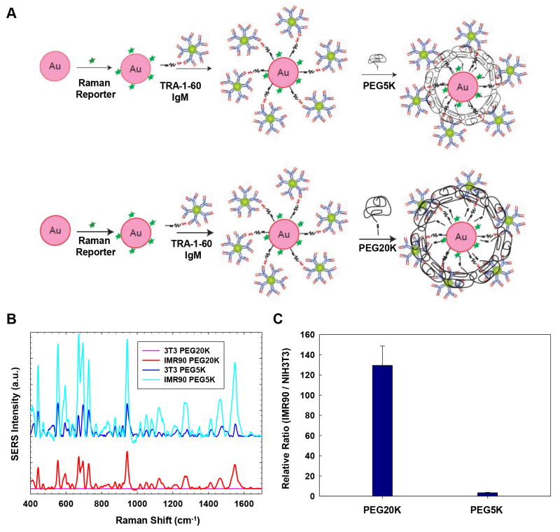
Figure 2.
Effect of the PEG-SH layer of TRA-1-60-conjugated nanoparticles on relative SERS signals. (A) Schematic of applying PEG-SH layers with a molecular weight of 5,000 Da (PEG5K) or 20,000 Da (PEG20K) onto TRA-1-60-conjugated nanoparticles. (B) SERS signals from PEG20K- and PEG5K-stabilized nanoparticles in IMR-90 iPSCs or NIH3T3 fibroblasts (3T3). Note: PEG20K-stabilized nanoparticles produced negligible SERS signals from NIH3T3 fibroblasts (pink spectra), while PEG5K-stabilized nanoparticles produced high background signals from NIH3T3 fibroblasts (dark blue spectra). (C) The relative SERS signals from PEG20K-stabilized and PEG5K-stabilized nanoparticles targeting IMR-90 iPSCs vs. NIH3T3 fibroblasts.

