



Abstract
Erythrocyte-derived microparticles (MPs) are sub-micrometer, biologically active vesicles shed by red blood cells as part of the biochemical changes that occur during storage. We hypothesized that MPs from stored red blood cells would activate endothelial cells. MPs from aged murine packed red blood cells (pRBCs) were isolated and used to treat confluent layers of cultured endothelial cells. Endothelial expression of leukocyte adhesion molecules, ELAM-1 and ICAM-1, and inflammatory mediator, IL-6, were evaluated at 0.5, 6, 12, and 24 hours of treatment. Healthy C57BL/6 mice were transfused with a MP suspension and lung sections were analyzed for adhesion molecules and sequestered interstitial leukocytes. Increased levels of ELAM-1 and ICAM-1 were found on cultured endothelial cells 6 hours after MP stimulation (6.91 vs 4.07 relative fluorescent intensity, RFI, p<0.01, and 5.85 vs 3.55 RFI, p=0.01, respectively). IL-6 in cell culture supernatants was increased after 12 hours of MP stimulation compared to controls (1.24 vs 0.73 ng/ml, p=0.03). In vivo experiments demonstrated that MP injection increased ELAM-1 and ICAM-1 expression at 1 hour (18.56 vs 7.08 RFI, p<0.01, and 23.66 vs 6.87 RFI, p<0.01, respectively) and caused increased density of pulmonary interstitial leukocytes by 4 hours of treatment (69.25 vs 29.25 cells/HPF, p<0.01). This series of experiments supports our hypothesis that erythrocyte-derived MPs are able to activate pulmonary endothelium, leading to the pulmonary sequestration of leukocytes following the transfusion of stored pRBCs.
INTRODUCTION
Hemorrhagic shock continues to be a major cause of death following traumatic injury.1 In this patient population, the transfusion of blood and blood products is lifesaving and one of the most commonly implemented therapies in modern medicine.2 Treatment with blood products, especially in large volume, have long been associated with adverse clinical outcomes including multi-system organ failure (MOF), immunosuppression, increased post-operative infections, and death.3–6 Other studies have identified a host of molecular and biochemical changes that occur during packed red blood cell (pRBC) storage, collectively known as the red blood cell storage lesion, as the etiology of massive transfusion related morbidity.6–8 Although these changes are documented in the literature, there is little understanding as to how agents in donor blood interacts with the transfusion recipient to cause harm.
Microparticles (MPs) are submicron vesicles that are bound by lipid membranes derived from their cell of origin.9 Initially presumed to be rudimentary debris from apoptotic cells, these particles have now been shown to be capable of intercellular signaling and have been implicated in many disease processes.10–13 We have previously shown that pRBC units demonstrate increased MP concentrations during storage and that these MPs are associated with increased lung inflammation. The mechanism of action of pRBC-derived microparticles on transfusion recipients is unknown.14
In the present study, we hypothesized that MPs generated by stored pRBC activate endothelial cells and promote leukocyte migration into the lung.
METHODS
Male C57BL/6 mice weighing 21 g to 30 g were purchased from Jackson Laboratories (Bar Harbor, ME), fed standard laboratory diet and water ad libitum, and acclimated for 1 week in a climate controlled room with a 12-hour light-dark cycle. All experiments were approved by the Institutional Animal Care and Use Committee of the University of Cincinnati.
Murine blood banking
Mice were anesthetized with 0.1 mg/g body weight intraperitoneal pentobarbital and whole blood was collected by open cardiac puncture. Citrate phosphate double dextrose (CP2D) anticoagulant was then added in a ratio of 1:7. Serial centrifugation was used to remove platelet-rich plasma and the leukocyte-rich buffy coat. The red blood cell pellet was resuspended in standard erythrocyte storage medium (additive solution-3, AS3) at a ratio of 2:9. These packed red blood cell units were stored at 4°C for 14 days with gentle agitation and protected from light. Previous studies have demonstrated that 14 days of storage in murine red blood cells is approximately equivalent to 42 days of storage in human red blood cells, which is the current FDA limit of storage for packed red blood cells in the United States.15 At the end of the storage period, the cellular portion of the pRBC was removed by centrifugation at 300 × g for 10 minutes. Cellular debris and platelets were removed by centrifugation at 10,000 × g for 10 minutes. MPs were washed with phosphate buffered saline and isolated using ultracentrifugation at 20,000 × g for 30 minutes.16, 17
In control experiments, isolated MPs were stained with antibodies (BD Biosciences, San Jose, CA) for Ter119 (a murine erythrocyte surface marker), CD45 (leukocyte marker) and CD41 (platelet marker) and analyzed with flow cytometry as previously described.18 This analysis indicated that the microparticles isolated from pRBC units were predominantly erythrocyte in origin, with less than 5% of leukocyte or platelet origin.
In vitro model
Murine endothelial cells were obtained from ATCC microbiology (hemangioendothelioma cells, CRL-2586) (Manassas, VA). Cells were grown to confluence in Dulbecco’s Modified Eagles Medium supplemented with 10% fetal bovine serum. Confluent layers of endothelial cells were treated at 37°C with MPs derived from 1 mL of murine pRBCs suspended in 1 ml of media. Cells treated with media alone and media with 20 ng/mL tumor necrosis factor-α (TNF-α) served as negative and positive controls respectively. Cell culture supernatants taken at 30 minutes, and 6, 12, and 24 hours were evaluated for interleukin-6 (IL-6) concentration using a pre-adsorbed sandwich ELISA kit from eBioscience (Santa Clara, CA). Immunofluorescence staining for endothelial-leukocyte adhesion molecule-1 (ELAM-1, E-selectin) and intercellular adhesion molecule-1 (ICAM-1) was performed on confluent layers of endothelial cells grown on glass cover slips after 30 minutes, 6, 12, and 24 hours of treatment.
In vivo model
In vivo experiments were conducted with healthy male C57BL/6 mice weighing 21 g to 30 g after induction of isoflurane anesthesia. MPs isolated from 1 mL of pRBCs suspended in 200 μL of lactated Ringers were transfused via penile vein injection to simulate the MP burden resulting from a massive transfusion, commonly defined as 10 units of pRBCs in 24 hours. Control mice treated with an equivalent volume of lactated Ringers served as controls. Mice were sacrificed at 1, 4, 8, and 24 hours and whole lungs were harvested and fixed immediately in neutral buffered formalin and embedded in paraffin. Thin cuts of pulmonary alveolar sections were then stained with monoclonal antibody to murine Ly-6g (eBioscience, San Diego, CA) to identify pulmonary interstitial immune cells which express murine myeloid antigen Gr-1, a marker for granulocytes. Quantification of leukocytes per high power field (HPF) was performed on sets of 4 mice per experimental group by an observer blinded to the experimental groups.
Histological analysis
Analysis of adhesion molecule expression was performed via immunofluorescent staining. After the exposure settings for each channel was normalized, a total of eight random captures of each slide was performed using imaging software ZEN 2012 version 1.1.2.0 on Axio Imager M2 microscope (Carl Zeiss AG, Jena, Germany). Images were taken at 10× magnification to maximize the number of cells per capture. The subchannel specific to the fluorophores representing each adhesion molecule was analyzed using the image analysis package ImageJ version 1.49v (Wayne Rasband, National Institutes of Health, USA).
Statistical analysis
In vitro experiments were conducted in triplicate. Animal experiments were performed with 4 mice in each experimental group and 4 mice in the control group. Results are reported as means and standard deviations where applicable. Two-tailed Students’ t-tests were performed and p-values less than 0.05 were deemed significant.
RESULTS
In initial experiments, we first sought to determine the effect of MPs isolated from stored pRBC units on endothelial cell activation. Treatment of confluent endothelial cells with pRBC-derived MPs resulted in an increased surface expression of ELAM-1 compared to control samples (Figure 1A and B). Within 6 hours of treatment with MPs, ELAM-1 expression was significantly increased (6.91 vs 4.07 relative fluorescent intensity, RFI, p<0.01; Figure 1C). This was similar to the increase observed in TNF-α stimulated cells compared to controls (7.16 vs 4.07 RFI, p<0.01; Figure 1C). Twelve hours after treatment with MPs, ELAM-1 expression was significantly greater than both TNF- α treatment and negative controls (10.49 vs 6.47 and 4.51 RFI respectively, p<0.01). This difference remained, but to a lesser degree, at 24 hours after treatment (5.60 vs 3.13 and 3.58 RFI, p<0.01).
Figure 1.

Immunofluorescence imaging of murine endothelial cells in confluent culture after 6 hour incubation with (A) Dulbecco’s Modified Eagles Media vs (B) DMEM with microparticles derived from 1 ml of murine packed red blood cells. Endothelial cells were labelled with fluorescent antibodies to ELAM-1 (red). Nuclei were counterstained with DAPI (blue) and actin filaments with FITC-phalloidin (green). (C) Quantification of red fluorescence intensity after 30 min, 6, 12 and 24 hours of incubation with plain media, microparticles, or TNF-α. * p<0.05 compared to control, n=4 at each time point.
Treatment with MPs also increased endothelial cell expression of ICAM-1 (Figure 2). Twelve hours after treatment with MPs, endothelial cell ICAM-1 expression was significantly increased above that of controls (4.79 vs 3.84 RFI, p<0.01). TNF-α induced ICAM-1 expression to a much greater extent than both media and MPs at both 12 and 24 hours (Figure 2).
Figure 2.

Immunofluorescence imaging of murine endothelial cells in confluent culture after 6 hour incubation with (A) Dulbecco’s Modified Eagles Media vs (B) DMEM with microparticles derived from 1 ml of murine packed red blood cells. Endothelial cells were labelled with a fluorescent antibody to ICAM-1 (red) Nuclei were counterstained with DAPI (blue) and actin filaments with FITC-phalloidin (green). (C) Quantification of red fluorescence intensity after 30 min, 6, 12 and 24 hours of incubation with plain media, microparticles and TNF-α. * p<0.05 compared to control, n=4 at each time point.
Endothelial cell expression of IL-6 was also increased by MPs over the course of 24 hours. IL-6 levels in cell culture supernatants were significantly increased within 12 hours of treatment with MPs compared to controls (1.24 vs 0.73 ng/ml, p<0.05). However, this increase was modest compared to the level of IL-6 induced by treatment with TNF- α (8.1 vs 1.2 ng/ml, p<0.01, Figure 3). Neither media nor MPs showed any detectable IL-6 levels prior to exposure to cultured cells (data not shown).
Figure 3.

IL-6 concentration in endothelial cell culture supernatant after 30 minutes or 6, 12 and 24 hours of stimulation with plain media, and media with red blood cell derived microparticles, or TNF-α. * p<0.05 compared to control, n=4 at each time point in each group.
To determine if the observations we made in vitro were operant in vivo, we next transfused mice with MPs and examined the pulmonary vascular endothelium for expression of ELAM-1 and ICAM-1. As shown in Figure 4, transfusion with MPs increased expression of ELAM-1 and ICAM-1 compared to control mice within 1 hour of transfusion (18.56 vs 7.08 RFI, p<0.01; and 23.66 vs 6.87 RFI, p<0.01, respectively). MP-induced ICAM-1 expression returned to control levels within 24 hours, whereas MP-induced increases in ELAM-1 expression remained significantly increased compared to controls at all time points (Figure 4).
Figure 4.

In vivo expression of ICAM-1 by murine pulmonary endothelium as determined by immunofluorescence 4 hours after injection of mice with (A) 200 μL lactated Ringer’s solution or (B) red cell derived microparticles. Sections were stained with red fluorescence labelled ICAM-1 and nuclei were counterstained with DAPI (blue). Quantification of (C) ICAM-1 and (D) ELAM-1 staining intensity at 1, 4, 8, and 24 hours after injection of red cell derived microparticles as compared to vehicle control. * p<0.05 compared to control, n=4 in each group.
Finally, we examined lung sections for leukocyte infiltration after transfusion of MPs. Within 1 hour of transfusion with MPs, the density of sequestered leukocytes in pulmonary tissue was increased compared to controls (69.25 vs 29.25 cells/HPF, p<0.01) and remained significantly elevated for 24 hours (Figure 5).
Figure 5.
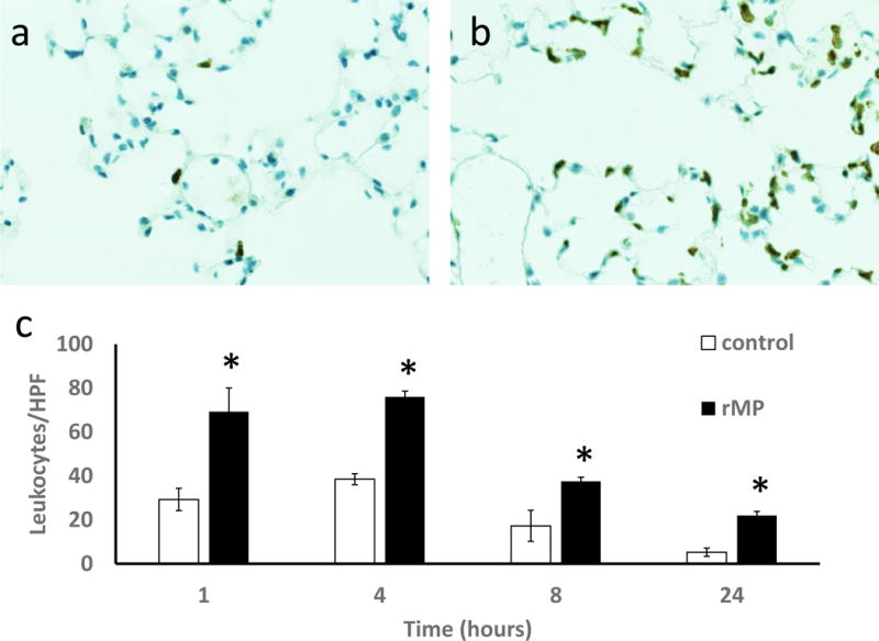
Immunohistochemical staining of thin lung sections 4 hours after injection of mice with (A) 200 μL lactated Ringer’s solution or (B) red blood cell derived microparticles. Sections were stained with Ly-6G/GR1 antibody to demonstrate leukocytes. (C) Quantitation of pulmonary leukocytes at 1, 4, 8, and 24 hours after microparticle injections as compared to vehicle controls. * p<0.05 compared to control, n=4 in each group.
DISCUSSION
In the present study, we examined the role of RBC-derived MPs on endothelial cell activation. Using both in vitro and in vivo model systems, we found that MPs derived from stored pRBC units induce endothelial cell expression of the leukocyte adhesion molecules, ELAM-1 and ICAM-1, and the cytokine, IL-6. The time course over which ELAM-1 is expressed on the plasma membrane of confluent microvascular endothelial cells is consistent with de novo synthesis. The lack of immunofluorescent staining observed on unstimulated cells is consistent with a quiescent endothelium. Likewise, stimulation with TNF-α confirms reactivity of the in vitro model to typical pro-inflammatory stimuli.
Patients suffering from traumatic injury continue to require lifesaving packed red blood cell transfusion, sometimes in large volumes, during resuscitation.19 During storage, pRBCs develop a series of biochemical and physical changes known as the red blood cell storage lesion.7 Although several previous studies have suggested that transfusion of older units may result in harm20, 21, clinical data has suggested that the storage age of pRBCs does not influence clinical outcome.22–24 One challenge in interpreting this literature is that the definition of “old” pRBCs varies between studies, ranging from 14 days25 to 35 days26 of storage. Critically ill, trauma, and complex surgical patients are the most vulnerable to adverse effects from the transfusion of older pRBC units.25–27 Due to the potential harm resulting from the transfusion of stored pRBCs, the development of strategies to mitigate the storage lesion, including novel storage solutions28 and cryopreservation29 remains an area of intense study. Our findings are important because they indicate that microparticles present in stored packed red blood cells lead to endothelial cell activation in the transfusion recipient.
Endothelial cell activation is prerequisite for leukocyte binding and transmigration, and is an integral step in the pathogenesis of lung injury. Activation of the endothelium involves expression of leukocyte adhesion molecules as well as development of a pro-inflammatory cytokine gradient. We validated our in vitro findings using a murine model in which MPs were intravenously transfused. In these experiments, we found that pulmonary endothelial activation results from the transfusion of pRBC-derived MPs, with associated sequestration of leukocytes in pulmonary tissues. Because leukocyte adhesion to the endothelium requires both selectins (ELAM-1) and integrins (ICAM-1), and we found that transfusion of MPs increased the expression of both surface proteins in vitro and in vivo, it is possible that MPs induce pulmonary inflammation via direct activation of the endothelium.
Several important points regarding potential harm from microparticles must be considered. The current studies utilized microparticles isolated from stored pRBC units. Although no clinical situation involves the transfusion of microparticles alone, previous studies from our laboratory indicate that microparticles isolated from stored red blood cell units exert similar biological effects as those present in stored pRBCs.14 In addition, previous work from our laboratory indicates that the microparticle concentration present in stored human pRBC units equals or exceeds the number of microparticles found in stored murine units.30 Additionally, previous studies have shown that higher MP concentrations increase their biological effects in a dose-response fashion.14 Thus, we suspect that potential harm from microparticles from stored pRBC units may become clinically relevant in patients who receive multiple units of pRBCs, especially in the setting of massive transfusion.
A second point addresses the clinical relevance of microparticles from stored pRBC units. Several studies have demonstrated an association between the age of transfused pRBCs and adverse clinical outcomes, such as deep vein thrombosis20, transfusion-related acute lung injury31, morbidity26, and mortality.20, 26, 32 An important goal of the current project has been to determine whether pRBC-derived microparticles could be responsible, at least in part, for these negative outcomes. Based on data from our and other laboratories, we have found that MPs are not present in fresh pRBCs, but accumulate gradually over the duration of the storage period.14, 33 Further research is necessary to determine the point during storage at which pRBC microparticles become potentially harmful, as well as potential methods of microparticle removal or neutralization.
One potential limitation of our experimental design is the focus on murine models. In the present study, we intentionally focused on murine cells for two reasons. First, we wished to remain consistent between our in vitro and in vivo models. Our primary endpoints of increased adhesion molecules and proinflammatory cytokines can vary between species, both in concentration and in time course, so we elected to remove this potentially confounding variable in our experimental design. Second, previous studies have suggested that the development of the RBC storage lesion, including microparticle formation, is significantly affected by poorly understood characteristics of the pRBC donor.34 Thus, use of a murine model minimizes human donor variability and maximizes our ability to understand the potential impact of microparticles on the inflammatory response. Another limitation relates to the injection of microparticle into naïve animals. Previous experiments from our laboratory have demonstrated that microparticle treatment in a hemorrhagic shock model can result in lung injury.14 In the present study, we sought to examine the effects of RBC-derived microparticles with minimized confounding factors, including hemorrhage and neutrophil activation. By removing these factors, we were able to investigate the interaction between microparticles and endothelial cells in a focused environment.
CONCLUSIONS
Microparticles generated during storage of pRBCs lead to increased endothelial cell expression of adhesion molecules as well as pulmonary leukocyte infiltration. Removal or neutralization of red blood cell derived microparticles may be beneficial to patients receiving a large volume of stored pRBCs.
Acknowledgments
This work was supported by research grants R01 GM107625 and T32 GM008478-23 from the National Institutes of Health.
ABBREVIATIONS
- AS3
additive solution-3
- CP2D
citrate phosphate double dextrose
- ELAM-1
endothelial-leukocyte adhesion molecule-1
- ELISA
enzyme-linked immunosorbent assay
- FDA
Food and Drug Administration
- HPF
high powered field
- ICAM-1
intercellular adhesion molecule-1
- IL-6
interleukin-6
- MOF
multisystem organ failure
- MPs
microparticles
- pRBCs
packed red blood cells
- RFI
relative fluorescent intensity
- TNF-α
tumor necrosis factor-alpha
References
- 1.Kauvar DS, Lefering R, Wade CE. Impact of hemorrhage on trauma outcome: an overview of epidemiology, clinical presentations, and therapeutic considerations. J Trauma. 2006;60:S3–11. doi: 10.1097/01.ta.0000199961.02677.19. [DOI] [PubMed] [Google Scholar]
- 2.Whitaker BL, Henry RA. 2011 national blood collection and utilization survey report [Internet] 2011 [cited 1 July 2016]. Available from: http://www.hhs.gov/ash/bloodsafety/2011-nbcus.pdf.
- 3.Hess JR, Thomas MJ. Blood use in war and disaster: lessons from the past century. Transfusion. 2003;43:1622–33. doi: 10.1046/j.1537-2995.2003.00576.x. [DOI] [PubMed] [Google Scholar]
- 4.Makley AT, Goodman MD, Belizaire RM, Friend LA, Johannigman JA, Dorlac WC, et al. Damage control resuscitation decreases systemic inflammation after hemorrhage. J Surg Res. 2012;175:e75–82. doi: 10.1016/j.jss.2011.11.1028. [DOI] [PMC free article] [PubMed] [Google Scholar]
- 5.Society of Thoracic Surgeons Blood Conservation Guideline Task F. Ferraris VA, Ferraris SP, Saha SP, Hessel EA, 2nd, Haan CK, et al. Perioperative blood transfusion and blood conservation in cardiac surgery: the Society of Thoracic Surgeons and The Society of Cardiovascular Anesthesiologists clinical practice guideline. Ann Thorac Surg. 2007;83:S27–86. doi: 10.1016/j.athoracsur.2007.02.099. [DOI] [PubMed] [Google Scholar]
- 6.Zimrin AB, Hess JR. Current issues relating to the transfusion of stored red blood cells. Vox Sang. 2009;96:93–103. doi: 10.1111/j.1423-0410.2008.01117.x. [DOI] [PubMed] [Google Scholar]
- 7.Hoehn RS, Jernigan PL, Chang AL, Edwards MJ, Pritts TA. Molecular mechanisms of erythrocyte aging. Biol Chem. 2015;396:621–31. doi: 10.1515/hsz-2014-0292. [DOI] [PMC free article] [PubMed] [Google Scholar]
- 8.Lagerberg JW, Truijens-de Lange R, de Korte D, Verhoeven AJ. Altered processing of thawed red cells to improve the in vitro quality during postthaw storage at 4 degrees C. Transfusion. 2007;47:2242–9. doi: 10.1111/j.1537-2995.2007.01453.x. [DOI] [PubMed] [Google Scholar]
- 9.Mullier F, Bailly N, Chatelain C, Chatelain B, Dogne JM. Pre-analytical issues in the measurement of circulating microparticles: current recommendations and pending questions. J Thromb Haemost. 2013;11:693–6. doi: 10.1111/jth.12171. [DOI] [PubMed] [Google Scholar]
- 10.Chironi GN, Boulanger CM, Simon A, Dignat-George F, Freyssinet JM, Tedgui A. Endothelial microparticles in diseases. Cell Tissue Res. 2009;335:143–51. doi: 10.1007/s00441-008-0710-9. [DOI] [PubMed] [Google Scholar]
- 11.Hargett LA, Bauer NN. On the origin of microparticles: From “platelet dust” to mediators of intercellular communication. Pulm Circ. 2013;3:329–40. doi: 10.4103/2045-8932.114760. [DOI] [PMC free article] [PubMed] [Google Scholar]
- 12.Italiano JE, Jr, Mairuhu AT, Flaumenhaft R. Clinical relevance of microparticles from platelets and megakaryocytes. Curr Opin Hematol. 2010;17:578–84. doi: 10.1097/MOH.0b013e32833e77ee. [DOI] [PMC free article] [PubMed] [Google Scholar]
- 13.Simak J, Gelderman MP. Cell membrane microparticles in blood and blood products: potentially pathogenic agents and diagnostic markers. Transfus Med Rev. 2006;20:1–26. doi: 10.1016/j.tmrv.2005.08.001. [DOI] [PubMed] [Google Scholar]
- 14.Belizaire RM, Prakash PS, Richter JR, Robinson BR, Edwards MJ, Caldwell CC, et al. Microparticles from stored red blood cells activate neutrophils and cause lung injury after hemorrhage and resuscitation. J Am Coll Surg. 2012;214:648–55. doi: 10.1016/j.jamcollsurg.2011.12.032. discussion 56–7. [DOI] [PMC free article] [PubMed] [Google Scholar]
- 15.Makley AT, Goodman MD, Friend LA, Johannigman JA, Dorlac WC, Lentsch AB, et al. Murine blood banking: characterization and comparisons to human blood. Shock. 2010;34:40–5. doi: 10.1097/SHK.0b013e3181d494fd. [DOI] [PMC free article] [PubMed] [Google Scholar]
- 16.Baron M, Boulanger CM, Staels B, Tailleux A. Cell-derived microparticles in atherosclerosis: biomarkers and targets for pharmacological modulation? J Cell Mol Med. 2012;16:1365–76. doi: 10.1111/j.1582-4934.2011.01486.x. [DOI] [PMC free article] [PubMed] [Google Scholar]
- 17.Shah MD, Bergeron AL, Dong JF, Lopez JA. Flow cytometric measurement of microparticles: pitfalls and protocol modifications. Platelets. 2008;19:365–72. doi: 10.1080/09537100802054107. [DOI] [PubMed] [Google Scholar]
- 18.Kasten KR, Tschop J, Goetzman HS, England LG, Dattilo JR, Cave CM, et al. T-cell activation differentially mediates the host response to sepsis. Shock. 2010;34:377–83. doi: 10.1097/SHK.0b013e3181dc0845. [DOI] [PubMed] [Google Scholar]
- 19.Holcomb JB, Tilley BC, Baraniuk S, Fox EE, Wade CE, Podbielski JM, et al. Transfusion of plasma, platelets, and red blood cells in a 1:1:1 vs a 1:1:2 ratio and mortality in patients with severe trauma: the PROPPR randomized clinical trial. JAMA. 2015;313:471–82. doi: 10.1001/jama.2015.12. [DOI] [PMC free article] [PubMed] [Google Scholar]
- 20.Spinella PC, Carroll CL, Staff I, Gross R, Mc Quay J, Keibel L, et al. Duration of red blood cell storage is associated with increased incidence of deep vein thrombosis and in hospital mortality in patients with traumatic injuries. Crit Care. 2009;13:R151. doi: 10.1186/cc8050. [DOI] [PMC free article] [PubMed] [Google Scholar]
- 21.Weinberg JA, McGwin G, Jr, Griffin RL, Huynh VQ, Cherry SA, 3rd, Marques MB, et al. Age of transfused blood: an independent predictor of mortality despite universal leukoreduction. J Trauma. 2008;65:279–82. doi: 10.1097/TA.0b013e31817c9687. discussion 82–4. [DOI] [PubMed] [Google Scholar]
- 22.Alexander PE, Barty R, Fei Y, Vandvik PO, Pai M, Siemieniuk RA, et al. Transfusion of fresher vs older red blood cells in hospitalized patients: a systematic review and meta-analysis. Blood. 2016;127:400–10. doi: 10.1182/blood-2015-09-670950. [DOI] [PubMed] [Google Scholar]
- 23.Lacroix J, Hebert PC, Fergusson DA, Tinmouth A, Cook DJ, Marshall JC, et al. Age of transfused blood in critically ill adults. N Engl J Med. 2015;372:1410–8. doi: 10.1056/NEJMoa1500704. [DOI] [PubMed] [Google Scholar]
- 24.Steiner ME, Ness PM, Assmann SF, Triulzi DJ, Sloan SR, Delaney M, et al. Effects of red-cell storage duration on patients undergoing cardiac surgery. N Engl J Med. 2015;372:1419–29. doi: 10.1056/NEJMoa1414219. [DOI] [PMC free article] [PubMed] [Google Scholar]
- 25.Spadaro S, Reverberi R, Fogagnolo A, Ragazzi R, Napoli N, Marangoni E, et al. Transfusion of stored red blood cells in critically ill trauma patients: a retrospective study. Eur Rev Med Pharmacol Sci. 2015;19:2689–96. [PubMed] [Google Scholar]
- 26.Goel R, Johnson DJ, Scott AV, Tobian AA, Ness PM, Nagababu E, et al. Red blood cells stored 35 days or more are associated with adverse outcomes in high-risk patients. Transfusion. 2016 doi: 10.1111/trf.13559. [DOI] [PubMed] [Google Scholar]
- 27.Horvath KA, Acker MA, Chang H, Bagiella E, Smith PK, Iribarne A, et al. Blood transfusion and infection after cardiac surgery. Ann Thorac Surg. 2013;95:2194–201. doi: 10.1016/j.athoracsur.2012.11.078. [DOI] [PMC free article] [PubMed] [Google Scholar]
- 28.Cancelas JA, Dumont LJ, Maes LA, Rugg N, Herschel L, Whitley PH, et al. Additive solution-7 reduces the red blood cell cold storage lesion. Transfusion. 2015;55:491–8. doi: 10.1111/trf.12867. [DOI] [PubMed] [Google Scholar]
- 29.Scott KL, Lecak J, Acker JP. Biopreservation of red blood cells: past, present, and future. Transfus Med Rev. 2005;19:127–42. doi: 10.1016/j.tmrv.2004.11.004. [DOI] [PubMed] [Google Scholar]
- 30.Hoehn RS, Jernigan PL, Japtok L, Chang AL, Midura EF, Caldwell CC, et al. Acid Sphingomyelinase Inhibition in Stored Erythrocytes Reduces Transfusion-Associated Lung Inflammation. Ann Surg. 2016 doi: 10.1097/SLA.0000000000001648. [DOI] [PMC free article] [PubMed] [Google Scholar]
- 31.Peters AL, van Hezel ME, Juffermans NP, Vlaar AP. Pathogenesis of non-antibody mediated transfusion-related acute lung injury from bench to bedside. Blood Rev. 2015;29:51–61. doi: 10.1016/j.blre.2014.09.007. [DOI] [PubMed] [Google Scholar]
- 32.Weinberg JA, McGwin G, Jr, Vandromme MJ, Marques MB, Melton SM, Reiff DA, et al. Duration of red cell storage influences mortality after trauma. J Trauma. 2010;69:1427–31. doi: 10.1097/TA.0b013e3181fa0019. discussion 31–2. [DOI] [PMC free article] [PubMed] [Google Scholar]
- 33.Kriebardis AG, Antonelou MH, Stamoulis KE, Economou-Petersen E, Margaritis LH, Papassideri IS. RBC-derived vesicles during storage: ultrastructure, protein composition, oxidation, and signaling components. Transfusion. 2008;48:1943–53. doi: 10.1111/j.1537-2995.2008.01794.x. [DOI] [PubMed] [Google Scholar]
- 34.Tzounakas VL, Georgatzakou HT, Kriebardis AG, Voulgaridou AI, Stamoulis KE, Foudoulaki-Paparizos LE, et al. Donor variation effect on red blood cell storage lesion: a multivariable, yet consistent, story. Transfusion. 2016;56:1274–86. doi: 10.1111/trf.13582. [DOI] [PubMed] [Google Scholar]