Abstract
The effect of diet on reproduction is well documented in a large number of organisms; however, much remains to be learned about the molecular mechanisms underlying this connection. The Drosophila ovary has a well described, fast and largely reversible response to diet. Ovarian stem cells and their progeny proliferate and grow faster on a yeast-rich diet than on a yeast-free (poor) diet, and death of early germline cysts, degeneration of early vitellogenic follicles and partial block in ovulation further contribute to the ~60-fold decrease in egg laying observed on a poor diet. Multiple diet-dependent factors, including insulin-like peptides, the steroid ecdysone, the nutrient sensor Target of Rapamycin, AMP-dependent kinase, and adipocyte factors mediate this complex response. Here, we describe the results of a visual screen using a library of green fluorescent protein (GFP) protein trap lines to identify additional factors potentially involved in this response. In each GFP protein trap line, an artificial GFP exon is fused in frame to an endogenous protein, such that the GFP fusion pattern parallels the levels and subcellular localization of the corresponding native protein. We identified 53 GFP-tagged proteins that exhibit changes in levels and/or subcellular localization in the ovary at 12-16 hours after switching females from rich to poor diets, suggesting them as potential candidates for future functional studies.
Keywords: diet, oogenesis, ovary, germline stem cells, GFP protein trap line, Drosophila
INTRODUCTION
Reproduction demands high levels of energy and resources to support oocyte growth and/or embryonic development. In addition, adequate food availability maximizes the chance of offspring survival. Millions of years of evolution have therefore ensured the tight coupling of nutrient availability and reproductive processes, including oogenesis (Ables et al., 2012; Laws and Drummond-Barbosa, 2015). In women, for example, malnutrition due to low food availability or eating disorders is associated with decreased fertility, whereas obesity is also linked to infertility and other reproductive disorders (Fontana and Della Torre, 2016). Much remains to be learned, however, about the cellular and molecular underpinnings of this connection.
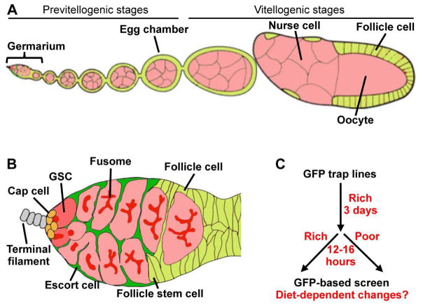
A well-described example of how the ovary responds to diet is seen in Drosophila melanogaster, a model organism highly amenable to genetic, molecular and cell biological analyses (Ables et al., 2012; Hudson and Cooley, 2014). The ovary is composed of 15 to 20 ovarioles, each of which contains an anterior germarium followed by developing egg chambers, or follicles (Fig.1A). The germarium houses germline stem cells (GSCs) and follicle stem cells (FSCs) that continuously produce germ cells and follicle cells, respectively (Fig. 1B). Two to three GSCs reside in a well-defined niche composed of cap cells, terminal filament cells and a subset of escort cells. Each GSC division yields a GSC and a cystoblast that undergoes four rounds of incomplete division to produce two-, four-, eight- and 16-cell cysts. Early germ cells have a unique membranous structure, called the fusome, which undergoes stereotypical morphological changes: in GSCs, the predominantly round fusome always abuts the interface with cap cells; as germline cyst divisions progress, the fusome becomes gradually more branched as it interconnects all of the cells in the cyst. In the 16-cell cyst, one of the cells is determined as the oocyte and undergoes meiosis, while the remaining 15 become supportive polyploid nurse cells. FSC-derived follicle cells envelop the 16-cell cyst to form a follicle that leaves the germarium and progresses through fourteen developmental stages. Follicle cells divide mitotically until stage 7, after which they undergo endoreplication. During stage 8, oocytes initiate yolk uptake, or vitellogenesis, and at stage 14, mature eggs are ready to be ovulated, fertilized, and laid (Hudson and Cooley, 2014; Spradling, 1993).
Fig. 1.
A visual screen for GFP protein trap lines showing diet-dependent changes in expression levels or subcellular localization in the Drosophila ovary. (A) Each ovariole contains a string of egg chambers formed in the germarium as 16-cell germline cysts become enveloped by follicle cells. One germline cell becomes the oocyte, whereas the others become nurse cells. Each egg chamber goes through fourteen developmental stages to form a mature oocyte. (B) In the germarium, GSCs reside in a niche composed of somatic cap cells, terminal filament cells, and a subset of escort cells. (C) For the GFP-based screen, young females were kept on a yeast-rich diet for 3 days, and then either maintained on a yeast-rich diet or switched to a yeast-poor diet for an additional 12–16 hours.
Drosophila oogenesis is highly regulated by diet at multiple steps, resulting in up to ~60-fold differences in rates of egg laying on protein-rich versus -poor diets. On a protein-rich diet, GSCs and FSCs have relatively high division rates, and their progeny proliferate and grow robustly. On a protein-poor diet, proliferation and growth slow down two- to four-fold, GSC loss increases, early germline cysts die more frequently within the germarium, follicles entering vitellogenesis degenerate at high rates, and ovulation is partially blocked (Drummond-Barbosa and Spradling, 2001). In addition, developing previtellogenic follicles undergo a rearrangement of the microtubule cytoskeleton and accumulation of ribonucleoproteins in large processing bodies under starvation (Burn et al., 2015; Shimada et al., 2011). This dietary response is fast and largely reversible; for example, upon switching from poor to rich diets, changes in proliferation and growth rates occur within less than 18 hours, while from rich to poor diets these rates change within less than 24 hours (Drummond-Barbosa and Spradling, 2001).
Multiple diet-dependent factors mediate the Drosophila ovarian response to diet. Insulin-like peptides directly modulate GSC proliferation (Hsu et al., 2008; LaFever and Drummond-Barbosa, 2005), and act on cap cells to control GSC maintenance (Hsu and Drummond-Barbosa, 2009; Hsu and Drummond-Barbosa, 2011; Yang et al., 2013). In addition, insulin signaling cell autonomously controls the growth of germline cysts and vitellogenesis (Hsu et al., 2008; LaFever and Drummond-Barbosa, 2005), and act through follicle cells to modulate the microtubule cytoskeleton and processing body formation in underlying germline cysts (Burn et al., 2015). The nutrient sensor Target of rapamycin (TOR) has multiple roles in the GSC and follicle stem cell lineages (LaFever et al., 2010; Sun et al., 2010), and its appropriate downregulation on a poor diet is important for survival of previtellogenic egg chambers (Wei et al., 2014). Ecdysone stimulates GSC responsiveness to niche signals, controls germline differentiation, and stimulates oocyte lipid accumulation (Ables et al., 2015; Ables and Drummond-Barbosa, 2010; Konig et al., 2011; Morris and Spradling, 2012; Sieber and Spradling, 2015). Adiponectin signaling (which in mammals is stimulated by the adipocyte proteohormone adiponectin) is also required for GSC maintenance (Laws et al., 2015), and amino acid sensing by adult adipocytes controls GSC numbers and ovulation (Armstrong et al., 2014). Here, to identify additional factors potentially involved in the ovarian response to diet, we screened 887 green fluorescent protein (GFP) protein trap lines. In these lines, an artificial GFP exon is inserted in frame into endogenous loci, and the resulting GFP fusion proteins therefore exhibit levels and subcellular localization patterns that reflect those of their native counterparts (Buszczak et al., 2007; Kelso et al., 2004; Morin et al., 2001; Quinones-Coello et al., 2007). For each line, we compared the ovarian GFP pattern of females kept on a rich diet to that of females switched to a poor diet for 12–16 hours, with the goal of capturing early changes in the response to diet. Of the lines we screened, 61 (corresponding to 53 proteins) exhibited distinct GFP fusion expression levels or subcellular localization patterns in the ovary on different diets. These lines identify candidates to be investigated in the future for potential regulatory or early effector roles in the ovarian response to diet.
MATERIALS AND METHODS
Drosophila GFP protein trap lines and culture conditions
Drosophila GFP protein trap lines from Yale University and Carnegie Institution for Science were maintained at room temperature on standard medium, consisting of cornmeal, yeast, molasses and agar. For the screen, ten pairs of two- to three-day-old flies from each GFP protein trap line were first cultured in plastic vials containing standard medium supplemented with wet yeast paste (yeast-rich diet) for 3 days. Half of the flies were maintained on a rich diet, whereas the other half was switched to a poor diet (an empty vial containing a Kimwipe soaked with 5% molasses in water) for 12–16 hours. GFP trap lines showing obvious and consistent diet-dependent differences in GFP levels or subcellular localization were selected as candidates, and results for each line were confirmed in three or more separate experiments.
Ovary GFP fluorescence microscopy
Ovaries were dissected, fixed with 4% formaldehyde at room temperature for 13 minutes, washed in phosphate buffered saline (PBS, pH 7.0) with 0.1% Triton X-100 (Sigma) (PBST) 10 minutes for three times, and stored with a few drops of Vectashield (Vector Labs) at 4°C until mounting. Ovaries from females on rich or poor diets for each line were mounted side by side on a glass slide, and imaged and analyzed using Zeiss LSM 510 or LSM 700 confocal microscopes. Note that confocal settings for images taken from well-fed and starved ovaries from the same line were identical. Images of egg chambers represent single optical sections either through the largest anterior-posterior diameter of the egg chamber (e.g. Fig. 4A,A’, Fig. 5A,A’,B,B’) or through the outer follicle cell layer of the egg chamber (e.g. Fig. 4B,B’,C,C’, Fig. 5C,C’).
Fig. 4.
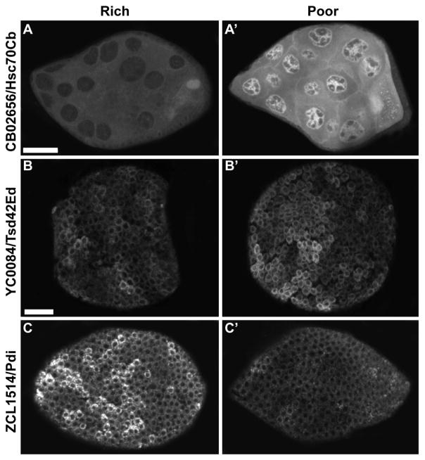
GFP-trap lines showing diet-dependent expression in egg chambers. Expression of Hsc70Cb, Tsd42Ed and Pdi in vitellogenic egg chambers of females maintained on a yeast-rich (A-D) versus -poor diet (A’–D’). Scale bars, 20 μm. A and A’, and B-D’ are shown at the same magnification.
Fig. 5.
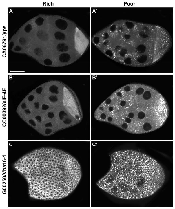
GFP protein trap lines showing diet-dependent punctate patterns in developing egg chambers. Expression of yps, eIF-4E, and Vha16–1 in vitellogenic egg chambers of females kept on a yeast-rich (A–D) versus -poor diet (A’–D’). Scale bar, 20 μm.
RESULTS AND DISCUSSION
Fifty-three GFP trap lines respond to diet within 12–16 hours
To uncover additional factors involved in the ovarian response to diet, we searched for GFP fusion proteins that undergo rapid changes in expression levels or subcellular localization upon switching females from yeast-rich to -poor diets. We screened 887 publically available GFP trap lines by initially placing females on a rich diet for three days and then switching them to a poor diet for 12–16 hours. We then compared the GFP pattern of dissected and fixed ovaries to those of control females maintained on a rich diet (Fig. 1C). We identified 61 lines corresponding to 53 distinct proteins whose expression intensity or subcellular localization was diet-dependent (Tables 1–4).
Table 1.
GFP protein trap lines showing diet-dependent changes in expression levels in germline and somatic cells within the germarium
| FlyTrap line | Gene | Increased GFP levels on a poor diet |
|---|---|---|
| CA07176 | CG5174 | GSCs, cystoblasts, germline cysts |
| CA06926 | CG5059 | GSCs, cystoblasts, germline cysts |
| CC00392 | Eukaryotic initiation factor 4E (eIF-4E) | GSCs, cystoblasts |
| CC01326 | polo | Germline cysts |
|
| ||
| FlyTrap line | Gene | Decreased GFP levels on a poor diet |
|
| ||
| CA06989 | Semaphorin 2a (Sema2a) | GSCs, cystoblasts |
| YB0057le | IGF-II mRNA-binding protein (Imp) | GSCs, cystoblasts, germline cysts |
| CB05282 | maternal expression at 31B (me31B) | Germline cysts |
| CC01684 | Surfeit 4 (Surf4) | Escort cells |
| YC0036/ CC01359 | failed axon connections (fax) | Escort cells |
| CC04973 | Elongation factor 2 (EF2) | Early follicle cells |
| CC00416 | tropomodulin (tmod) | Muscle sheath cells |
Table 4.
Widely expressed GFP-protein trap lines with diet-regulated expression levels
| FlyTrap line | Gene | GFP levels on a poor diet |
|---|---|---|
| CA07176 | CG5174 | Increased* |
| CA06914 | archipelago (ago1) | Increased |
| CC01311 | Cdc42-interacting protein 4 (Cip4) | Increased |
| CC00758 | Aconitase (Acon) | Increased |
| CC00814/ZCL2988 | Calmodulin (Cam) | Increased |
| ZCL2696/CC00869 | belle (bel) | Increased |
| YB0039 | Decondensation factor 31 (Df31) | Increased |
| YC0063 | β-Tubulin at 56D (βTub56D) | Increased |
| YD0788 | Bristle (Bl) | Increased |
| YC0014 | La related protein (larp) | Increased |
| YD0623 | Rm62 | Increased |
| CA07717 | Rab11 | Decreased |
| CA06506 | 14-3-3ε | Decreased |
| CA07004 | visgun (vsg) | Decreased |
| CB04973 | TER94 | Decreased |
| CB03223 | CG6330 | Decreased |
| CC01995 | mushroom-body expressed (mub) | Decreased |
| YB0112 | Tetratricopeptide repeat protein 2 (Trp2) | Decreased |
| YB0059 | Lasp | Decreased |
CG5174 is listed on Tables 1 and 4 because it shows increased expression in all ovarian cells, but particularly elevated levels in GSCs and cystoblasts on a poor diet.
GFP trap lines with diet-regulated expression in germ cells within the germarium
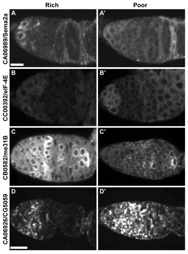
Among our 61 GFP trap candidate lines, seven were expressed in germ cells within the germarium and showed distinct GFP patterns in females on rich versus poor diets (Fig. 2 and Table 1). For instance, a Semaphorin 2a fusion (Sema2a::GFP) was well expressed in GSCs and cystoblasts on a rich diet, but its expression was reduced upon the poor diet switch (Fig. 2A and A’). Sema2a is a secreted semaphorin and regulates neural guidance (Ayoob et al., 2006). In C. elegans, germ granules maintain germline identity (Ciosk et al., 2006); compromising germ granules causes germ cells to express neuronal and muscle markers and extend neurite-like projections (Updike et al., 2014). It would be interesting to investigate if Sema2a might be somehow involved in this process. The expression of an Eukaryotic initiation factor 4E fusion (eIF-4E::GFP) was elevated on a poor diet (Fig. 2B and B’). eIF-4E, eIF-4A and eIF-4G form a protein complex to control ribosome assembly during translation (Svitkin et al., 2001), and eIF-4A has been demonstrated to control Drosophila female GSC self-renewal by regulating the translation of E-cadherin, which mediates the attachment of GSCs to their niche (Shen et al., 2009). We observe an increase in eIF4-E::GFP levels in GSCs and cystoblasts on a poor diet (Fig. 2B,B’), which might reflect a feedback mechanism to counteract other mechanisms that lead to E-cadherin reduction (Hsu and Drummond-Barbosa, 2009) and thereby prevent excessive GSC loss on a poor diet. A maternal expression at 31B fusion (me31B::GFP) was highly expressed in germline cysts (including developing oocytes) on a yeast-rich diet (Fig. 2C), but had markedly decreased levels under a poor diet (Fig. 2C’). me31B, which belongs to a DEAD box helicase family, is a component of the oskar ribonucleoprotein complex, and plays an essential role in translational silencing of oocyte-localized mRNAs during their transport to the oocyte (Nakamura et al., 2001). It has been shown that the oskar ribonucleoprotein complex is transported from nurse cells to the oocyte during vitelogenesis (Shimada et al., 2011). Under nutrient stress, the oskar ribonucleoprotein complex accumulates in large processing bodies in nurse cells while this accumulation is reversed by re-feeding or exogenous addition of insulin (Shimada et al., 2011). The reduced expression of me31B:GFP in germline cysts under starvation might accompany reduced rate of transport into the oocyte. CG5059 encodes the ortholog of mammalian BNIP3 (Low et al., 2013), which belongs to the Bcl-2 family and is involved in necrosis, apoptosis and autophagy (Burton and Gibson, 2009; Low et al., 2013). CG5059::GFP shows increased expression in subcellular structures within cystoblasts and germline cysts of females on a poor relative to rich diet (Fig. 1D and D’). It will be interesting to investigate whether CG5059 plays a role in the death of early germline cysts that is observed in response to a poor diet (Drummond-Barbosa and Spradling, 2001).
Fig. 2.
GFP trap lines with diet-regulated expression in early germ cells. Expression of Sema2a, eIF-4E, me31B, and CG5059 in germaria of females on a yeast-rich (A–D) versus –poor (A’–D’) diet. Scale bars, 10 μm. A– C’ are shown at the same magnification. D and D’ are shown at the same magnification.
GFP trap lines with diet-dependent expression in somatic cells within the germarium
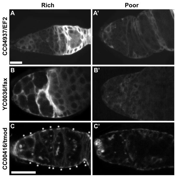
Several populations of somatic cells, including terminal filament cells, cap cells and escort cells, play important roles in controlling GSC behavior and the development of their early dividing progeny, while follicle cells envelop fully formed 16-cell cysts to give rise to individual follicles (Spradling, 1993). In addition, a sheath of mononucleate muscle cells surrounds each ovariole (Hudson et al., 2008). We identified four genes that were expressed in somatic cells in the germarium and responded to diet (Fig. 3 and Table 1). An Elongation factor 2 fusion (EF2::GFP) was strongly expressed in follicle cells on a rich diet (Fig. 3A) and but nearly absent from follicle cells on a poor diet (Fig. 3A’). EF2 is a translational elongation factor (Grinblat et al., 1989); it is therefore possible that it might play a role in controlling follicle cell growth and/or proliferation in response to diet. failed axon connections (fax) has unknown molecular function but is involved in axonogenesis and neurogenesis (Hill et al., 1995), and fax::GFP was previously shown to be expressed in ovarian escort cells and testicular cyst cells (Decotto and Spradling, 2005). In our screen, we also found that fax::GFP was expressed in the cytoplasm of escort cells (Fig. 3B), but its expression was reduced on a poor diet (Fig. 3B’). This diet-dependent response in escort cells suggest a possible role for fax in modulating escort cell-early germ cell interactions in response to diet. tropomodulim (tmod) regulates cytoskeleton arrangement by binding to actin (Weber et al., 1994), and it also a fusome component in Drosophila (Lighthouse et al., 2008). We found that, in addition to been expressed in the fusome, tmod::GFP was also expressed in muscle sheath cells of females on a rich diet (Fig. 3C, asterisks), although its expression levels decreased in response to a poor diet (Fig. 3C’). It is conceivable that tmod modulates ovariole contraction in response to diet.
Fig. 3.
GFP trap lines with diet-regulated expression in early somatic cells. Expression of EF2, fax, and tmod in germaria of females on a yeast-rich (A–C) versus –poor (A’–C’) diet. Scale bars, 10 μm. A–B’ are shown at the same magnification. Asterisks in C and C’ indicate muscle sheath cells.
Diet-dependent GFP-trap lines expressed in egg chambers
Vitellogenic egg chambers undergo apoptosis when nutrients are limiting (Drummond-Barbosa and Spradling, 2001), and insulin, TOR and ecdysone signaling are involved in this process (Drummond-Barbosa and Spradling, 2001; Hsu et al., 2008; LaFever and Drummond-Barbosa, 2005; LaFever et al., 2010; Pritchett and McCall, 2012; Sieber and Spradling, 2015).We identified 17 GFP-trap lines corresponding to 14 proteins that were expressed in the germ cells or follicle cells of vitellogenic egg chambers and whose expression levels change in response to diet (Fig. 4 and Table 2). Hsc70Cb is a heat shock-induced protein that exhibits chaperone activity (Folk et al., 2006); we observed that Hsc70Cb::GFP is weakly expressed in the ovaries of well-fed females, but its expression is dramatically increased in the nuclei of nurse cells in vitellogenic egg chambers on a poor diet (Fig. 4A and A’). We speculate that Hsc70Cb might protect germ cells in egg chambers from starvation stress by stabilizing proteins. The function of Tetraspanin 42Ed (Tsp42Ed) is not well understood, but Tetraspanin is known as a highly conserved protein with four transmembrane domains (Charrin et al., 2014). Tetraspanins are proposed to act as scaffolding proteins, anchoring multiple proteins to one area of the cell membrane, and may be involved in cell adhesion, activation or proliferation (Charrin et al., 2014). Tsp42Ed::GFP was expressed more strongly in the cytoplasm of follicle cells surrounding egg chambers of starved flies compared those of well-fed flies (Fig. 4B and B’). Protein disulfide isomerase (Pdi) is predicted to regulate protein folding (McKay et al., 1995), and conversely, Pdi::GFP was strongly expressed in the cytoplasm of follicle cells of egg chambers in females fed on a protein-rich diet (Fig. 4C) but dramatically decreased on a yeast-poor diet (Fig. 4C’).
Table 2.
GFP protein trap lines showing diet-dependent changes in expression levels in developing egg chambers
| FlyTrap line | Gene | Increased GFP levels on a poor diet |
|---|---|---|
| CB02656 | Hsc70Cb | Nurse cells |
| CC01326 | polo | Nurse cells |
| CC00277 | Alhambra (Alh) | Oocytes |
| CC01941 | bazooka (baz) | Follicle cells |
| CC01936/ YC0005 | discs large 1 (dlg1) | Follicle cells |
| YC0084 | Tetraspanin 42Ed (Tsp42Ed) | Follicle cells |
| ZCL3012 | CG10927 | Follicle cells |
| CC03751/ YD0523 | Ornithine decarboxylase antizyme ( Oda) | Nurse cells |
|
| ||
| Flytrap line | Gene | Decreased GFP levels on a poor diet |
|
| ||
| CA07674 | Secretion-associated Ras-related 1 (Sar1) | Follicle cells |
| CA06526 | Protein disulfide isomerase (Pdi) | Follicle cells |
| CB02647 | NAD-dependent methylenetetrahydrofolate dehydrogenase (Nmdmc) | Follicle cells |
| YB0057le | IGF-II mRNA-binding protein (Imp) | Follicle cells |
| YB0164 | Innexin 7 (Inx7) | Follicle cells |
| CC01359/ YC0036 | failed axon connections (fax) | Follicle cells |
GFP-trap lines showing punctate patterns in developing egg chambers on a poor diet
An additional response of developing egg chambers to nutrient deprivation is the rapid accumulation of ribonucleoproteins, including ypsilon schachtel (yps), me31B, and eIF-4E, in large processing bodies in germ cells (Burn et al., 2015; Shimada et al., 2011). Our screen identified 6 fusion proteins that exhibited punctate GFP signals in germ cells in females on a poor diet (Fig. 5 and Table 3). These included GFP-tagged Yps (Fig. 5A and A’), me31B, and eIF-4E (Fig. 5B and B’), as had been previously described (Shimada et al., 2011), validating our screen strategy. In addition, we observed that GFP-tagged Elf (Ef1α-like factor) also formed puncta in both nurse cells and oocytes under starvation; however, further investigation is needed to establish whether or not Elf is present in processing bodies and to determine its role in the response to diet. We also detected GFP-tagged Vha16-1 (Vacuolar H+ ATPase 6kD subunit 1) (Fig. 5C and C’) in small puncta within follicle cells on a rich diet, and these puncta become larger on a poor diet. Vacuolar H+-ATPases pump protons across the plasma membrane, and thus acidify intracellular organelles (Finbow and Harrison, 1997), an essential process for cargo trafficking, sorting, and degradation during endocytosis and autophagy (Forgac, 2007). In addition, Vacuolar H+-ATPase-mediated acidification of secretory vesicles is necessary for secretion and for neurotransmitter release in neurons (Wang and Hiesinger, 2013). Recent studies have shown that Vha16-1 is involved in lysosomal biogenesis (Tognon et al., 2016), and it also triggers activation of mTOR at the lysosomal surface in response to growth signaling in an amino-acid-dependent manner (Zoncu et al., 2011). The biological significance of the localization of Vha16-1::GFP to large puncta under starvation, therefore, merits further investigation.
Table 3.
GFP protein trap lines showing punctate expression in egg chambers on a poor diet
| Flytrap line | Gene | Punctate GFP signals |
|---|---|---|
| CB05282 | maternal expression at 31B (me31B) | Nurse cells, oocytes |
| CA06515 | Ef1α-like factor (Elf) | Nurse cells, oocytes |
| CA07674 | Secretion-associated Ras-related 1 (Sar1) | Oocytes |
| CC00392 | Eukaryotic initiation factor 4E (eIF-4E) | Nurse cells, oocytes |
| CC06791 | ypsilon schachtel (yps) | Nurse cells, oocytes |
| CA06780/ G00007/ ZCL2017 | Vacuolar H+ ATPase 16kD subunit 1 (Vha16-1) | Follicle cells |
Widely expressed GFP fusion proteins with diet-dependent expression levels
In our screen, we also identified 19 GFP-tagged proteins that are expressed in all ovarian cells and show diet-dependent changes in expression levels (Table 4). With the exception of 14-3-3ε, which belongs to a highly conserved acidic protein family (Mhawech, 2005), none of them is a known component of the insulin signaling pathway or has been previously shown to control diet-dependent processes. For example, Rm62, an ATP-dependent RNA helicase DEAD-box, cooperates with chromatin remodeling factors to silence gene transcription (Boeke et al., 2011; Buszczak and Spradling, 2006; Lamiable et al., 2010). Under starvation, Rm62::GFP expression is dramatically increased in all ovarian cells, suggesting a potential role in saving energy by reducing gene expression. Lasp, an actin-binding protein, controls localization of Oskar protein to the posterior pole to determine primordial germ cell number (Suyama et al., 2009). It has also been proposed that Lasp interacts with integrin to anchor male GSCs to the niche (Lee et al., 2008). Under starvation, expression of Lasp is reduced in all ovarian cells, suggesting that actin or integrin-dependent interaction might be disrupted. The roles of these widely expressed, diet-dependent proteins in oogenesis remain to be further investigated.
Acknowledgments
We thank L. Cooley and A. Spradling for providing GFP protein trap lines. This work was supported by American Cancer Society RSG-07-182-01 and National Institutes of Health R01 GM069875.
Footnotes
Publisher's Disclaimer: This is a PDF file of an unedited manuscript that has been accepted for publication. As a service to our customers we are providing this early version of the manuscript. The manuscript will undergo copyediting, typesetting, and review of the resulting proof before it is published in its final citable form. Please note that during the production process errors may be discovered which could affect the content, and all legal disclaimers that apply to the journal pertain.
References
- Ables ET, Bois KE, Garcia CA, Drummond-Barbosa D. Ecdysone response gene E78 controls ovarian germline stem cell niche formation and follicle survival in Drosophila. Dev Biol. 2015;400:33–42. doi: 10.1016/j.ydbio.2015.01.013. [DOI] [PMC free article] [PubMed] [Google Scholar]
- Ables ET, Drummond-Barbosa D. The steroid hormone ecdysone functions with intrinsic chromatin remodeling factors to control female germline stem cells in Drosophila. Cell Stem Cell. 2010;7:581–92. doi: 10.1016/j.stem.2010.10.001. [DOI] [PMC free article] [PubMed] [Google Scholar]
- Ables ET, Laws KM, Drummond-Barbosa D. Control of adult stem cells in vivo by a dynamic physiological environment: diet-dependent systemic factors in Drosophila and beyond. Wiley Interdiscip Rev Dev Biol. 2012;1:657–74. doi: 10.1002/wdev.48. [DOI] [PMC free article] [PubMed] [Google Scholar]
- Armstrong AR, Laws KM, Drummond-Barbosa D. Adipocyte amino acid sensing controls adult germline stem cell number via the amino acid response pathway and independently of Target of Rapamycin signaling in Drosophila. Development. 2014;141:4479–88. doi: 10.1242/dev.116467. [DOI] [PMC free article] [PubMed] [Google Scholar]
- Ayoob JC, Terman JR, Kolodkin AL. Drosophila Plexin B is a Sema-2a receptor required for axon guidance. Development. 2006;133:2125–2135. doi: 10.1242/dev.02380. [DOI] [PubMed] [Google Scholar]
- Boeke J, Bag I, Ramaiah MJ, Vetter I, Kremmer E, Pal-Bhadra M, Bhadra U, Imhof A. The RNA Helicase Rm62 Cooperates with SU(VAR)3-9 to Re-Silence Active Transcription in Drosophila melanogaster. PLoS One. 2011;6:e20761. doi: 10.1371/journal.pone.0020761. [DOI] [PMC free article] [PubMed] [Google Scholar]
- Burn KM, Shimada Y, Ayers K, Lu F, Hudson AM, Cooley L. Somatic insulin signaling regulates a germline starvation response in Drosophila egg chambers. Developmental Biology. 2015;398:206–217. doi: 10.1016/j.ydbio.2014.11.021. [DOI] [PMC free article] [PubMed] [Google Scholar]
- Burton TR, Gibson SB. The role of Bcl-2 family member BNIP3 in cell death and disease: NIPping at the heels of cell death. Cell Death Differ. 2009;16:515–23. doi: 10.1038/cdd.2008.185. [DOI] [PMC free article] [PubMed] [Google Scholar]
- Buszczak M, Paterno S, Lighthouse D, Bachman J, Planck J, Owen S, Skora AD, Nystul TG, Ohlstein B, Allen A, et al. The carnegie protein trap library: a versatile tool for Drosophila developmental studies. Genetics. 2007;175:1505–31. doi: 10.1534/genetics.106.065961. [DOI] [PMC free article] [PubMed] [Google Scholar]
- Buszczak M, Spradling AC. The Drosophila P68 RNA helicase regulates transcriptional deactivation by promoting RNA release from chromatin. Genes & Development. 2006;20:977–989. doi: 10.1101/gad.1396306. [DOI] [PMC free article] [PubMed] [Google Scholar]
- Charrin S, Jouannet S, Boucheix C, Rubinstein E. Tetraspanins at a glance. J Cell Sci. 2014;127:3641–8. doi: 10.1242/jcs.154906. [DOI] [PubMed] [Google Scholar]
- Ciosk R, DePalma M, Priess JR. Translational regulators maintain totipotency in the Caenorhabditis elegans germline. Science. 2006;311:851–3. doi: 10.1126/science.1122491. [DOI] [PubMed] [Google Scholar]
- Decotto E, Spradling AC. The Drosophila ovarian and testis stem cell niches: similar somatic stem cells and signals. Dev Cell. 2005;9:501–10. doi: 10.1016/j.devcel.2005.08.012. [DOI] [PubMed] [Google Scholar]
- Drummond-Barbosa D, Spradling AC. Stem Cells and Their Progeny Respond to Nutritional Changes during Drosophila Oogenesis. Developmental Biology. 2001;231:265–278. doi: 10.1006/dbio.2000.0135. [DOI] [PubMed] [Google Scholar]
- Finbow ME, Harrison MA. The vacuolar H+-ATPase: a universal proton pump of eukaryotes. Biochem J. 1997;324( Pt 3):697–712. doi: 10.1042/bj3240697. [DOI] [PMC free article] [PubMed] [Google Scholar]
- Folk DG, Zwollo P, Rand DM, Gilchrist GW. Selection on knockdown performance in Drosophila melanogaster impacts thermotolerance and heat-shock response differently in females and males. J Exp Biol. 2006;209:3964–73. doi: 10.1242/jeb.02463. [DOI] [PubMed] [Google Scholar]
- Fontana R, Della Torre S. The Deep Correlation between Energy Metabolism and Reproduction: A View on the Effects of Nutrition for Women Fertility. Nutrients. 2016;8:87. doi: 10.3390/nu8020087. [DOI] [PMC free article] [PubMed] [Google Scholar]
- Forgac M. Vacuolar ATPases: rotary proton pumps in physiology and pathophysiology. Nat Rev Mol Cell Biol. 2007;8:917–29. doi: 10.1038/nrm2272. [DOI] [PubMed] [Google Scholar]
- Grinblat Y, Brown NH, Kafatos FC. Isolation and characterization of the Drosophila translational elongation factor 2 gene. Nucleic Acids Res. 1989;17:7303–14. doi: 10.1093/nar/17.18.7303. [DOI] [PMC free article] [PubMed] [Google Scholar]
- Hill KK, Bedian V, Juang JL, Hoffmann FM. Genetic interactions between the Drosophila Abelson, Abl, tyrosine kinase and failed axon connections (Fax), a novel protein in axon bundles. Genetics. 1995;141:595–606. doi: 10.1093/genetics/141.2.595. [DOI] [PMC free article] [PubMed] [Google Scholar]
- Hsu HJ, Drummond-Barbosa D. Insulin levels control female germline stem cell maintenance via the niche in Drosophila. Proc Natl Acad Sci U S A. 2009;106:1117–21. doi: 10.1073/pnas.0809144106. [DOI] [PMC free article] [PubMed] [Google Scholar]
- Hsu HJ, Drummond-Barbosa D. Insulin signals control the competence of the Drosophila female germline stem cell niche to respond to Notch ligands. Dev Biol. 2011;350:290–300. doi: 10.1016/j.ydbio.2010.11.032. [DOI] [PubMed] [Google Scholar]
- Hsu HJ, LaFever L, Drummond-Barbosa D. Diet controls normal and tumorous germline stem cells via insulin-dependent and -independent mechanisms in Drosophila. Dev Biol. 2008;313:700–12. doi: 10.1016/j.ydbio.2007.11.006. [DOI] [PMC free article] [PubMed] [Google Scholar]
- Hudson AM, Cooley L. Methods for studying oogenesis. Methods. 2014;68:207–17. doi: 10.1016/j.ymeth.2014.01.005. [DOI] [PMC free article] [PubMed] [Google Scholar]
- Hudson AM, Petrella LN, Tanaka AJ, Cooley L. Mononuclear muscle cells in Drosophila ovaries revealed by GFP protein traps. Dev Biol. 2008;314:329–40. doi: 10.1016/j.ydbio.2007.11.029. [DOI] [PMC free article] [PubMed] [Google Scholar]
- Kelso RJ, Buszczak M, Quinones AT, Castiblanco C, Mazzalupo S, Cooley L. Flytrap, a database documenting a GFP protein-trap insertion screen in Drosophila melanogaster. Nucleic Acids Res. 2004;32:D418–20. doi: 10.1093/nar/gkh014. [DOI] [PMC free article] [PubMed] [Google Scholar]
- Konig A, Yatsenko AS, Weiss M, Shcherbata HR. Ecdysteroids affect Drosophila ovarian stem cell niche formation and early germline differentiation. EMBO J. 2011;30:1549–62. doi: 10.1038/emboj.2011.73. [DOI] [PMC free article] [PubMed] [Google Scholar]
- LaFever L, Drummond-Barbosa D. Direct control of germline stem cell division and cyst growth by neural insulin in Drosophila. Science. 2005;309:1071–3. doi: 10.1126/science.1111410. [DOI] [PubMed] [Google Scholar]
- LaFever L, Feoktistov A, Hsu HJ, Drummond-Barbosa D. Specific roles of Target of rapamycin in the control of stem cells and their progeny in the Drosophila ovary. Development. 2010;137:2117–26. doi: 10.1242/dev.050351. [DOI] [PMC free article] [PubMed] [Google Scholar]
- Lamiable O, Rabhi M, Peronnet F, Locker D, Decoville M. Rm62, a DEAD-box RNA helicase, complexes with DSP1 in Drosophila embryos. genesis. 2010;48:244–253. doi: 10.1002/dvg.20609. [DOI] [PubMed] [Google Scholar]
- Laws KM, Sampson LL, Drummond-Barbosa D. Insulin-independent role of adiponectin receptor signaling in Drosophila germline stem cell maintenance. Dev Biol. 2015;399:226–236. doi: 10.1016/j.ydbio.2014.12.033. [DOI] [PMC free article] [PubMed] [Google Scholar]
- Laws KM, Drummond-Barbosa D. AMP-activated protein kinase has diet-dependent and -independent roles in Drosophila oogenesis. Developmental Biology. 2016 doi: 10.1016/j.ydbio.2016.10.006. [DOI] [PMC free article] [PubMed] [Google Scholar]
- Lee S, Zhou L, Kim J, Kalbfleisch S, Schock F. Lasp anchors the Drosophila male stem cell niche and mediates spermatid individualization. Mech Dev. 2008;125:768–76. doi: 10.1016/j.mod.2008.06.012. [DOI] [PubMed] [Google Scholar]
- Lighthouse DV, Buszczak M, Spradling AC. New components of the Drosophila fusome suggest it plays novel roles in signaling and transport. Dev Biol. 2008;317:59–71. doi: 10.1016/j.ydbio.2008.02.009. [DOI] [PMC free article] [PubMed] [Google Scholar]
- Low P, Varga A, Pircs K, Nagy P, Szatmari Z, Sass M, Juhasz G. Impaired proteasomal degradation enhances autophagy via hypoxia signaling in Drosophila. BMC Cell Biol. 2013;14:29. doi: 10.1186/1471-2121-14-29. [DOI] [PMC free article] [PubMed] [Google Scholar]
- McKay RR, Zhu L, Shortridge RD. A Drosophila gene that encodes a member of the protein disulfide isomerase/phospholipase C-alpha family. Insect Biochemistry and Molecular Biology. 1995;25:647–654. doi: 10.1016/0965-1748(95)00001-c. [DOI] [PubMed] [Google Scholar]
- Mhawech P. 14-3-3 proteins--an update. Cell Res. 2005;15:228–36. doi: 10.1038/sj.cr.7290291. [DOI] [PubMed] [Google Scholar]
- Morin X, Daneman R, Zavortink M, Chia W. A protein trap strategy to detect GFP-tagged proteins expressed from their endogenous loci in Drosophila. Proc Natl Acad Sci U S A. 2001;98:15050–5. doi: 10.1073/pnas.261408198. [DOI] [PMC free article] [PubMed] [Google Scholar]
- Morris LX, Spradling AC. Steroid signaling within Drosophila ovarian epithelial cells sex-specifically modulates early germ cell development and meiotic entry. PLoS One. 2012;7:e46109. doi: 10.1371/journal.pone.0046109. [DOI] [PMC free article] [PubMed] [Google Scholar]
- Nakamura A, Amikura R, Hanyu K, Kobayashi S. Me31B silences translation of oocyte-localizing RNAs through the formation of cytoplasmic RNP complex during Drosophila oogenesis. Development. 2001;128:3233–42. doi: 10.1242/dev.128.17.3233. [DOI] [PubMed] [Google Scholar]
- Pritchett TL, McCall K. Role of the insulin/Tor signaling network in starvation-induced programmed cell death in Drosophila oogenesis. Cell Death Differ. 2012;19:1069–79. doi: 10.1038/cdd.2011.200. [DOI] [PMC free article] [PubMed] [Google Scholar]
- Quinones-Coello AT, Petrella LN, Ayers K, Melillo A, Mazzalupo S, Hudson AM, Wang S, Castiblanco C, Buszczak M, Hoskins RA, et al. Exploring strategies for protein trapping in Drosophila. Genetics. 2007;175:1089–104. doi: 10.1534/genetics.106.065995. [DOI] [PMC free article] [PubMed] [Google Scholar]
- Shen R, Weng C, Yu J, Xie T. eIF4A controls germline stem cell self-renewal by directly inhibiting BAM function in the Drosophila ovary. Proc Natl Acad Sci U S A. 2009;106:11623–8. doi: 10.1073/pnas.0903325106. [DOI] [PMC free article] [PubMed] [Google Scholar]
- Shimada Y, Burn KM, Niwa R, Cooley L. Reversible response of protein localization and microtubule organization to nutrient stress during Drosophila early oogenesis. Developmental Biology. 2011;355:250–262. doi: 10.1016/j.ydbio.2011.04.022. [DOI] [PMC free article] [PubMed] [Google Scholar]
- Sieber MH, Spradling AC. Steroid Signaling Establishes a Female Metabolic State and Regulates SREBP to Control Oocyte Lipid Accumulation. Curr Biol. 2015;25:993–1004. doi: 10.1016/j.cub.2015.02.019. [DOI] [PMC free article] [PubMed] [Google Scholar]
- Spradling A. The Development of Drosophila melanogaster. Cold Spring Harbor, New York: Cold Spring Harbor Laboratory Press; 1993. Developmental genetics of oogenesis. [Google Scholar]
- Sun P, Quan Z, Zhang B, Wu T, Xi R. TSC1/2 tumour suppressor complex maintains Drosophila germline stem cells by preventing differentiation. Development. 2010;137:2461–9. doi: 10.1242/dev.051466. [DOI] [PubMed] [Google Scholar]
- Suyama R, Jenny A, Curado S, Pellis-van Berkel W, Ephrussi A. The actin-binding protein Lasp promotes Oskar accumulation at the posterior pole of the Drosophila embryo. Development. 2009;136:95–105. doi: 10.1242/dev.027698. [DOI] [PubMed] [Google Scholar]
- Svitkin YV, Pause A, Haghighat A, Pyronnet S, Witherell G, Belsham GJ, Sonenberg N. The requirement for eukaryotic initiation factor 4A (elF4A) in translation is in direct proportion to the degree of mRNA 5' secondary structure. RNA. 2001;7:382–94. doi: 10.1017/s135583820100108x. [DOI] [PMC free article] [PubMed] [Google Scholar]
- Tognon E, Kobia F, Busi I, Fumagalli A, De Masi F, Vaccari T. Control of lysosomal biogenesis and Notch-dependent tissue patterning by components of the TFEB-V-ATPase axis in Drosophila melanogaster. Autophagy. 2016;12:499–514. doi: 10.1080/15548627.2015.1134080. [DOI] [PMC free article] [PubMed] [Google Scholar]
- Updike DL, Knutson AK, Egelhofer TA, Campbell AC, Strome S. Germ-granule components prevent somatic development in the C. elegans germline. Curr Biol. 2014;24:970–5. doi: 10.1016/j.cub.2014.03.015. [DOI] [PMC free article] [PubMed] [Google Scholar]
- Wang D, Hiesinger PR. The vesicular ATPase: a missing link between acidification and exocytosis. J Cell Biol. 2013;203:171–3. doi: 10.1083/jcb.201309130. [DOI] [PMC free article] [PubMed] [Google Scholar]
- Weber A, Pennise CR, Babcock GG, Fowler VM. Tropomodulin caps the pointed ends of actin filaments. J Cell Biol. 1994;127:1627–35. doi: 10.1083/jcb.127.6.1627. [DOI] [PMC free article] [PubMed] [Google Scholar]
- Wei Y, Reveal B, Reich J, Laursen WJ, Senger S, Akbar T, Iida-Jones T, Cai W, Jarnik M, Lilly MA. TORC1 regulators Iml1/GATOR1 and GATOR2 control meiotic entry and oocyte development in Drosophila. Proc Natl Acad Sci U S A. 2014;111:E5670–7. doi: 10.1073/pnas.1419156112. [DOI] [PMC free article] [PubMed] [Google Scholar]
- Yang SA, Wang WD, Chen CT, Tseng CY, Chen YN, Hsu HJ. FOXO/Fringe is necessary for maintenance of the germline stem cell niche in response to insulin insufficiency. Dev Biol. 2013;382:124–35. doi: 10.1016/j.ydbio.2013.07.018. [DOI] [PubMed] [Google Scholar]
- Zoncu R, Bar-Peled L, Efeyan A, Wang S, Sancak Y, Sabatini DM. mTORC1 senses lysosomal amino acids through an inside-out mechanism that requires the vacuolar H(+)-ATPase. Science. 2011;334:678–83. doi: 10.1126/science.1207056. [DOI] [PMC free article] [PubMed] [Google Scholar]


