Abstract
We describe a case of postoperative galactorrhea following the use of a pedicled pectoralis major myocutaneous flap for reconstruction of a pharyngolaryngeal defect in a woman with squamous cell carcinoma. We believe this to be unique in the literature, and an important complication to be reported, due to the similarities in appearance of galactorrhoea and postoperative aerodigestive tract/cutaneous fistula.
Keywords: Galactorrhoea, Pectoralis major myocutaneous flap, Head and neck reconstruction
The pedicled pectoralis major myocutaneous flap was first described by Ariyan in 1979 as an alternative to the deltopectoral flap for reconstruction of head and neck defects.1,2 It has since become a workhorse flap for the reconstruction of head and neck defects. Numerous different modifications have been described. As a musculocutaneous flap, the skin paddle can be orientated vertically, obliquely or horizontally. Due to the lie of the muscle, it is often necessary to take skin with breast tissue to maintain cutaneous perfusion from the muscle perforators.
Galactorrhoea is the spontaneous flow of milk from the breast unassociated with childbirth or nursing. It is a common condition, affecting an estimated 20%–25% of women,3 and is usually benign. Causes include hormonal dysregulation, medications such as methyldopa, risperidone, H2 antagonists, proton-pump inhibitors, opioids and serotonin reuptake inhibitors, or excessive stimulation of the breast or chest wall.
We describe a case of postoperative galactorrhoea discharging through a neck wound from a pedicled pectoralis major myocutaneous flap that was used for reconstruction of a pharyngolaryngeal defect following squamous cell carcinoma. We believe this to be unique in the literature and an important complication to be reported, due to the similarities in appearance of galactorrhoea and pus in the postoperative patient.
Case presentation
A 69-year-old non-smoking female underwent salvage pharyngolaryngectomy and bilateral modified radical neck dissections for recurrent supraglottic SCC. Owing to the risk of postoperative leak in a previously radiotherapy-treated neck, a double-layer closure was necessary and a pedicled pectoralis major myocutaneous flap selected. The skin paddle was designed in the inframammary fold, and the pectoralis major was dissected in the standard fashion off the overlying breast tissue. While raising the flap, it was noted that there was yellow-grey fluid in the breast tissue, which was assumed to be mammary duct ectasia. Appropriate peri- and postoperative antibiotics were given to cover both aerodigestive tract and breast flora.
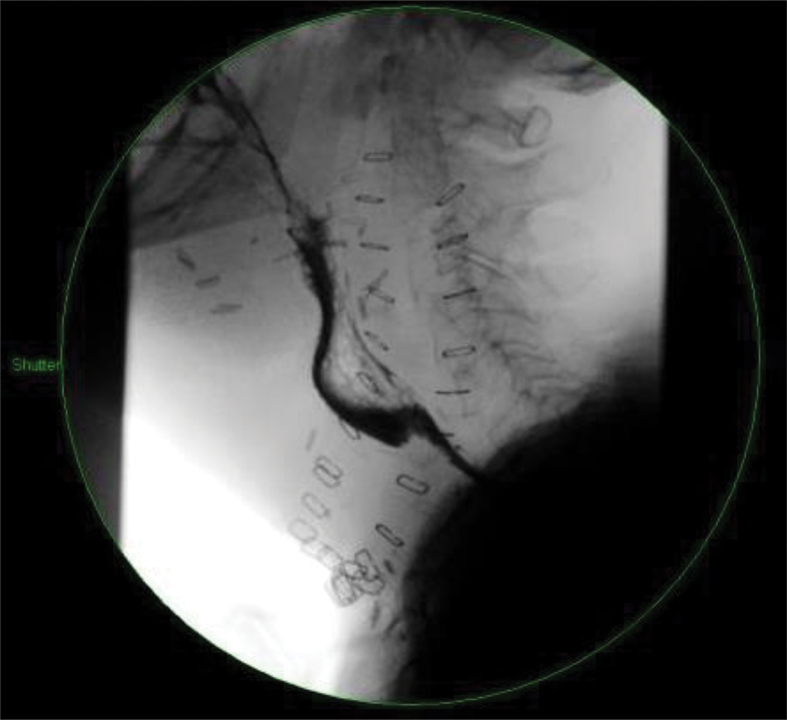
On postoperative day 8, the patient was noted to have a pus-like discharge from a previous drain site on the right side of the neck. A contrast swallow study showed that there was no leak or obstruction (Fig 1). Swabs were sent, which cultured extended-spectrum beta-lactamase Escherichia coli, and the patient was started on meropenem.
Figure 1.
Contrast swallow study
Over the following 2 weeks, the right neck drain site continued to intermittently discharge a creamy pus-like fluid. Further swabs did not culture any bacteria. Contrast swallow studies were repeated on postoperative days 13 and 16 but, again, they showed no leak or obstruction. The patient remained clinically well and was entirely asymptomatic for what was presumed to be a postoperative collection of pus in the neck.
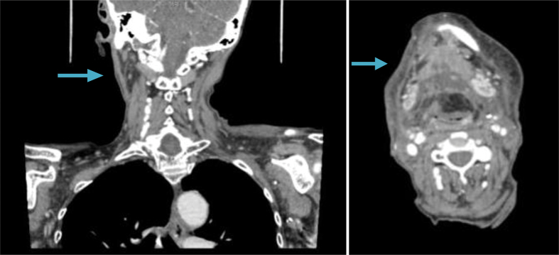
A computed tomography scan of the neck was performed on postoperative day 23, as the patient had developed a further small collection in the neck. The scan showed several small collections of fluid bilaterally (Fig 2). As the patient remained asymptomatic, these were not treated operatively and underwent observation. By postoperative day 28, the discharge had completely dried up and the patient made an uneventful recovery.
Figure 2.
Computed tomography of the neck on postoperative day 23
On further specimen review with the microbiologists, it was felt that the discharge may represent galactorrhoea, rather than pus, as no further micro-organisms were grown and the patient exhibited no signs or symptoms of an inflammatory reaction to these wound collections. The exudate was also sent to the histopathology laboratory, which showed the presence of fat globules. Immunohistochemical staining confirmed that the exudate contained fat. The pathologist, in concordance with the literature,4 reported that the combination of these two features was suggestive of galactorrhoea.
Discussion
Galactorrhoea is a relatively common condition in the female breast; however, this represents, to our knowledge, the first such case reported in the literature following the use of a pedicled pectoralis major myocutaneous flap for reconstruction of a pharyngolaryngeal defect. While this was an unusual presentation of galactorrhoea, it is important to consider all potential diagnoses when faced with postoperative complications. The pectoralis major myocutaneous flap is frequently used in head and neck reconstruction, and thus we feel it important to report this case. The diagnosis of galactorrhoea should be considered in patients with similar presentations of asymptomatic neck collections, as it may reduce both the number of unnecessary investigations and protracted hospital stays in these often high-risk cases.
Acknowledgements
None.
Conflict of interests
None.
References
- 1.Ariyan S. The pectoralis major myocutaneous flap. A versatile flap for reconstruction in the head and neck. Plast Reconstr Surg 1979; : 73–81. [DOI] [PubMed] [Google Scholar]
- 2.Ariyan S. Further experiences with the pectoralis major myocutaneous flap for the immediate repair of defects from excisions of head and neck cancers. Plast Reconstr Surg 1979; : 605–612. [PubMed] [Google Scholar]
- 3.Peña KS, Rosenfeld JA. Evaluation and treatment of galactorrhea. Am Fam Physician 2001; : 1,763–1,770. [PubMed] [Google Scholar]
- 4.Leung AK, Pacaud D. Diagnosis and management of galactorrhea. Am Fam Physician 2004; : 543–550. [PubMed] [Google Scholar]


